Editorial
. 2022 Aug 3;10(8):920-931.
doi: 10.1158/2326-6066.CIR-21-0865.

cDC1 Vaccines Drive Tumor Rejection by Direct Presentation Independently of Host cDC1

Affiliations
Editorial

cDC1 Vaccines Drive Tumor Rejection by Direct Presentation Independently of Host cDC1

Stephen T Ferris et al. Cancer Immunol Res. .

Abstract

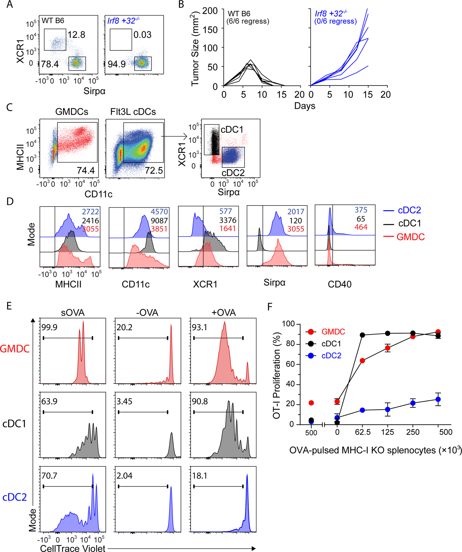
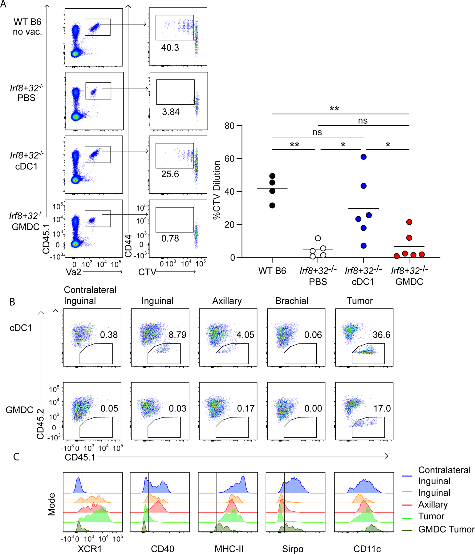
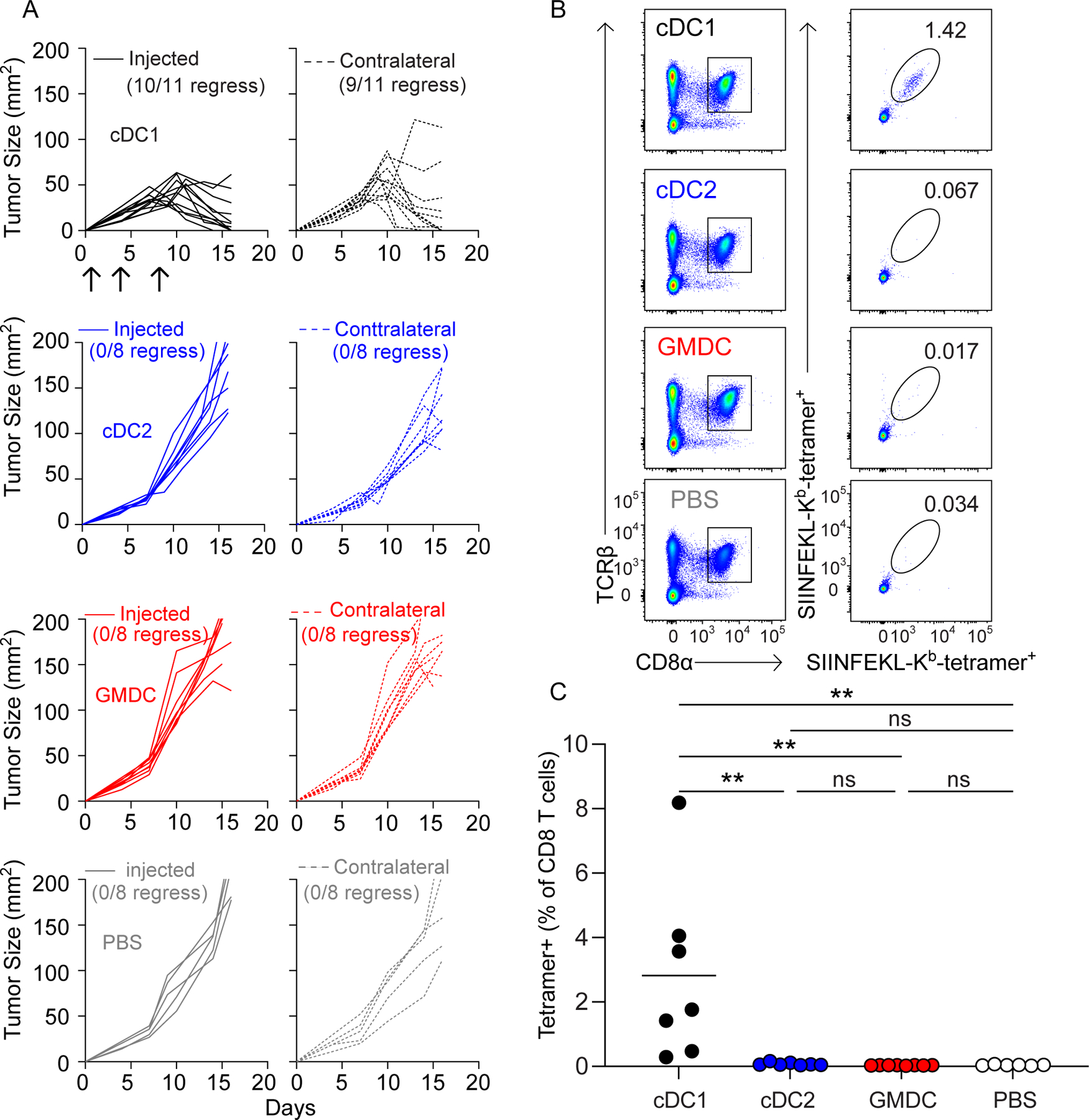
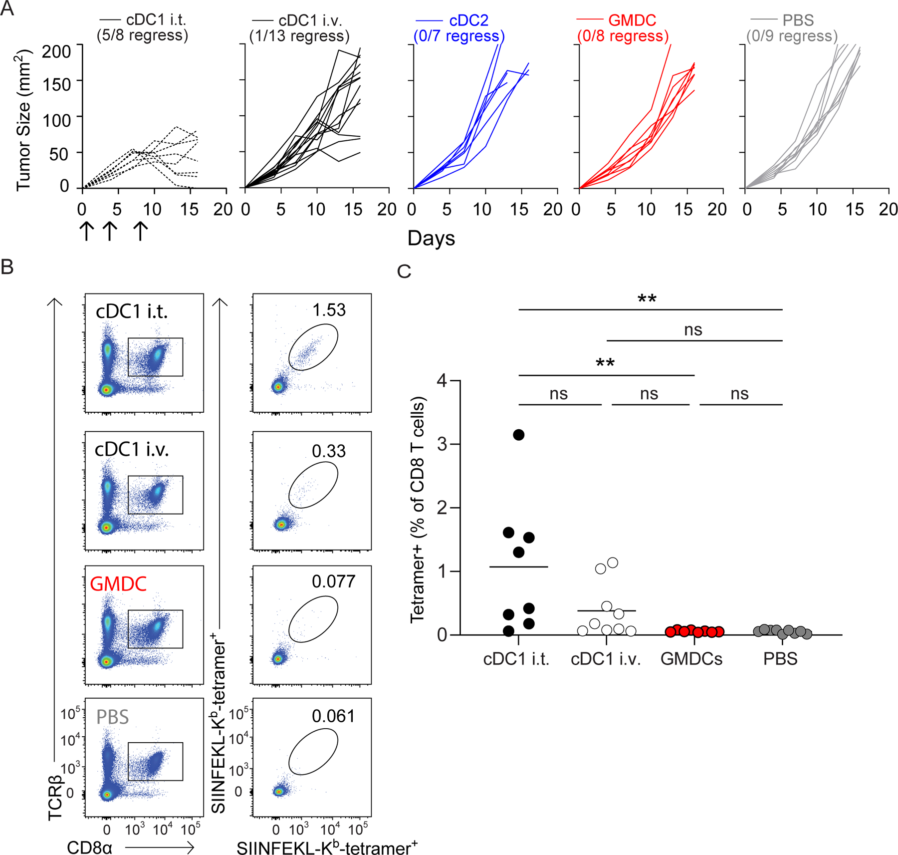
As a cell-based cancer vaccine, dendritic cells (DC), derived from peripheral blood monocytes or bone marrow (BM) treated with GM-CSF (GMDC), were initially thought to induce antitumor immunity by presenting tumor antigens directly to host T cells. Subsequent work revealed that GMDCs do not directly prime tumor-specific T cells, but must transfer their antigens to host DCs. This reduces their advantage over strictly antigen-based strategies proposed as cancer vaccines. Type 1 conventional DCs (cDC1) have been reported to be superior to GMDCs as a cancer vaccine, but whether they act by transferring antigens to host DCs is unknown. To test this, we compared antitumor responses induced by GMDCs and cDC1 in Irf8 +32-/- mice, which lack endogenous cDC1 and cannot reject immunogenic fibrosarcomas. Both GMDCs and cDC1 could cross-present cell-associated antigens to CD8+ T cells in vitro. However, injection of GMDCs into tumors in Irf8 +32-/- mice did not induce antitumor immunity, consistent with their reported dependence on host cDC1. In contrast, injection of cDC1s into tumors in Irf8 +32-/- mice resulted in their migration to tumor-draining lymph nodes, activation of tumor-specific CD8+ T cells, and rejection of the tumors. Tumor rejection did not require the in vitro loading of cDC1 with antigens, indicating that acquisition of antigens in vivo is sufficient to induce antitumor responses. Finally, cDC1 vaccination showed abscopal effects, with rejection of untreated tumors growing concurrently on the opposite flank. These results suggest that cDC1 may be a useful future avenue to explore for antitumor therapy. See related Spotlight by Hubert et al., p. 918.


Conflict of interest statement

Conflict of Interest: RDS is a co-founder, paid consultant and stockholder of Jounce Therapeutics and Asher Biotherapeutics and paid consultant and stockholder of A2 Biotherapeutics, Arch oncology, Codiak Biosciences, NGM Biotherapeutics, Sensei Biotherapeutics, and GlaxoSmithKline. KMM is a paid member of the scientific advisory board of Harbour BioMed.

Figures

Figure 1.
Figure 1.. GMDCs and Flt3L-cDC1 cross-present cell-associated antigen in vitro.
(A) Representative flow plots of 3 WT B6 (left) and 3 Irf8 +32−/− (right) splenic DCs gated as B220CD11c+MHCII+. (B) WT B6 (black, n=6) and Irf8+32−/− (blue, n=6) mice were injected with 106 1956 mOVA cells and followed for tumor growth. (C) Gating strategy for GMDCs, cDC1, and cDC2. (D) Surface expression of in vitro GMDCs (red), Flt3L-cDC1 (black), and Flt3L-cDC2 (blue). (E) Proliferation of OVA-specific OT-I CD8+ T cells in response to GMDCs (red), Flt3L-cDC1 (black), or Flt3L-cDC2 (blue) and either soluble OVA (sOVA), control PBS-loaded MHCI TKO splenocytes (-OVA), or OVA-loaded MHCI TKO splenocytes (+OVA). Shown are representative histograms of cell trace violet (CTV) intensity on OT-I cells gated as CD45.1+TCRβ+Vα2+CD44+ after 3 days of culture. (F) Proliferation of OVA-specific OT-I CD8+ T cells in response to OVA-loaded splenocytes presented by GMDCs (red), Flt3L-cDC1 (black), or Flt3L-cDC2 (blue). Representative data from one of three independent experiments are shown. Error bars represent S.D.
Figure 2.
Figure 2.. Vaccination with cDC1, but not GMDCs, induces anti-tumor immunity independently of host cDC1.
(A) Scheme of WT B6 bone marrow culture for DC injections on day 1, 4, and 8 after tumor inoculation. (B) Irf8+32−/− mice were injected with 106 1956 mOVA cells, intratumorally injected with 106 Flt3L-cDC1 (black, n=11), 106 Flt3L-cCD2 (blue, n=7), 106 GMDC (red, n=7), or PBS (grey, n=6) as indicated in (A) and followed for tumor growth. (C) Irf8+32−/− mice were injected with 106 1956 mOVA cells, and spleens were stained for the presence of SIINFEKL-Kb tetramer+CD8+ T cells on day 15 after tumor inoculation. Left, representative flow plots of percentages of tetramer+CD8+ T cells. Right, tetramer+CD8+ T cells as a percentage of all CD8+ T cells. Data represent pooled biologically independent samples from three independent experiments (n=7 for Flt3L-cDC1, n=3 for Flt3L-cDC2, n=7 for GMDCs, and n=3 for PBS). **P≤0.01, *P≤0.05, one-way ANOVA.
Figure 3.
Figure 3.. cDC1, but not GMDCs, migrate to lymph nodes to activate T cells.
(A) WT B6 and Irf8+32−/− mice were injected with 106 1956 mOVA, and OT-II cells were transferred intravenously (i.v.) on day 2 after tumor inoculation. Left, representative flow plots of proliferating OT-II T cells three days after transfer. Right, percentages of proliferating OT-II cells transferred. Data are pooled biologically independent samples from two independent experiments (n=5 for PBS Irf8+32−/−, n=4 for WT B6, and n=6 for all other groups). **P≤0.01, *P≤0.05, one-way ANOVA. (B) CD45.2+ Irf8+32−/− mice were injected with 106 1956 mOVA. On day 4 after tumor inoculation, 107 CD45.1+ Flt3L-cDC1 (top panels) or 107 CD45.1+ GMDC (bottom panels) were injected intratumorally. 40 hours after DC injection, the skin draining lymph nodes and tumors were isolated and stained for presence of injected DCs. (C) Surface expression of injected Flt3L-cDC1 in inguinal (orange), axillary (red), tumor (light green), and injected GMDCs isolated from the tumor (dark green). Contralateral lymph node DCs gated on endogenous cDC2 (blue) are shown as a comparison.
Figure 4.
Figure 4.. Flt3L-cDC1 induce abscopal tumor rejection in Irf8+32−/− mice that lack endogenous cDC1.
(A) Irf8+32−/− mice were injected in both flanks with 5 × 10⁵ 1956 mOVA and one flank received intratumoral injections of PBS (grey, n=8) or 106 GMDCs (red, n=8), Flt3L-cDC1 (black, n=11), or Flt3L-cDC2 (blue, n=8)and the other flank received no DC injections Tumor growth curves of DC-injected tumors are solid lines (left), and tumor growth curves of uninjected tumors are dashed lines (right). (B-C) Irf8+32−/− mice were injected in both flanks with 5 × 10⁵ 1956 mOVA and one flank received intratumoral injections of 106 DCs as in (A). Splenocytes were stained for the presence of SIINFEKL-Kb tetramer+CD8+ T cells on day 15 after tumor inoculation. (B) Representative flow plots of percentages of tetramer+CD8+ T cells. (C) Tetramer+CD8+ T cells as a percentage of all CD8+ T cells. Data represent pooled biologically independent samples from three independent experiments (n=7 for Flt3L-cDC1, n=7 for Flt3L-cDC2, n=9 for GMDCs, and n=6 for PBS). **P≤0.01, *P≤0.05, one-way ANOVA.
Figure 5.
Figure 5.. Intravenous vaccination with cDC1 does not induce anti-tumor immunity.
(A) Irf8+32−/− mice were injected with 106 1956 mOVA cells and followed for tumor growth. Tumor growth curves of 1956 mOVA in Irf8+32−/− mice intratumorally injected with 106 Flt3L-cDC1 (dashed black, n=8) or intravenously injected with 106 Flt3L-cDC1 (black, n=11), 106 Flt3L-cCD2 (blue, n=7), 106 GMDC (red, n=8), or PBS (grey, n=9). (B-C) Irf8+32−/− mice were injected with 106 1956 mOVA cells, and splenocytes were stained for the presence of SIINFEKL-Kb tetramer+CD8+ T cells on day 15 after tumor inoculation. (B) Representative flow plots of percentages of tetramer+CD8 T cells. (C) Tetramer+CD8+ T cells as a percentage of all CD8+ T cells. Data represent pooled biologically independent samples from three independent experiments (n = 8 for IT cDC1, n = 9 for IV cDC1, n = 8 for GMDC, and n = 9 for PBS). **P≤0.01, one-way ANOVA.

Comment in

Comment on

References

    1. Markowicz S and Engleman EG. Granulocyte-macrophage colony-stimulating factor promotes differentiation and survival of human peripheral blood dendritic cells in vitro. J Clin.Invest 1990;85:955–961. - PMC - PubMed
    1. Sims RB. Development of sipuleucel-T: autologous cellular immunotherapy for the treatment of metastatic castrate resistant prostate cancer. Vaccine 2012;30:4394–4397. - PubMed
    1. Helft J, Bottcher J, Chakravarty P, Zelenay S, Huotari J, Schraml BU et al. GM-CSF Mouse Bone Marrow Cultures Comprise a Heterogeneous Population of CD11c(+)MHCII(+) Macrophages and Dendritic Cells. Immunity 2015;42:1197–1211. - PubMed
    1. Briseno CG, Haldar M, Kretzer NM, Wu X, Theisen DJ, KC W et al. Distinct Transcriptional Programs Control Cross-Priming in Classical and Monocyte-Derived Dendritic Cells. Cell Rep 2016;15:2462–2474. - PMC - PubMed
    1. Inaba K, Inaba M, Romani N, Aya H, Deguchi M, Ikehara S et al. Generation of large numbers of dendritic cells from mouse bone marrow cultures supplemented with granulocyte/macrophage colony-stimulating factor. J Exp.Med 1992;176:1693–1702. - PMC - PubMed