. 2022;34(7-8):200-218.
doi: 10.1080/08958378.2022.2081386. Epub 2022 Jun 1.

Pulmonary toxicity and gene expression changes in response to whole-body inhalation exposure to multi-walled carbon nanotubes in rats

Affiliations

Pulmonary toxicity and gene expression changes in response to whole-body inhalation exposure to multi-walled carbon nanotubes in rats

Tina M Sager et al. Inhal Toxicol. 2022.

Abstract

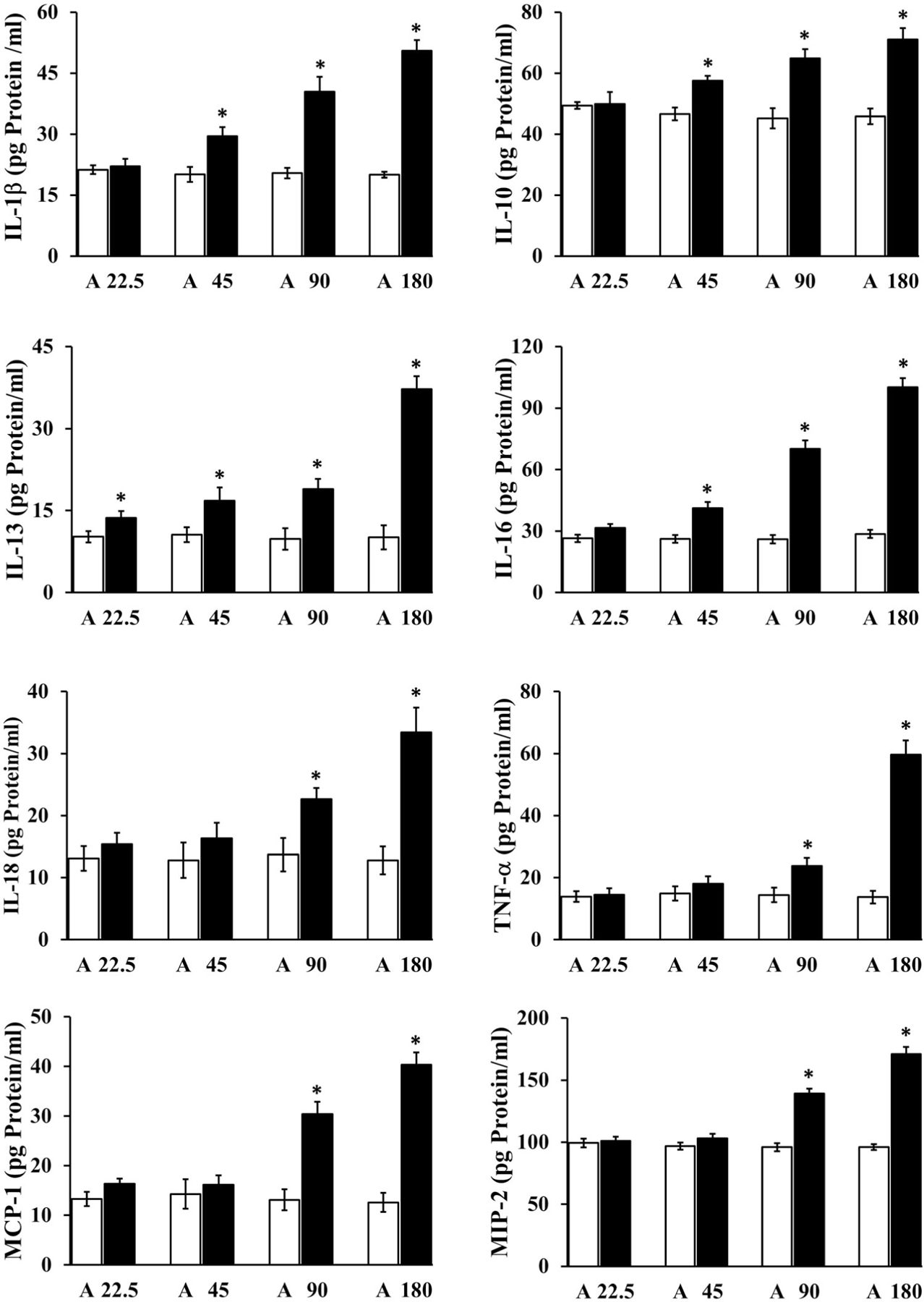
Purpose: To investigate the molecular mechanisms underlying the pulmonary toxicity induced by exposure to one form of multi-walled carbon nanotubes (MWCNT-7).Materials and methods: Rats were exposed, by whole-body inhalation, to air or an aerosol containing MWCNT-7 particles at target cumulative doses (concentration x time) ranging from 22.5 to 180 (mg/m3)h over a three-day (6 hours/day) period and toxicity and global gene expression profiles were determined in the lungs.Results: MWCNT-7 particles, associated with alveolar macrophages (AMs), were detected in rat lungs following the exposure. Mild to moderate lung pathological changes consisting of increased cellularity, thickening of the alveolar wall, alveolitis, fibrosis, and granuloma formation were detected. Bronchoalveolar lavage (BAL) toxicity parameters such as lactate dehydrogenase activity, number of AMs and polymorphonuclear leukocytes (PMNs), intracellular oxidant generation by phagocytes, and levels of cytokines were significantly (p < 0.05) increased in response to exposure to MWCNT-7. Global gene expression profiling identified several significantly differentially expressed genes (fold change >1.5 and FDR p value <0.05) in all the MWCNT-7 exposed rats. Bioinformatic analysis of the gene expression data identified significant enrichment of several diseases/biological function categories (for example, cancer, leukocyte migration, inflammatory response, mitosis, and movement of phagocytes) and canonical pathways (for example, kinetochore metaphase signaling pathway, granulocyte and agranulocyte adhesion and diapedesis, acute phase response, and LXR/RXR activation). The alterations in the lung toxicity parameters and gene expression changes exhibited a dose-response to the MWCNT exposure.Conclusions: Taken together, the data provided insights into the molecular mechanisms underlying the pulmonary toxicity induced by inhalation exposure of rats to MWCNT-7.

Keywords: Multi-walled carbon nanotubes; fibrosis; inflammation; lung toxicity; molecular mechanisms.


Conflict of interest statement

Disclosure statement

No potential conflict of interest was reported by the author(s). The findings and conclusions in this report are those of the authors and do not necessarily represent the official position of the National Institute for Occupational Safety and Health, Centers for Disease Control and Prevention. The Next Generation Sequence data discussed in this publication have been deposited in NCBI’s Gene Expression Omnibus (GEO) and are accessible through GEO Series accession number GSE148869.

Figures

Figure 1.
Figure 1.
Size distribution and morphology of MWCNT-7 particles in the aerosol generated for rat whole-body inhalation exposure. An aerosol containing MWCNT-7 particles was generated and used for inhalation exposure of rats. The aerosol generated was analyzed for particle size distribution using a MOUDI (A) and for particle size using an electron microscope (B) as described in the text. DAE: aerodynamic diameter; dC: concentration on each MOUDI stage; C: total concentration on all MOUDI stages.
Figure 2.
Figure 2.
Deposition of MWCNT-7 particles in rat lungs and uptake by alveolar macrophages. Rats were exposed by whole body inhalation to air, or an aerosol containing MWCNT-7 and euthanized as described in detail in the text. Sections of unlavaged right lung lobes prepared were observed under a light microscope to detect MWCNT-7 particles deposited in the lungs. Similarly, microscope slides containing BAL cells obtained following lavage of the left lungs were observed under a light microscope to detect MWCNT-7 particles (indicated by arrows in figure D). The lung sections and BAL samples obtained from rats exposed to air or 90 (mg/m3)h MWCNT are presented as representatives.
Figure 3.
Figure 3.
Photomicrographs of lung sections from the control (air) and MWCNT-7 exposed rats. Lung sections prepared following euthanasia of the air or MWCNT-7 exposed rats were stained with hematoxylin and eosin and observed under a light microscope. Lungs of the air exposed rats showed normal histology (A). Except for occasional accumulation of pigmented nanoparticles in the alveolar lumina or inside macrophages (asterisk), no parenchymal changes were seen with the lower dose group of 45 (mg/m3)h (B). With higher doses [90 (mg/m3)h (C) and 180 (mg/m3)h (D)], early alveolitis, seen as mild alveolar wall thickening and increased cellularity was noted (arrows), together with the presence of very early intra-alveolar granulomas (g) and numerous deposits of black pigmented MWCNT-7 particles (asterisk). The parenchymal changes were more obvious in the highest dose group (D). In all panels, bar = 50 μm and magnification = 40×.
Figure 4.
Figure 4.
Bronchoalveolar lavage parameters of toxicity in the rat lungs. The lungs of the control and MWCNT-7 exposed rats were lavaged and BAL cells and BAL fluid were isolated and analyzed for BAL parameters of toxicity, viz. LDH (I), total BAL cells (II), macrophages (III) and (PMN (IV).The filled and open bars correspond to the MWCNT-7 exposed and the respective control (air) groups of rats. The asterisk represents the statistical significance of the difference (p < 0.05) between the MWCNT-7 exposed and the corresponding control groups (n = 12).
Figure 5.
Figure 5.
Intracellular oxidants generated by the lung phagocytes in rats. The BAL cells obtained from the air or MWCNT-7 exposed rat lungs, following euthanasia, were analyzed for chemiluminescence representing the generation of intracellular reactive oxidants. Intracellular oxidants generated by the AMs only (I) and AMs and PMNs (II) are presented in the MWCNT exposed (filled bar) and corresponding control groups (A, open bar). The asterisk represents the statistical significance of the difference (p < 0.05) between the MWCNT-7 exposed and the corresponding control groups (n = 12).
Figure 6.
Figure 6.
Cytokine analysis in the lungs of the rats. The BAL fluid samples obtained from the air or MWCNT exposed lungs, following euthanasia of the rats, were analyzed and the protein corresponding to the individual cytokines were quantified by ELISA. The filled and open bars represent the MWCNT-7 exposed and the corresponding air exposed control (A) groups of rats, respectively. Data is presented as mean ± S.E. (n = 5 or 6) and the asterisks represent the statistical significance (p < 0.05) of the data between the MWCNT-7 exposed and the corresponding air exposed groups of rats.
Figure 7.
Figure 7.
Gene expression profile in rat lungs in response to exposure to MWCNT-7. Total RNA isolated from the control and MWCNT exposed lungs obtained from the euthanized rats was analyzed for global gene expression profile and the significantly differentially expressed (total, up-regulated, and down-regulated) genes were identified as described in the Materials and Methods section.
Figure 8.
Figure 8.
Biological functions and canonical pathways enriched in the lungs of the rats. Total RNA isolated from the air or MWCNT-7 exposed lungs obtained from the euthanized rats were analyzed for global gene expression profile and the SDEGs were identified as described in the Materials and Methods section. The SDEGs were used as input in the Ingenuity Pathway Analysis program and the classification categories significantly enriched in response to MWCNT-7 exposure in the rats were identified. Ten of the significantly enriched biological functions (A) and canonical pathways (B) and the number of SDEGs belonging to those categories are presented.

References

    1. Alfadda AA, Fatma S, Chishti MA, Al-Naami MY, Elawad R, Mendoza CD, Jo H, Lee YS. 2012. Orosomucoid serum concentrations and fat depot-specific mRNA and protein expression in humans. Mol Cells 33(1):35–41. - PMC - PubMed
    1. Andrews S. 2010. FastQC: a quality control tool for high throughput sequence data http://www.bioinformatics.babraham.ac.uk/projects/fastqc.
    1. Anjilvel S, Asgharian B. 1995. A multiple-path model of particle deposition in the rat lung. Fundam Appl Toxicol 28(1):41–50. - PubMed
    1. Asakura M, Sasaki T, Sugiyama T, Takaya M, Koda S, Nagano K, Arito H, Fukushima S. 2010. Genotoxicity and cytotoxicity of multi-wall carbon nanotubes in cultured Chinese hamster lung cells in comparison with chrysotile A fibers. J Occup Health 52(3):155–166. - PubMed
    1. Bai T, Zhao Y, Liu Y, Cai B, Dong N, Li B. 2019. Effect of KNL1 on the proliferation and apoptosis of colorectal cancer cells. Technol Cancer Res Treat 18:1533033819858668. - PMC - PubMed

Publication types

Substances