. 2022 Jun 14;119(24):e2203176119.
doi: 10.1073/pnas.2203176119. Epub 2022 Jun 1.

Co-component signal transduction systems: Fast-evolving virulence regulation cassettes discovered in enteric bacteria

Affiliations

Co-component signal transduction systems: Fast-evolving virulence regulation cassettes discovered in enteric bacteria

Lisa N Kinch et al. Proc Natl Acad Sci U S A. .

Abstract

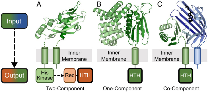
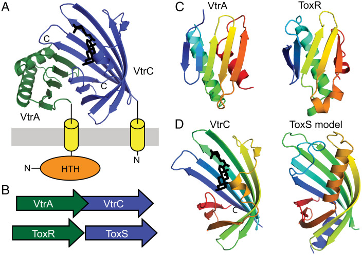
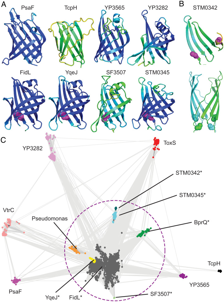
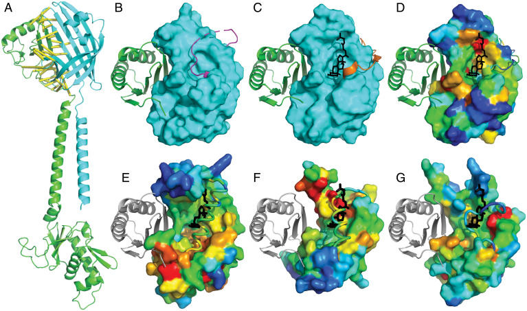
Bacterial signal transduction systems sense changes in the environment and transmit these signals to control cellular responses. The simplest one-component signal transduction systems include an input sensor domain and an output response domain encoded in a single protein chain. Alternatively, two-component signal transduction systems transmit signals by phosphorelay between input and output domains from separate proteins. The membrane-tethered periplasmic bile acid sensor that activates the Vibrio parahaemolyticus type III secretion system adopts an obligate heterodimer of two proteins encoded by partially overlapping VtrA and VtrC genes. This co-component signal transduction system binds bile acid using a lipocalin-like domain in VtrC and transmits the signal through the membrane to a cytoplasmic DNA-binding transcription factor in VtrA. Using the domain and operon organization of VtrA/VtrC, we identify a fast-evolving superfamily of co-component systems in enteric bacteria. Accurate machine learning–based fold predictions for the candidate co-components support their homology in the twilight zone of rapidly evolving sequences and provide mechanistic hypotheses about previously unrecognized lipid-sensing functions.

Keywords: co-component signal transduction system; enteric bacteria; protein sequence evolution; protein structure prediction; virulence transcription regulation.


Conflict of interest statement

The authors declare no competing interest.

Figures

Fig. 1.
Fig. 1.
Three transmembrane signaling systems found in enteric bacteria. Extracellular inputs (Top, cartoon color scale from dark N terminus to light C terminus) and cytoplasmic outputs (Bottom) are depicted for three representative types of bacterial signaling systems. (A) Two-component PhoQ (Protein Data Bank (PDB): 1yax) periplasmic input sensor domain (green) is attached through a TMH to an intracellular histidine kinase that signals to a second component, the PhoP output response regulator. (B) One-component CadC (PDB: 3ly7) periplasmic domain (green) is attached through a TMH to the output HTH domain. (C) Co-component VtrA/VtrC (PDB: 5kew) periplasmic input domains (green/blue), with VtrC attached through a TMH to the output HTH domain.
Fig. 2.
Fig. 2.
VtrA/VtrC T3SS2 virulence regulon is homologous to ToxR/ToxS. (A) The VtrA/VtrC structure (PDB: 5kew) depicted in cartoon adopts an obligate heterodimer of the periplasmic VtrA (green) and VtrC (blue) that transmits an input signal from VtrC-bound bile acid (black stick) through the membrane (gray) via the TMH (yellow cylinder) attached to the N-terminal VtrA DNA-binding HTH (orange sphere). (B) The VtrC gene (green arrow) encoding the coregulator is downstream from the VtrA gene (blue arrow) encoding the transcription factor in an overlapping operon that is arranged similarly to the genes of the ToxR (green arrow) and ToxS (blue arrow) operon. (C) The labeled VtrA periplasmic domain, colored in rainbow from the N terminus (blue) to the C terminus (red), adopts the same fold as the labeled ToxR periplasmic domain (PDB: 6utc). (D) The labeled VtrC periplasmic domain that binds the bile acid TDC (black stick) adopts the same fold as the predicted ToxS structure model.
Fig. 3.
Fig. 3.
Lipocalin-like folds unite diverse VtrC-like members. (A) Models for VtrC-like representatives are colored in rainbow scale from blue (highest confidence) to red (lowest confidence; note the confidence is too high to see any red), with Cys disulfides in magenta sphere. (B) STM0342 monomer (Top, depicted as in A) and dimer model (Bottom, cyan and green) with Cys residues (sphere). (C) VtrC-like sequences (nodes, colored according to family and labeled) are clustered with CLANS in two dimensions. Connecting lines denote similarity between nodes (< 0.0001 BLAST E-value cutoff). Families marked with (*) can be linked by sequence using PSI-BLAST and are circled with a dotted magenta line.
Fig. 4.
Fig. 4.
Functional implications of VtrA/VtrC superfamily fold prediction. (A) GrvA (green cartoon) complex model with FidL (cyan cartoon). Confidently predicted residue–residue contacts are connected by yellow bars (residue–residue distance in structure model ≤ 8 Å and predicted aligned error ≤ 4 Å). (B) VtrA (green cartoon) experimental structure bound to ligand-free VtrC (cyan surface), with a mobile loop (magenta) covering the lipid binding site. (C) VtrA/VtrC experimental structure bound to TDC (black stick) displaces the mobile loop (orange). (D) VtrC surface is colored in rainbow by family-based residue conservation from low (blue) to high (red). (E) ToxS, (F) YP3282, and (G) BprQ structure models (rainbow conservation) are superimposed with VtrC to highlight relative positions of VtrA (gray cartoon) and TDC (black stick). The corresponding mobile loops (cartoon tubes) cover the potential lipid binding sites as in the apo VtrA/VtrC structure.
Fig. 5.
Fig. 5.
VtrA periplasmic domain evolution. (A–D) Periplasmic domain SSEs are in rainbow cartoon from the N terminus (blue) to the C terminus (red), with disulfides in black sphere. (A) CadC periplasmic domain has an insertion (magenta) with respect to (B) the VtrA periplasmic domain, with the CadC insertion replaced by an N-terminal extension (slate). (C) Candidate co-component VtrA-like models are colored by the core SSEs in CadC and VtrA. (D) A conserved disulfide (*) links YqeI and TcpP, which includes a C-terminal extension (gray). (E) Representative operon ORFs (labeled by gene) with conserved C-terminal Cys codon (red) overlapping with start codon from input component. (F) Distance matrix of all-against-all structure comparisons colored by distance in red (distant)-white-blue (identical) color scale. Components that interact with confident sequence-related VtrC-like domains are bolded, and their distances are boxed.

References

    1. Matilla M. A., Ortega Á., Krell T., The role of solute binding proteins in signal transduction. Comput. Struct. Biotechnol. J. 19, 1786–1805 (2021). - PMC - PubMed
    1. Park C., Hazelbauer G. L., Mutations specifically affecting ligand interaction of the Trg chemosensory transducer. J. Bacteriol. 167, 101–109 (1986). - PMC - PubMed
    1. Cho U. S., et al. , Metal bridges between the PhoQ sensor domain and the membrane regulate transmembrane signaling. J. Mol. Biol. 356, 1193–1206 (2006). - PubMed
    1. Groisman E. A., The pleiotropic two-component regulatory system PhoP-PhoQ. J. Bacteriol. 183, 1835–1842 (2001). - PMC - PubMed
    1. Hoch J. A., Two-component and phosphorelay signal transduction. Curr. Opin. Microbiol. 3, 165–170 (2000). - PubMed

Publication types

MeSH terms

LinkOut - more resources