. 2021 Dec 15;29(4):581-605.
doi: 10.38212/2224-6614.3377.

Medicarpin isolated from Radix Hedysari ameliorates brain injury in a murine model of cerebral ischemia

Affiliations

Medicarpin isolated from Radix Hedysari ameliorates brain injury in a murine model of cerebral ischemia

Chang-Ming Chern et al. J Food Drug Anal. .

Abstract

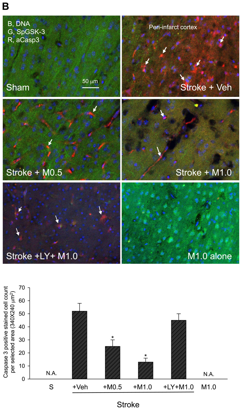
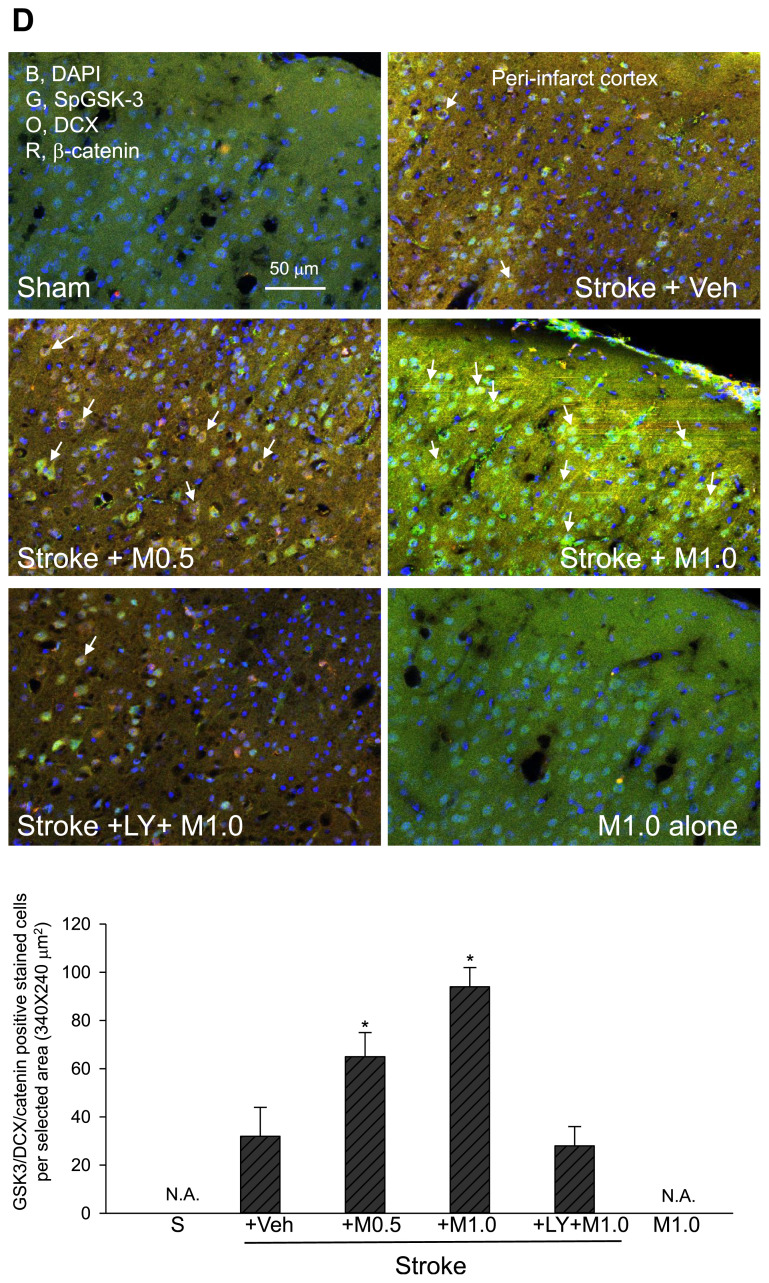
The development of effective post-stroke therapy is highly demanded. Medicarpin is a key active component of a famous Chinese herbal prescription used for post-stroke treatment in Taiwan; however, little is known about its biological effects and mechanisms of action. Herein, we implemented a murine model of cerebral ischemic/reperfusional injury-related stroke to elucidate medicarpin's neuroprotective effect. In male ICR mice 24 h after stroke induction, treatment with medicarpin (0.5 and 1.0 mg/kg, i.v.) markedly enhanced the survival rates, improved moving distance and walking area coverage, reduced brain infarction, and preserved the blood-brain barrier, supporting medicarpin's protective effect on stroke-induced injury. Immunohistochemistry analysis further revealed that medicarpin treatment decreased the expression/activation of p65NF-κB and caspase 3, especially near the infarct cortex, while promoting the expression of neurogenesis-associated proteins, including doublecortin (DCX), brain-derived neurotrophic factor (BDNF), and tyrosine receptor kinase B (TrkB). These changes of expression levels were accompanied by GSK-3 inactivation and β-catenin upregulation. Notably, pretreatment with LY294002, a PI3K inhibitor, abolished the aforementioned beneficial effects of medicarpin, illustrating an essential role of PI3K/Akt activation in medicarpin's neuroprotective and reparative activities. In vitro studies revealed that medicarpin displayed strong anti-inflammatory activity by reducing nitric oxide (NO) production in lipopolysaccharide-stimulated microglial cells (BV2) with an IC50 around 5 ±1 (μM) and anti-apoptotic activity in neuronal cells (N2A) subjected to oxygen-glucose deprivation with an IC50 around 13 ± 2 (μM). Collectively, this is the first report to demonstrate that medicarpin, isolated from Radix Hedysari, ameliorates ischemic brain injury through its anti-inflammatory microglia/NO), anti-apoptotic (neuronal cells/OGD) and neuroprotective effects by activating the PI3K/Akt-dependent GSK-3 inactivation for upregulating β-catenin, which in turn decreases the expression/activation of p65NF-κB and caspase 3 and promotes the expression of neurogenic (DCX, BDNF, TrkB) and neuroprotective (Bcl2) factors in the brain.


Conflict of interest statement

Conflict of interest

The authors declare that they have no conflict of interest.

Figures

Fig. 1
Fig. 1
(A) General experimental protocol and (B) Chemical structure of medicarpin (MW: 270.3).
Fig. 2
Fig. 2
Effect of medicarpin on the changes in survival rates (over 3 days) after cerebral ischemia-reperfusional injury (stroke). Survival curves were assessed from stroke animals divided into various sub-groups as described below using the Log–Rank test followed by the Holm-Sidak method for all pairwise multiple comparisons (n = 10 for each group in the beginning). *,†p < 0.05, as compared with corresponding vehicle-treated stroke group (Stroke + Veh) or Sham. Animal sub-groups: (1 and 2) a sham-operated group (sham) and a medicarpin (at 1.0 mg/kg, i.v.)-treated alone without stroke induction (M1.0 alone), (3 and 4) two medicarpin (at 0.5 mg/kg or 1.0 mg/kg, 2 h after stroke induction, i.v.)-treated groups with stroke (Stroke + M0.5 (or M1.0)), and (5 and 6) a vehicle (normal saline with 0.1% of dimethyl sulfoxide (DMSO))-treated group with stroke (Stroke + Veh) or a LY294002 (at 1.0 mg/kg, i.p., 30 min before medicarpin (at 1.0 mg/kg)) plus medicarpin (at 1.0 mg/kg, 2 h after stroke, i.v.)-treated group with stroke (Stroke + LY + M1.0).
Fig. 3
Fig. 3
Effects of medicarpin on the changes in neurological deficits (at 24 h) after cerebral ischemia-reperfusional injury (stroke). Upper panel, typical animal tracking (moving) profiles within 3 min for evaluation of neurological deficits; statistical results of the neurological deficits analysis were yielded by measuring the moving distance (in meter (m)) (middle panel) and moving area coverage (A%) (lower pannel). *,†p < 0.05, compared with the corresponding vehicle-treated stroke (Stroke + Veh) or Sham group by one-way ANOVA followed by S–N–K t-test. Animal sub-groups are described in Fig. 2.
Fig. 4
Fig. 4
Effects of medicarpin on the changes in brain infarction 24 h after cerebral ischemia-reperfusional injury (stroke). Upper panel, typical brain infarction analysis (by TTC stains); lower panel, statistical summary. N.A., data not available. *p < 0.05, compared with the corresponding vehicle-treated stroke (Stroke + Veh) group by one-way ANOVA followed by S–N–K t-test. Animal sub-groups are described in Fig. 2.
Fig. 5
Fig. 5
Effects of medicarpin on the changes in the expression levels of protein markers 24 h after cerebral ischemia-reperfusional injury (stroke) by confocal image analysis. (A) BBB damage and inflammation. Upper panel, adopted from the ipsilateral ischemic hemisphere (bregma −1.5 to −1.9 mm) for assessing BBB and tissue damage (loss of occludin, green, G), inflammation (p65NF-κB expression (red, R, as arrows indicated), and DAPI (blue, B, nuclei); square boxes indicate peri-infarct cortex and dentate gyrus (DG) of hippocampus, respectively. Lower panel, statistical summary. (B) GSK-3 inactivation and apoptosis. Upper panel, adopted from peri-infarct cortex for detecting GSK-3 inactivation by the levels of phospho-S9-GSK-3β (SpGSK-3, green, G), apoptosis by the levels of activated caspase 3 (aCasp3, red, R, as arrows indicated). DAPI staining (blue, B) indicates the position of cell nuclei. Lower panel, statistical summary. (C) Neurogenesis marker. Upper panel, adopted from dentate gyrus (DG) of hippocampus for examining neurogenesis (doublecortin (DCX), orange, O, as arrows indicated) and colocalization of GSK-3 inactivation (phospho-S9-GSK-3β (SpGSK-3), green, G), β-catenin expression (red, R), and DAPI (blue, B, nuclei). Lower panel, statistical summary. (D) GSK-3 inactivation, β-catenin expression and neurogenesis. Upper panel, adopted from peri-infarct cortex for colocalization (as arrows indicated) of GSK-3 inactivation (phospho-S9-GSK-3β (SpGSK-3), green, G), β-catenin expression (red, R), neurogenesis (doublecortin (DCX), orange, O), and DAPI (blue, B, nuclei). Lower panel, statistical summary. (E) TrkB expression. Upper, TrkB (green, G), β-catenin (orange, O), activated caspase 3 (aCasp3, red, R), and DAPI (blue, B, nuclei); stars indicated strong staining and colocalization of TrkB and β-catenin. Lower, statistical summary. Animal sub-groups are described in Fig. 2.
Fig. 5
Fig. 5
Effects of medicarpin on the changes in the expression levels of protein markers 24 h after cerebral ischemia-reperfusional injury (stroke) by confocal image analysis. (A) BBB damage and inflammation. Upper panel, adopted from the ipsilateral ischemic hemisphere (bregma −1.5 to −1.9 mm) for assessing BBB and tissue damage (loss of occludin, green, G), inflammation (p65NF-κB expression (red, R, as arrows indicated), and DAPI (blue, B, nuclei); square boxes indicate peri-infarct cortex and dentate gyrus (DG) of hippocampus, respectively. Lower panel, statistical summary. (B) GSK-3 inactivation and apoptosis. Upper panel, adopted from peri-infarct cortex for detecting GSK-3 inactivation by the levels of phospho-S9-GSK-3β (SpGSK-3, green, G), apoptosis by the levels of activated caspase 3 (aCasp3, red, R, as arrows indicated). DAPI staining (blue, B) indicates the position of cell nuclei. Lower panel, statistical summary. (C) Neurogenesis marker. Upper panel, adopted from dentate gyrus (DG) of hippocampus for examining neurogenesis (doublecortin (DCX), orange, O, as arrows indicated) and colocalization of GSK-3 inactivation (phospho-S9-GSK-3β (SpGSK-3), green, G), β-catenin expression (red, R), and DAPI (blue, B, nuclei). Lower panel, statistical summary. (D) GSK-3 inactivation, β-catenin expression and neurogenesis. Upper panel, adopted from peri-infarct cortex for colocalization (as arrows indicated) of GSK-3 inactivation (phospho-S9-GSK-3β (SpGSK-3), green, G), β-catenin expression (red, R), neurogenesis (doublecortin (DCX), orange, O), and DAPI (blue, B, nuclei). Lower panel, statistical summary. (E) TrkB expression. Upper, TrkB (green, G), β-catenin (orange, O), activated caspase 3 (aCasp3, red, R), and DAPI (blue, B, nuclei); stars indicated strong staining and colocalization of TrkB and β-catenin. Lower, statistical summary. Animal sub-groups are described in Fig. 2.
Fig. 6
Fig. 6
Effects of medicarpin on the changes in the expression levels of proteins in neurogenesis-promoting-associated signaling 24 h after stroke injury. (A) Upper, typical Western blot analysis using tissues from the ischemic (ipsilateral) brain hemisphere was conducted to show the changes in the levels of occludin, phospho-Akt (pAkt), phospho-S9-GSK-3β (SpGSK-3), β-catenin, Bcl-2, and BDNF. β-actin was included as a reference for normalization. Lower, statistical results from the densitometric measurements after normalization against β-actin. Animal sub-groups are described in Fig. 2.
Fig. 7
Fig. 7
A schematic illustration showing the key pathways regulated by medicarpin for achieving its neuroprotective and neurogenesis-promoting effects. Ischemic stroke induced PI3K/Akt inhibition due to excess release of glutamate (excitotoxicity). Medicarpin triggers the activation of PI3K/Akt to inactivate GSK-3 for upregulating β-catenin, which then promotes neurogenesis by stimulating the expression of neuroprotective and neurogenesis proteins, including occludin, Bcl-2, DCX, BDNF, and TrkB.
None
None
None
None

References

    1. Katan M, Luft A. Global burden of stroke. Semin Neurol. 2018;38:208–11. - PubMed
    1. Zhou Z, Lu J, Liu WW, Manaenko A, Hou X, Mei Q, et al. Advances in stroke pharmacology. Pharmacol Ther. 2018;191:23–42. - PubMed
    1. Pena ID, Borlongan C, Shen G, Davis W. Strategies to extend thrombolytic time window for ischemic stroke treatment: an unmet clinical need. J Stroke. 2017;19:50–60. - PMC - PubMed
    1. Wang YH, Chern CM, Liou KT, Kuo YH, Shen YC. Ergostatrien-7,9(11),22-trien-3beta-ol from Antrodia camphorata ameliorates ischemic stroke brain injury via downregulation of p65NF-kappa-B and caspase 3, and activation of Akt/GSK3/catenin-associated neurogenesis. Food Funct. 2019;10:4725–38. - PubMed
    1. Hou Y, Wang K, Wan W, Cheng Y, Pu X, Ye X. Resveratrol provides neuroprotection by regulating the JAK2/STAT3/PI3K/AKT/mTOR pathway after stroke in rats. Genes Dis. 2018;5:245–55. - PMC - PubMed

MeSH terms