

Imaging Dopaminergic Neurotransmission in Neurodegenerative Disorders
- PMID: 35649651
- PMCID: PMC9165729
- DOI: 10.2967/jnumed.121.263197
Imaging Dopaminergic Neurotransmission in Neurodegenerative Disorders
Abstract
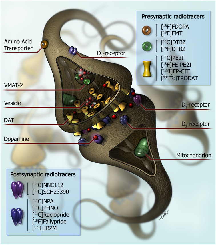
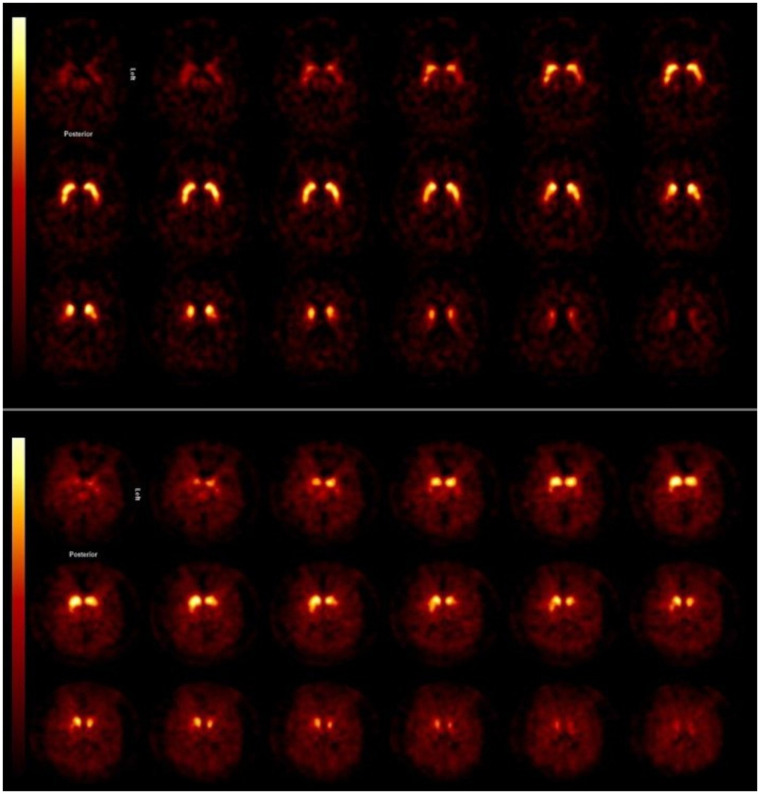
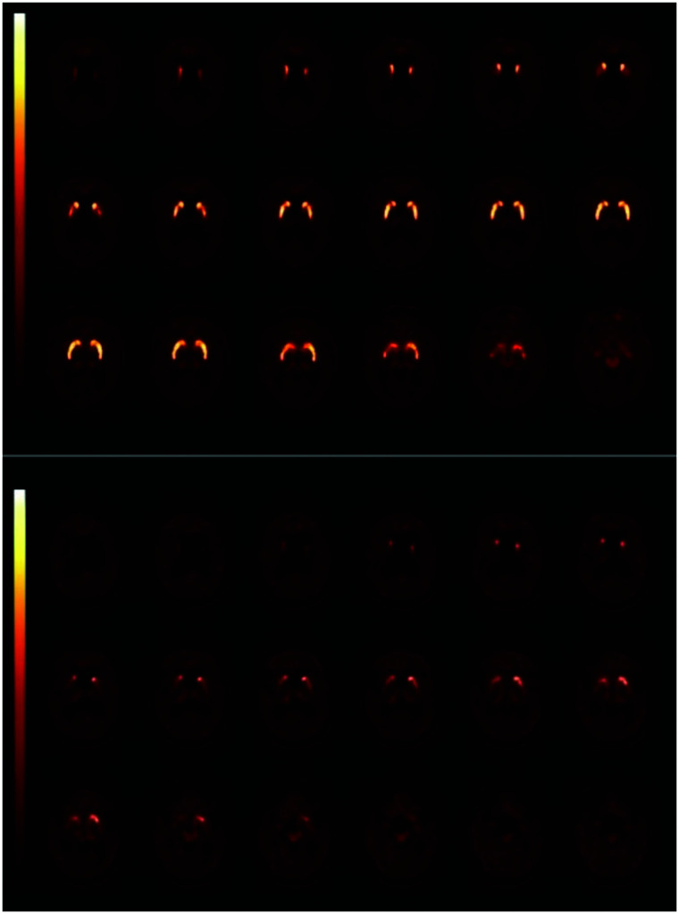
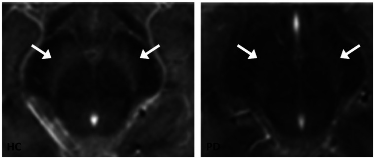
Imaging of dopaminergic transmission in neurodegenerative disorders such as Parkinson disease (PD) or dementia with Lewy bodies plays a major role in clinical practice and in clinical research. We here review the role of imaging of the nigrostriatal pathway, as well as of striatal receptors and dopamine release, in common neurodegenerative disorders in clinical practice and research. Imaging of the nigrostriatal pathway has a high diagnostic accuracy to detect nigrostriatal degeneration in disorders characterized by nigrostriatal degeneration, such as PD and dementia with Lewy bodies, and disorders of more clinical importance, namely in patients with clinically uncertain parkinsonism. Imaging of striatal dopamine D2/3 receptors is not recommended for the differential diagnosis of parkinsonian disorders in clinical practice anymore. Regarding research, recently the European Medicines Agency has qualified dopamine transporter imaging as an enrichment biomarker for clinical trials in early PD, which underlines the high diagnostic accuracy of this imaging tool and will be implemented in future trials. Also, imaging of the presynaptic dopaminergic system plays a major role in, for example, examining the extent of nigrostriatal degeneration in preclinical and premotor phases of neurodegenerative disorders and to examine subtypes of PD. Also, imaging of postsynaptic dopamine D2/3 receptors plays a role in studying, for example, the neuronal substrate of impulse control disorders in PD, as well as in measuring endogenous dopamine release to examine, for example, motor complications in the treatment of PD. Finally, novel MRI sequences as neuromelanin-sensitive MRI are promising new tools to study nigrostriatal degeneration in vivo.
Keywords: PET; Parkinson; SPECT; dopamine; neurodegeneration.
© 2022 by the Society of Nuclear Medicine and Molecular Imaging.
Figures
References
-
- Piggott MA, Marshall EF, Thomas N, et al. . Striatal dopaminergic markers in dementia with Lewy bodies, Alzheimer’s and Parkinson’s diseases: rostrocaudal distribution. Brain. 1999;122:1449–1468. - PubMed
-
- Kish SJ, Shannak K, Hornykiewicz O. Uneven pattern of dopamine loss in the striatum of patients with idiopathic Parkinson’s disease: pathophysiologic and clinical implications. N Engl J Med. 1988;318:876–880. - PubMed
-
- Kraemmer J, Kovacs GG, Perju-Dumbrava L, Pirker S, Traub-Weidinger T, Pirker W. Correlation of striatal dopamine transporter imaging with post mortem substantia nigra cell counts. Mov Disord. 2014;29:1767–1773. - PubMed
-
- Walker L, Stefanis L, Attems J. Clinical and neuropathological differences between Parkinson’s disease, Parkinson’s disease dementia and dementia with Lewy bodies: current issues and future directions. J Neurochem. 2019;150:467–474. - PubMed
-
- Kaasinen V, Vahlberg T. Striatal dopamine in Parkinson disease: a meta-analysis of imaging studies. Ann Neurol. 2017;82:873–882. - PubMed
MeSH terms
Substances
LinkOut - more resources
Full Text Sources
Medical