Review
. 2022 Nov:91:101090.
doi: 10.1016/j.preteyeres.2022.101090. Epub 2022 May 29.

Corneal stromal repair and regeneration

Affiliations
Review

Corneal stromal repair and regeneration

Rajiv R Mohan et al. Prog Retin Eye Res. 2022 Nov.

Abstract

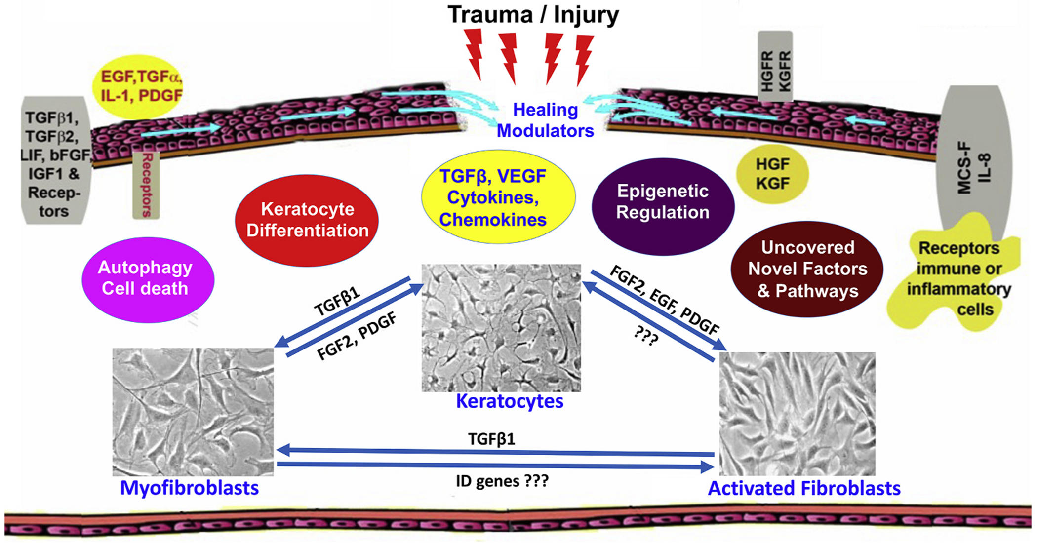
The cornea is a specialized, transparent, avascular, immune-privileged, and heavily innervated tissue that affords 2/3rd of refraction to the eye. Ocular injuries, infections, and genetic factors affect corneal function and cause vision impairment. Presently, a variety of laser/non-laser surgeries, immunosuppressants, and/or corneal transplants are predominantly used to revive sight in human patients. The development of novel, precision-guided, and tissue-targeted non-surgical therapies promoting corneal repair and regeneration based on mechanistic understanding is of paramount importance to reduce the impact of global blindness. Research over the past decade revealed that modulation of pathological signaling pathways and factors by a variety of therapeutic delivery methods effectively treats corneal disorders including corneal scar/haze, inflammation, and angiogenesis in various pre-clinical animal models and are primed for human translation. This review discusses recent advances in the areas of corneal repair, restoration, and regeneration. Herein, we provide an overview of evolving approaches and therapeutic modalities that have shown great promise in reviving corneal transparency and function through the use of small drug molecules, gene therapy, nanomedicine, stem cells, trophic factors, exosomes, stromal equivalents, bioengineered stromal scaffolds, tissue adhesives, and 3D bioprinting.

Keywords: Cornea; Corneal gene therapy; Corneal wound healing; Emerging therapies; Keratocytes; Stromal regeneration; Stromal remodeling.


Conflict of interest statement

Declaration of competing interest None.

Figures

Fig. 1.
Fig. 1.
Schematic diagram showing human corneal anatomy. The cornea is avascular and consists of the outer epithelium made up of superficial cells, wing cells, and basal cells. Epithelium forms 10% of the cornea and protects the eye. Below the epithelium are the Bowman’s layer and stroma, which forms 85–90% of the cornea and contains collagen fibrils, ECM components, keratocytes, and nerve fibers. Stroma plays an important role in corneal homeostasis, repair, and transparency maintenance. Bowman’s layer is an acellular layer with ECM. Posterior to the stroma are Descemet’s membrane and a single layer of endothelium. Descemet’s membrane provides a resting structure for the endothelial cells. The endothelium controls corneal hydration and helps maintain corneal transparency.
Fig. 2.
Fig. 2.
Representative in vivo confocal microscopy images of the corneal stroma of human subjects show the presence of only healthy quiescent keratocytes in clear/normal cornea (A), and activation, migration, proliferation, and differentiation of quiescent keratocytes, presence of activated keratocytes, fibroblasts, and myofibroblasts in hazy/opaque cornea (B–F). Corneal keratocytes are critical for stromal repair and corneal transparency.
Fig. 3.
Fig. 3.
Schematic diagram showing events related to corneal repair, remodeling, and regeneration during wound healing after ocular trauma/injury. The cornea contains an array of cytokines, chemokines, and growth factors and their receptors which facilitate stromal repair and regeneration in an injured cornea. Keratocyte apoptosis, activation, proliferation, migration, and transdifferentiation to myofibroblast are controlled by many mechanisms to facilitate stromal repair, regeneration, and restoration. Myofibroblasts are a major cell type to perform these functions. Amount and timing of appearance/disappearance of myofibroblasts during/after wound repair dictate the pathological and physiological status of the cornea.
Fig. 4.
Fig. 4.
Representative images showing involvement of epigenetic mechanism in corneal fibrosis inhibition. TSA, a well-known epigenetic modifier, significantly decreased SMA and fibronectin expression in vitro (A–C) and PRK-induced corneal haze in vivo in rabbit cornea (D–M). SMA (green), fibronectin (red), and DAPI (blue), adapted from (Sharma et al., 2009).
Fig. 5.
Fig. 5.
Representative images showing bench-to-bedside potential of SAHA, an FDA-approved epigenetic modifier. A single treatment of SAHA (25 μM) after PRK on rabbit eyes significantly prevented the development of PRK-induced corneal haze in vivo (A–F) adapted from (Tandon et al., 2012), and the underlying mechanism used by SAHA for anti-fibrotic response (G–J) adapted from (Gronkiewicz et al., 2016b). Arrows show α-SMA (green), and f-actin (red). Western blot results show MAPKs and MMPs.
Fig. 6.
Fig. 6.
Representative images profiling SAHA versus MMC efficacy (A–F) and long-term safety (G–R) in vivo in rabbits after 1-month and 4 months post-PRK. Myofibroblasts (arrows), α-SMA (green); TUNEL positive cells = red (arrows), adapted from (Anumanthan et al., 2017).
Fig. 7.
Fig. 7.
Representative data displaying efficacy and safety of SAHA, MMC, or SAHA + MMC treatment on multidrug resistance proteins and limbal stem/progenitor cells derived from donor corneas, corneoscleral rims, and lenticules collected from human subjects. Representative FACS plots and quantification of ABCG2 (multidrug resistance protein) expression after SAHA, MMC, or SAHA + MMC treatment on cultured limbal epithelial cells differentiated to corneal lineage (A–G). Western blotting and quantification of the expression of the CK3/CK12, ΔNP63, COLL4, αSMA, BCl2 and GAPDH proteins after SAHA, MMC, or SAHA + MMC on limbal cornea epithelial cells isolated from corneoscleral rims of human subjects (H–I), adapted from (Shetty et al., 2021).
Fig. 8.
Fig. 8.
Representative clinical Slit-lamp images of the clear cornea 12 months post-PRK (A), grade 2 subepithelial corneal haze 12 months post-PRK (B), densitometry mapping of corneal haze by Oculus Pentacam (C–D) in human subjects, and microarray analysis of the pooled mRNA samples from haze predisposed and control groups (E–F). In this, corneal epithelium from patients was collected at the time of surgery and grouped into those who developed post-surgical haze at 12 months (A) compared to those that healed without any complications (B). Transcriptomic and ontological analyses found 1100 genes upregulated and 1780 genes downregulated in the haze predisposed group with changes in pathways regulating inflammation, oxidative stress, nerve functions, extra cellular matrix remodeling, and Wnt signaling. Factors like PREX1, SOX17, GABRA1, WNT3A, and PXDN showing significantly altered expression in haze predisposed subjects than with those of active haze subjects provoked us to conclude their pro-fibrotic role in corneal stromal wound healing and haze development, adapted from (Kumar et al., 2019).
Fig. 9.
Fig. 9.
Representative data showing the promise of a strategy involving selective sequestering of TGF-β signaling in corneal fibrosis/haze treatment in vitro and in vivo. Significantly reduced keratocyte/fibroblast transdifferentiation to myofibroblast in vitro and in vivo (A–L) and fibroblast migration in vitro (M–R) was observed in these experiments, adapted from (Fink et al., 2015; Gupta et al., 2020b; Sharma et al., 2012; Tandon et al., 2013).
Fig. 10.
Fig. 10.
Representative data showing effects of single and combined targeting of profibrotic Smads (Smad-2, -3, or -4) and antifibrotic Smads (Smad7) on corneal fibroblast differentiation. Both, single and combined, Smad targeting suppressed corneal fibroblast differentiation but combined targeting of Smads did not improve anti-fibrotic response in equine corneal fibrosis in vitro model, adapted from (Marlo et al., 2018).
Fig. 11.
Fig. 11.
Representative data exhibiting that decorin gene overexpression in human corneal fibroblasts intercepts TGF-β-induced myofibroblast formation in vitro (A–D), adapted from (Mohan et al., 2010), and corneal haze/fibrosis in rabbit cornea in vivo (E–L) adapted from (Mohan et al., 2011b). These studies suggested that the corneal wound healing process can be easily targeted for stromal repair/regeneration and used to develop novel therapeutics to restore corneal functions. Green = SMA positive cells. E-I = representative stereomicroscope images.
Fig. 12.
Fig. 12.
Representative images showing induction of corneal neovascularization by VEGF, and its inhibition by targeted decorin gene transfer into stroma in rabbits in vivo. A controlled time-dependent in-growth of blood vessels and neovascularization increase density in the avascular cornea was observed after VEGF-pellet implanted in the stroma (A, C, E) and rabbit corneas administration of decorin gene in stroma significantly reduced corneal neovascularization (A, C, E) by modifying stromal ECM. The decorin-delivered corneas in H & E staining showed vividly recovered corneal histology (G, H), reduced expression of CD31, an angiogenic marker, protein and mRNA (I, J), and rerecovered balance in pro-and anti-angiogenic genes (K), adapted from (Mohan et al., 2011d).
Fig. 13.
Fig. 13.
Representative images detecting KCa3.1 gene expression in human corneal epithelial, fibroblast, and endothelial cells by RT-PCR (A) and donor human cornea by immunofluorescence (B). KCa3.1 deficient mice showing distinctly reduced corneal haze post alkali insult compared to wild type in a time-dependent manner (C) indicated expression and functional role of KCa3.1 in corneal wound healing, adapted from (Anumanthan et al., 2018).
Fig. 14.
Fig. 14.
Representative phase-contrast microscopic data showing therapeutic promise of KCa3.1 controlling corneal fibrosis by pharmacological agent, TRAM-34 (a selective inhibitor of KCa3.1). Human corneal fibroblasts (HCF) grown in±of TRAM-34 and TGF-β1 and the effects of TRAM-34 were evaluated on fibroblast migration (A–F) and differentiation to myofibroblast (G–K). Treatment of TRAM-34 to HCFs demonstrated reduced fibroblast migration (A–F) and α-SMA (a fibrotic marker) levels in immunostaining (green; G-I) and western blotting (J, K), adapted from (Anumanthan et al., 2018).
Fig. 15.
Fig. 15.
Representative transmission electron microscopy showing assembly, alignment, and packing of collagen fibrils in normal (A) and injured (B) mouse corneal stroma. A characteristic distribution, arrangement, and packing of collagen fibrils specific to corneal stroma were observed in normal cornea (A). On the other hand, the injured cornea demonstrated significantly altered assembly, distribution, and packing of collagen fibrils (B). Precise collagen fibrils organization is vital for maintaining corneal shape and optical property.
Fig. 16.
Fig. 16.
Quantitative real-time PCR and transmission electron microscopy analyses comparing expression of collagen (A) and fibrosis (B) related genes and collagen fibrillogenesis (C, D) in normal and diabetic pig corneas. Age-matched normal and diabetic corneas of Ossabaw mini pig, a Type 2 diabetic animal model with a “thrifty genotype” were used in the investigation. None of the pig corneas showed any clinically relevant corneal haze. Nonetheless, detection of an altered expression of wound healing genes and mild change in stromal collagen fibrillogenesis divulges the vulnerability of the cornea to a diabetic condition, adapted from (Gupta et al., 2022).
Fig. 17.
Fig. 17.
Transmission electron microscopy showing the role of decorin in corneal stromal collagen fibrillogenesis in decorin deficient transgenic mice ± injury. The injured corneal stroma showed altered collagen fibrils arrangement and packing than the uninjured corneas. Violin graphs show inter-fibril distances (D), adapted from (Gupta et al., 2022).
Fig. 18.
Fig. 18.
Schematic depicting the role of the immune cell machinery in corneal wound healing. Upon injury, the immediate response begins with mast cell degranulation and immediate activation of neutrophils and the pro-inflammatory M1 macrophages. During the early phase, neutrophils, macrophages, and γδ-T cells migrate to the injury site and continue to reduce during the reparative phase. At this time, the wound is being actively healed via tissue remodeling. During this entire process, the dendritic cells are also activated and further interact with various immune cells during both the initial response and resolution phases. The anti-inflammatory M2 macrophages appear during the resolution phase and are important for adequate wound closure and completion of the healing process and return of corneal clarity. The imbalance within these cellular players as well as those not depicted here is often a driving factor for corneal scarring and fibrosis. Therefore, immunomodulation could play a critical role in optimal corneal wound healing.
Fig. 19.
Fig. 19.
En face images of corneas from human subjects affected by fibrosis/haze of diverse etiologies. (A) Corneal Keratitis scar, (B) Scarring post corneal repair after trauma, (C) Diffuse corneal opacity with limbal stem cell deficiency post alkali burns, (D) Opacification of transplanted graft, (E) Corneal hydrops in advanced Keratoconus, (F) Corneal scarring in chronic sequelae of Stevens-Johnson syndrome, (G) Post PRK scar, and (H) Post keratoplasty suture scar.
Fig. 20.
Fig. 20.
Current therapies and emerging novel treatment strategies for corneal stromal repair and regeneration.
Fig. 21.
Fig. 21.
Representative data showing 6-month toxicity profiling of AAV5-decorin gene therapy to the eye in a rabbit model. Representative images of in vivo stereo microscopy (A, B), slit-lamp microscopy (C, D), H & E staining (E, F), and in vivo confocal microscopy in the corneal epithelium, stroma and endothelium (G–P), suggested that targeted AAV5-decorin gene therapy to of cornea is safe and tolerable at least up to 6-month in vivo in rabbits, adapted from (Mohan et al., 2021a).
Fig. 22.
Fig. 22.
Representative data showing effects of BMP7+HGF nanomedicine in controlling corneal fibrosis in vivo in a rabbit model. The corneas with the delivery of BMP7+HGF nanomedicine showed significant abrogation of alkali-induced corneal fibrosis/haze and restoration of corneal transparency in live animals in slit-lamp examination (B, D, F) after 21 days compared to no therapy given corneas (A, C, E). Histological H & E staining and immunofluorescence for α-SMA (green) supported the anti-fibrotic effects of BMP7+HGF nanomedicine (J, K) as significantly improved corneal health and reduced α-SMA (green) were observed compared to untreated corneas (G, H). Double immunofluorescence analysis of α-SMA+ (green) and TUNEL + cells (red) showed a prospective underlying mechanism employed by BMP + HGF, adapted from (Gupta et al., 2018, 2021).
Fig. 23.
Fig. 23.
Representative data showing 7-month-long tolerability of BMP7+HGF nanomedicine in vivo in rabbit eyes. In vivo confocal images of rabbit corneal epithelium, stroma, and endothelial layers in naïve (A–E), naked vector delivered (F–J) and BMP-HGF (K–O) suggest that BMP7+HGF nanomedicine is safe and nontoxic to rabbit eyes. Line graphs showing time-dependent analyses of central corneal thickness (CCT) with pachymetry (P), tear flow with Schirmer Tear Test Strips (Q) and intraocular pressure (IOP) with tonometry (R) highlight the safety and tolerability of BMP + HGF nanomedicine in rabbit eyes up to 7 months, adapted from (Gupta et al., 2021).

References

    1. Agarwal S, Srinivasan B, Gupta R, Iyer G, 2020. Allogenic simple limbal epithelial transplantation versus amniotic membrane grafting in the early management of severe-grade ocular chemical injuries-A retrospective comparative study. Am. J. Ophthalmol 217, 297–304. - PubMed
    1. Aghaji AE, Duke R, Aghaji UCW, 2019. Inequitable coverage of vitamin A supplementation in Nigeria and implications for childhood blindness. BMC Publ. Health 19, 282. - PMC - PubMed
    1. Ahmed F, House RJ, Feldman BH, 2015. Corneal abrasions and corneal foreign bodies. Prim Care 42, 363–375. - PubMed
    1. Alio Del Barrio JL, Arnalich-Montiel F, De Miguel MP, El Zarif M, Alio JL, 2021a. Corneal stroma regeneration: preclinical studies. Exp. Eye Res 202, 108314. - PubMed
    1. Alio Del Barrio JL, Bhogal M, Ang M, Ziaei M, Robbie S, Montesel A, Gore DM, Mehta JS, Alio JL, 2021b. Corneal transplantation after failed grafts: options and outcomes. Surv. Ophthalmol 66, 20–40. - PubMed

Publication types