. 2022 Jun 1;13(1):3057.
doi: 10.1038/s41467-022-30710-z.

The androgen receptor is a therapeutic target in desmoplastic small round cell sarcoma

Affiliations

The androgen receptor is a therapeutic target in desmoplastic small round cell sarcoma

Salah-Eddine Lamhamedi-Cherradi et al. Nat Commun. .

Abstract

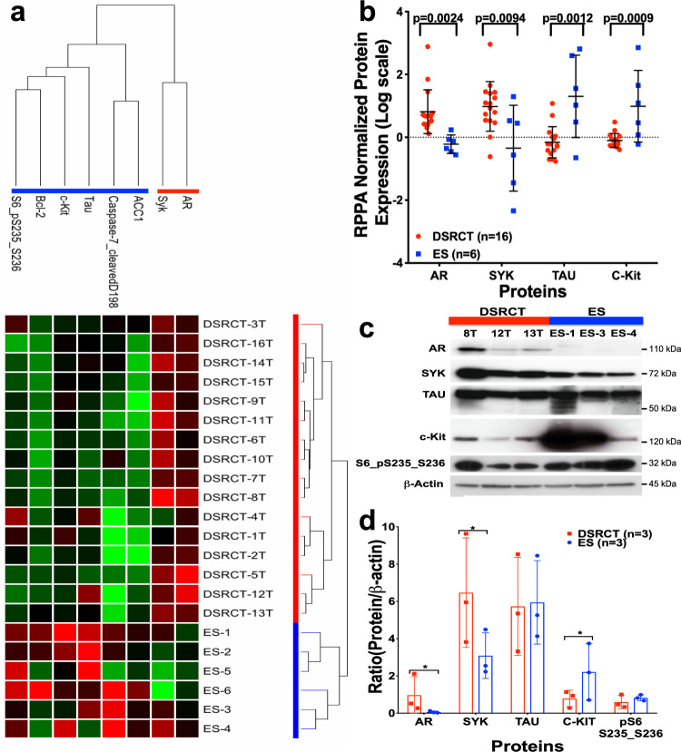
Desmoplastic small round cell tumor (DSRCT) is an aggressive, usually incurable sarcoma subtype that predominantly occurs in post-pubertal young males. Recent evidence suggests that the androgen receptor (AR) can promote tumor progression in DSRCTs. However, the mechanism of AR-induced oncogenic stimulation remains undetermined. Herein, we demonstrate that enzalutamide and AR-directed antisense oligonucleotides (AR-ASO) block 5α-dihydrotestosterone (DHT)-induced DSRCT cell proliferation and reduce xenograft tumor burden. Gene expression analysis and chromatin immunoprecipitation sequencing (ChIP-seq) were performed to elucidate how AR signaling regulates cellular epigenetic programs. Remarkably, ChIP-seq revealed novel DSRCT-specific AR DNA binding sites adjacent to key oncogenic regulators, including WT1 (the C-terminal partner of the pathognomonic fusion protein) and FOXF1. Additionally, AR occupied enhancer sites that regulate the Wnt pathway, neural differentiation, and embryonic organ development, implicating AR in dysfunctional cell lineage commitment. Our findings have direct clinical implications given the widespread availability of FDA-approved androgen-targeted agents used for prostate cancer.


Conflict of interest statement

A.R.M. is an employee and shareholder of Ionis Pharmaceuticals. The remaining authors declare no competing interests.

Figures

Fig. 1
Fig. 1. Proteomic comparison of DSRCT and ES.
a The protein lysates from DSRCT (red; n = 16) and ES (blue; n = 6) were subjected to RPPA analysis for 151 proteins and phosphoproteins (red, increased signal; green, decreased signal). Unsupervised double-hierarchical clustering using the Pearson correlation distance metric between proteins (rows) and Centroid linkage (a clustering method) separated the 22 samples into two groups by tumor type (columns). Of the 22 proteins, 8 had expression that differed significantly between ES and DSRCT (p ≤ 0.05; fold-change ≥2). b The mean expression intensity values of the 8 proteins associated with DSRCT or ES and their statistical significance after normalization for global protein expression by median centering across 151 antibodies in the RPPA panel. c Western blotting was used to validate the proteins identified by RPPA as being differentially expressed between DSRCT and ES. d Normalized protein expression is relative to β-actin. Data points in b and d represent mean ± SD. n is the number of samples analyzed for each sarcoma subtype.
Fig. 2
Fig. 2. DSRCT TMA and frozen specimen profiling for AR and PSA expression.
a A histogram showing the AR IHC expression levels of 60 human DSRCT tumors grouped by intensity (low, moderate, and high). The demographic data, including the corresponding gender (red: male or green: female), the age at diagnosis, and the pre/post-chemotherapy treatment to the surgery of each primary or metastatic resected tumor patient, are displayed at the left of each histogram. b AR expression level interpretation on DSRCT TMA IHC-stain and percentage scoring of tumoral labeling (positive (>50%), low positive (10–50%), focal (1–10%), and negative (0–1%)). c Western blotting analyses of AR expression in 11 DSRCT snap-frozen primary tumors. AR expression: P = positive, N = negative, or M = moderate. d Relative AR levels across samples shown in c. Bars show mean ± SD. e The principal components analysis plot performed on gene expression from prostate cancer (PC), DSRCT, and additional type of sarcomas samples. f Boxplot for the AR gene expression level across DSRCT, prostate cancer, and four other sarcoma types. The Wilcoxon rank-sum test performed to compare the AR levels between DSRCT (n = 22) and each of the other cancer types. PC = prostate cancer (n = 12); CS = chondrosarcoma (n = 7); OS = osteosarcoma (n = 47); WDLPS = well-differentiated liposarcoma (n = 7), and DDLPS = dedifferentiated liposarcoma (n = 10). ***p value < 0.001, **p value < 0.01, and *p value < 0.05.
Fig. 3
Fig. 3. Double-hierarchical clustering of PC (n = 12), DSRCT (n = 22), and other sarcoma subtypes (n = 71).
The top 1500 most variable genes across all samples were used to compare DSRCT to PC and other sarcoma subtypes. Unsupervised double-hierarchical clustering placed DSRCT next to PC on branch 1. Other sarcoma subtypes clustered on branch 2. Distinct blocks within the heatmap indicate genes overexpressed (A) or underexpressed (B) in PC and DSRCT compared to other sarcoma subtypes. Within the branch 1, some genes were upregulated strictly in PC (C) compared to DSRCT (D). PC = prostate cancer (n = 12); OS = osteosarcoma (n = 47); CS = chondrosarcoma (n = 7); WDLPS = well-differentiated liposarcoma (n = 7), and DDLPS = dedifferentiated liposarcoma (n = 10).
Fig. 4
Fig. 4. In vitro stimulation and inhibition of DSRCT proliferation via AR.
a JN-DSRCT, TC71, LNCaP, and PC3 cell proliferation assays after treating them with an AR agonist hormone, dihydrotestosterone (DHT) in a dose-dependent manner. n is the number of experimental replicates. b Profiling JN-DSRCT, TC71, and LNCaP cells for their AR expression by western blotting and histogram presentation of relative AR levels across each cell line. c Profiling of JN-DSRCT cells for AR protein expression (green) by immunofluorescence analysis with DAPI-labeled nuclei (blue), d Quantitative scatter plot representation of the ratio Nuclear/Cytoplasmic AR mean intensity reported within a single cell at 0, 5 and 24 h of DHT post-treatment. e JN-DSRCT cells are relatively less sensitive to enzalutamide than (f) AR antisense oligonucleotides treatment, as shown by the in vitro WST1-Proliferation cell-based assay. g Western blot analysis of AR expression in JN-DSRCT cells untreated or after Control-ASO and AR-ASO treatments. Histogram presentation of relative AR levels across each cell line after GAPDH normalization. Data points in a, e, and f represent mean ± SEM using three experimental replicates for each cell line. Data in b (bottom panel), d and g (bottom panel) represent mean ± standard deviation. P values calculated by unpaired two-tailed t-test.
Fig. 5
Fig. 5. Preclinical efficacy of AR antisense-based therapy for the treatment of DSRCT.
ac Therapeutic effect of AR blockade in JN-DSRCT xenografts done in three replicates. Tumor-bearing mice volumes and survival were reported after treatment with enzalutamide (25 mg/kg, orange), AR-ASO (25 mg/kg, regular red; 50 mg/kg, dotted red), control ASO (gray), or placebo (black). The top panel (a) shows the individual data for each mouse; the middle panel (b) shows a smoothed grouped median of relative tumor volumes; the lower panel (c) shows the survival Kaplan–Meier curves of each treated group of mice. df Similar data is shown for a DSRCT PDX treated with the same agents. d Individual PDX data, e smoothed PDX data, and f Kaplan–Meier curves for the DSRCT PDX. P values reported for the smoothed tumor growth curves (b and e) were calculated by a two-tailed unpaired t-test. Kaplan–Meier P values were calculated by the log-rank (Mantel–Cox) test. n is the number of mice treated in each treatment group.
Fig. 6
Fig. 6. Proteomic evaluation of AR expression in JN-DSRCT and PDX tumors after AR-based antisense therapy.
a The principal components analysis plot and reverse-phase protein lysate array (RPPA) evaluations of JN-DSRCT and PDX tumors after therapies, separated the 32 samples into four groups and identified 37 proteins statistically significantly associated with the treatment at a false discovery rate (FDR) of 0.05. b Immunoblotting evaluation of JN-DSRCT xenograft and PDX-DSRCT tumors after AR-ASO treatment. c AR normalization relative to GAPDH within the preclinical tumor samples. AR biomarker was significantly reduced in mice treated with AR-ASO compared to the control ASO group (p = 0.01). d Representative AR immunofluorescence confocal microscopy quantification of the preclinical JN-DSRCT and PDX tumor samples, within the single cell or e the averaged treated samples (placebo, control ASO, and AR-ASO). f Immunohistochemical evaluation images of preclinical JN-DSRCT and PDX1 tumor samples. IHC stains for AR in primary tumors of JN-DSRCT and PDX-DSRCT mice after treatment with AR-ASO, control ASO, and placebo. 100 μm scale bars are shown. g Representative IHC AR mean intensity quantification of the preclinical JN-DSRCT and PDX tumor samples, within the single cell or h the averaged treated samples (placebo, control ASO, and AR-ASO). All tumors analyzed by RPPA were collected at tumor progression or the experiment’s conclusion, except for the AR-ASO PD group, which was collected 10 days after initiating therapy to enable pharmacodynamic analysis. Data in c, d, e, g, and h represent mean ± standard deviation. P values calculated by unpaired two-tailed t-test.
Fig. 7
Fig. 7. AR binding in JN-DSRCT cells.
a Heatmaps (left panels) and average intensity curves (right panels) of ChIP-seq reads (RPKM; reads per kilobase of transcript per million mapped reads) for AR binding regions. AR binding sites are shown in a 10-kb window (centered on the middle of the binding site) in Control ASO, AR-ASO, DHT + Control ASO, and DHT + AR-ASO samples. b Venn diagram showing the overlap of all AR peaks between Control ASO, DHT + Control ASO, and DHT + AR-ASO samples to identify the AR-unique or shared binding sites. c List of enriched transcription factor (TF) motifs in AR-specific binding sites. Motifs are identified using HOMER (Binomial test). d Dot plot showing significantly enriched pathways for AR-specific binding sites. Dot size represents gene ratio, and colors represent adjusted p values (Fisher’s exact test). e IGV images showing enrichment of AR peaks around WT1, SOX2, CTNNB1, GATA6, FOXF1, and GLI2 genes using aggregate ChIP-seq profiles of Control ASO, DHT + Control ASO, and DHT + AR-ASO samples.
Fig. 8
Fig. 8. Enhancer reprogramming by AR in JN-DSRCT cells.
a Heatmaps (left panels) and average intensity curves (right panels) of ChIP-seq reads (RPKM; reads per kilobase of transcript per million mapped reads) for typical enhancer regions. Enhancer regions are shown in a 10-kb window (centered on the middle of the binding site) in Control ASO, AR-ASO, DHT + Control ASO, and DHT + AR-ASO samples. b Venn diagram showing the overlap of all enhancer peaks between Control ASO, AR-ASO, DHT + Control ASO, and DHT + AR-ASO samples to identify the AR-unique or shared enhancer reprogramming. c Venn diagram showing the overlap of annotated genes for AR-specific gained enhancer peaks and upregulated gene list for DSRCT tumors vs. other sarcoma tumors to identify the AR-unique enhancer reprogramming associated transcription upregulation. d Bar plot showing significantly enriched pathways for AR-specific enhancer reprogramming associated transcription upregulation. Bar length represents gene numbers, and colors represent adjusted p values (Fisher’s exact test). e IGV images showing enrichment of H3K27Ac peaks around AGRE2, AXIN2, CDK6, and MYH10 genes using aggregate ChIP-seq profiles of Control ASO, DHT + Control ASO, and DHT + AR-ASO samples.

References

    1. Honore, C. et al. Abdominal desmoplastic small round cell tumor: multimodal treatment combining chemotherapy, surgery, and radiotherapy is the best option. Ann. Surg. Oncol.22, 1073–1079. 10.1245/s10434-014-4123-6 (2015). - PubMed
    1. Chang F. Desmoplastic small round cell tumors: cytologic, histologic, and immunohistochemical features. Arch. Pathol. Lab. Med. 2006;130:728–732. doi: 10.5858/2006-130-728-DSRCTC. - DOI - PubMed
    1. Gerald WL, Rosai J, Ladanyi M. Characterization of the genomic breakpoint and chimeric transcripts in the EWS-WT1 gene fusion of desmoplastic small round cell tumor. Proc. Natl Acad. Sci. USA. 1995;92:1028–1032. doi: 10.1073/pnas.92.4.1028. - DOI - PMC - PubMed
    1. Gerald WL, et al. Clinical, pathologic, and molecular spectrum of tumors associated with t(11;22)(p13;q12): desmoplastic small round-cell tumor and its variants. J. Clin. Oncol. 1998;16:3028–3036. doi: 10.1200/JCO.1998.16.9.3028. - DOI - PubMed
    1. Lettieri CK, Garcia-Filion P, Hingorani P. Incidence and outcomes of desmoplastic small round cell tumor: results from the surveillance, epidemiology, and end results database. J. cancer Epidemiol. 2014;2014:680126. doi: 10.1155/2014/680126. - DOI - PMC - PubMed

Publication types