. 2022 Jun 1;13(1):3040.
doi: 10.1038/s41467-022-30826-2.

De novo biosynthesis of rubusoside and rebaudiosides in engineered yeasts

Affiliations

De novo biosynthesis of rubusoside and rebaudiosides in engineered yeasts

Yameng Xu et al. Nat Commun. .

Abstract

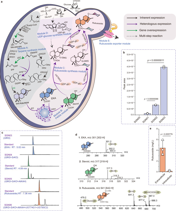
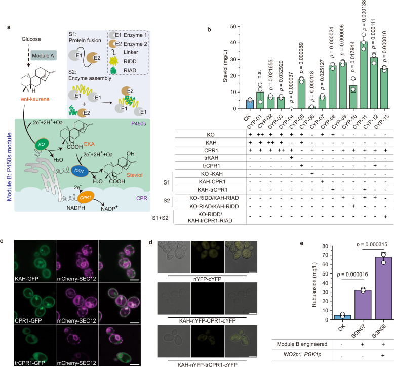
High-sugar diet causes health problems, many of which can be addressed with the use of sugar substitutes. Rubusoside and rebaudiosides are interesting molecules, considered the next generation of sugar substitutes due to their low-calorie, superior sweetness and organoleptic properties. However, their low abundance in nature makes the traditional plant extraction process neither economical nor environmental-friendly. Here we engineer baker's yeast Saccharomyces cerevisiae as a chassis for the de novo production of rubusoside and rebaudiosides. In this process, we identify multiple issues that limit the production, including rate-liming steps, product stress on cellular fitness and unbalanced metabolic networks. We carry out a systematic engineering strategy to solve these issues, which produces rubusoside and rebaudiosides at titers of 1368.6 mg/L and 132.7 mg/L, respectively. The rubusoside chassis strain here constructed paves the way towards a sustainable, large-scale fermentation-based manufacturing of diverse rebaudiosides.


Conflict of interest statement

The authors declare no competing interests.

Figures

Fig. 1
Fig. 1. Systematic engineering of yeast metabolism for de novo biosynthesis of rubusoside.
a Illustration of the modularized platform for producing and exporting rubusoside. Module A (terpene synthesis module) incorporates modifications designed to divert carbon flux to diterpene metabolic and brought ent-kaurene biosynthesis. Engineering yeast into the efficient platform to produce rubusoside by introducing Module B (P450s module) and Module C (rubusoside synthesis module). Module D (UDP-glucose synthesis module) provides glycoside ligands for producing rubusoside. Module E (rubusoside exporter module) is a possible exportation system of rubusoside. ERG10 acetyl-CoA C-acetyltransferase, ERG13 hydroxymethylglutaryl-CoA synthase, HMG1 hydroxymethylglutaryl-CoA reductase, tHMG1 truncated hydroxymethylglutaryl-CoA reductase, ERG12 mevalonate kinase, ERG8 phosphomevalonate kinase, IDI1 isopentenyl diphosphate delta-isomerase, ERG20 bifunctional (2E,6E)-farnesyl diphosphate, BST1 farnesyltranstransferase, KS kaurene synthase, KO ent-kaurene oxidase, KAH kaurenoic acid 13α-hydroxylase, UGT74G1 UDP-glycosyltransferase 74G1, UGT85C2 UDP-glycosyltransferase 85C2. FPSF112A mutant farnesyl pyrophosphate synthase. Glc-6-P glucose-6-phosphate, Acetyl-CoA acetyl coenzyme A, IPP isopentenyl diphosphate, GPP Geranyl diphosphate, FPP farnesyl diphosphate, GGPP geranylgeranyl pyrophosphate, DMAPP dimethylallyl diphosphate, EKA ent-kaurenoic acid, 13-SMG (5ξ,8α,9ξ,10α,13α)-13-(β-D-Glucopyranosyloxy) kaur-16-en-18-säure, 19-SMG 1-O-[(5ξ,8α,9ξ,10α,13α)-13-Hydroxy-18-oxokaur-16-én-18-yl]-β-D-glucopyranose. All the heterologous genes were controlled by GAL promoters. b Increased ent-kaurene biosynthesis by eliminating the rate-limiting steps in MVA pathway (overexpressed tHMG1 and IDI1, SGN02) and avoiding competition for FPP with the monoterpene synthesis pathway (introduced FPSF112A, SGN03). All the heterologous genes were controlled by GAL promoters. c HPLC spectra of ent-kaurenoic acid (EKA), steviol, rubusoside, and their standards. RT retention time. d LC-MS analysis results of EKA, steviol, and rubusoside in negative ion mode. Source data are provided as a Source Data file. e The rubusoside titer difference in the intracellular and extracellular of the SGN06 strain. b, e Data are presented as mean values ± SD from three independent biological replicates (n = 3), the circles represent individual data points. Significance (p-value) was evaluated by two-sided t-test.
Fig. 2
Fig. 2. Eliminating the rate-limiting steps in the P450s module.
a Illustration of different strategies for relieving the rate-limiting steps. S1 (Strategy1), proteins fused by a short protein linker (GGGGS3), S2 (Strategy2), enzymes fused with a pair of short peptide tags (RIAD and RIDD). NADPH nicotinamide adenine dinucleotide phosphate, NADP+ nicotinamide adenine dinucleotide phosphate. b Changes of steviol titer by modifying the P450s. S1, proteins fused by a linker (GGGGS3), S2, enzymes fused with a pair of short peptide tags (RIAD and RIDD). CK presents the SGN05 strain. c Visualized analysis of the subcellular localization of the P450s KAH and reductase CPR1. The fluorescence images in the first row are KAH-GFP (left, green) and mCherry-SEC12 (middle, magenta) and merge images (right). The fluorescence images in the second row are CPR1-GFP (left, green), mCherry-SEC12 (middle, magenta), and merge images (right). The fluorescence images in second row are trCPR1-GFP (left, green), mCherry-SEC12 (middle, magenta), and merge images (right). Bar = 5 μm. d BiFC of KAH fused with nYFP, and CPR1/trCPR1 fused with cYFP. Confocal images of the cells expressing nYFP-cYFP (top), KAH-nYFP-CPR1-cYFP (middle), and KAH-nYFP-trCPR1-cYFP (bottom). cYFP C-terminal 186–250 amino acid residues, nYFP N‐terminal 1‐185 amino acid residues. Bar = 5 μm. e Increased rubusoside biosynthesis by eliminating the rate-limiting steps in P450s module (Module B), and overexpressing the ER regulator INO2 by replacing it promoter (INO2p) with a stronger one PGK1p. CK (the SGN06 strain). b, e Data are presented as mean values ± SD from three independent biological replicates (n = 3), the circles represent individual data points. Significance (p-value) was evaluated by two-sided t-test, no significance (n.s.) presents p > 0.05. b, c Image analysis was carried out on the Leica LAS X software package and the ImageJ 1.53k software.
Fig. 3
Fig. 3. Improvement of the yeast adaptation to rubusoside.
a Changes of rubusoside titer after deleting the ABC exporters in the PM and the pressure response regulators. Cells were cultured for 108 h; CK presents SGN08. b Changes of the rubusoside production in the ABC transporter and the pressure responsive factor MSN4 overexpression strains. CK presents the strain SGN08. c The biomass of the ABC transporter and the pressure responsive factor MSN4 overexpressed strains. d FESEM pictures of the yeast samples. (1). the original strain S. cerevisiae CEN.PK2-1C; (2). the strain SGN08; (3). the strain SGN08-Δpdr11; (4). the strain SGN08-Δmsn4. (5). the strain SGN10 (overexpressed the ABC transporter PDR11); (6). the strain of SGN12 (overexpressed stress-respond regulator MSN4). Strains were cultivated in shake-flask, and cells were collected after fermentation 72 h. Bar = 5 μm. ac Data are presented as mean values ± SD from three independent biological replicates (n = 3). a, b The circles represent individual data points. Significance (p-value) was evaluated by two-sided t-test, n.s. presents p > 0.05.
Fig. 4
Fig. 4. Redistribution and optimization of the metabolic networks based on in silico prediction tools.
a Schematic illustration of the in silico prediction process. b Metabolic interventions predicted using OptKnock for rubusoside overproduction. GAL7 galactose-1-phosphate uridyl transferase, ABZ2 aminodeoxychorismate lyase, ALT1 alanine transaminase, ALT2 alanine transaminase, ARO8 aromatic aminotransferase I, PGM1 phosphoglucomutase, PGM2 phosphoglucomutase, UGP1 UTP (uridine triphosphate) glucose-1-phosphate uridylyltransferase. G6P glucose-6-phosphate, G1P glucose-1-phosphate, F6P fructose-6-phosphate, F1,6P fructose-1,6-bisphosphate, PPP pathway, GA3P glyceraldehyde-3-phosphate, 1,3BPG 1,3-Bisphospho-D-glycerate, 3PG 3-Phospho-D-glycerate, 2PG 2-Phospho-D-glycerate, PEP phosphoenolpyruvate, PYR pyruvate, E4P erythrose 4-phosphate, DAHP 3-deoxy-arabino-heptulonate 7-phosphate, EPSP 5-O-(1-carboxyvinyl)-3-phosphoshikimate, CHA Chorismite, PRE prephenate, ADC 4-Amino-4-deoxychorismate, ABEE 4-Aminobenzoate, PPA phenylpyruvate, L-Phe L-Phenylalanine, 4-HPPA 4-Hydroxyphenylpyruvate, L-Tyr L-tyrosine, GL1 alpha-D-Galactose-1-phosphat, UTP Uridine triphosphate, UDP-Glc UDP-glucose. c The changes of rubusoside titer via deleting the target genes predicted by OptKnock (SGN14–SGN18), and engineering the UDP-glucose synthesis module (SGN19–SGN24). Cells were cultured for 108 h. Data are presented as mean values ± SD from three independent biological replicates (n = 3), the circles represent individual data points. Significance (p-value) was evaluated by two-sided t-test. d Fed-batch fermentation of strain SGN23 to produce rubusoside.
Fig. 5
Fig. 5. The de novo biosynthesis platform of rebaudiosides.
a Schematic illustration of rebaudiosides biosynthesis based on the rubusoside producing strain. Module F1: the metabolic pathways were reported to exist in S. rebaudiana, but not be detected; Module F2: the metabolic pathways were reported to exist in S. rebaudiana, and the metabolites were hunted in the M23. UGT74G1 UDP-glycosyltransferase 74G1, UGT85C2 UDP-glycosyltransferase 85C2, EUGT11 UDP-glycosyltransferase 91C1, UGT76G1-MUT UDP-glycosyltransferase 76G1. 13-SMG (5ξ,8α,9ξ,10α,13α)-13-(β-D-Glucopyranosyloxy) kaur-16-en-18-säure, 19-SMG 1-O-[(5ξ,8α,9ξ,10α,13α)-13-Hydroxy-18-oxokaur-16-én-18-yl]-β-D-glucopyranose, 1,2-bioside (5β,8α,9β,10α,13α)-13-{ [2-O-(β-D-Glucopyranosyl)-β-D-glucopyran-osyl]oxy} kaur-16-en-18-oic acid, Reb A rebaudioside A, Reb B rebaudioside B, Reb C rebaudioside C, Reb D rebaudioside D, Reb E rebaudioside E, Reb G rebaudioside G, Reb I rebaudioside I, Reb M rebaudioside M, Reb N rebaudioside N, Reb Q rebaudioside Q. b LC-MS analysis results of stevioside, Reb A, Reb D, and Reb M in negative ion mode, analyzed by comparison with the standard product. c The titer changes of rebaudiosides by modifying the EUGT11 (M07), fine-turning the UGT76G1-MUT expression time (M23-0 h, M23-6 h, M23-12 h, M23-24 h, and M23-48 h) by inducing it expression an 0, 6, 12, 24, and 48 h after fermentation, and strengthening the stress-respond regulator MSN4 (M24). d 1,2-bioside, Reb B, Reb E, and Reb N in negative ion mode, identified by Progenesis QI v2.4 software. QI software data (Supplementary Data 1) are provided as a Source Data file. e Fed-batch fermentation of strain M24 to produce rebaudiosides. The UGT76G1-MUT expressed before the genes controlled by GAL promoters. All the data represent the mean of n = 3 biologically independent samples.

Similar articles

Cited by

References

    1. World Health Organization. Global action plan for the prevention and control of noncommunicable diseases 2013–2020. (World Health Organization, 2013).
    1. Yang T, et al. Hydrophobic recognition allows the glycosyltransferase UGT76G1 to catalyze its substrate in two orientations. Nat. Commun. 2019;10:3214. doi: 10.1038/s41467-019-11154-4. - DOI - PMC - PubMed
    1. Zhang J, et al. Catalytic flexibility of rice glycosyltransferase OsUGT91C1 for the production of palatable steviol glycosides. Nat. Commun. 2021;12:7030. doi: 10.1038/s41467-021-27144-4. - DOI - PMC - PubMed
    1. Zhang Y, et al. Efficient biocatalytic preparation of rebaudioside KA: highly selective glycosylation coupled with UDPG regeneration. Sci. Rep. 2020;10:6230. doi: 10.1038/s41598-020-63379-9. - DOI - PMC - PubMed
    1. Zhang J, Chou G, Liu Z, Liu M. Employing rubusoside to improve the solubility and permeability of antitumor compound betulonic acid. Nanomedicine. 2016;11:2829–2844. - PubMed

Publication types

MeSH terms

LinkOut - more resources