

Genetic architecture of band neutrophil fraction in Iceland
- PMID: 35650273
- PMCID: PMC9160026
- DOI: 10.1038/s42003-022-03462-1
Genetic architecture of band neutrophil fraction in Iceland
Abstract
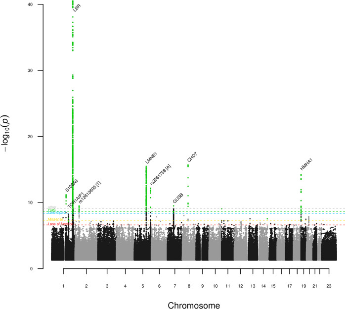
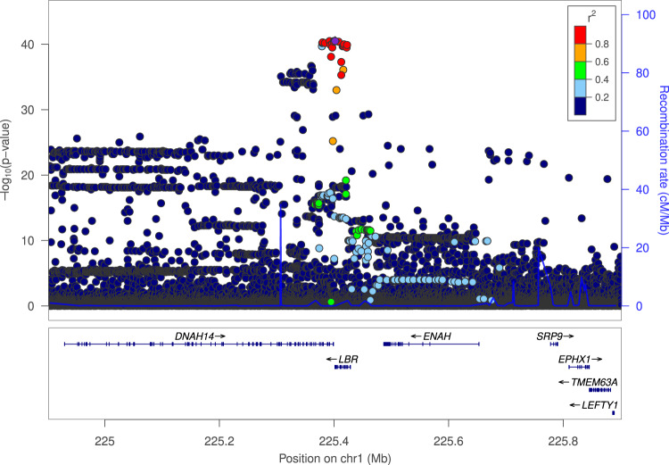
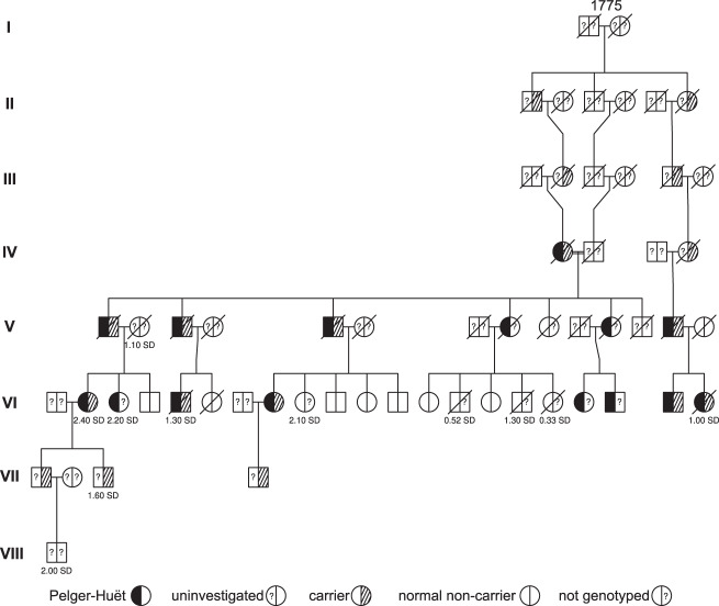
The characteristic lobulated nuclear morphology of granulocytes is partially determined by composition of nuclear envelope proteins. Abnormal nuclear morphology is primarily observed as an increased number of hypolobulated immature neutrophils, called band cells, during infection or in rare envelopathies like Pelger-Huët anomaly. To search for sequence variants affecting nuclear morphology of granulocytes, we performed a genome-wide association study using band neutrophil fraction from 88,101 Icelanders. We describe 13 sequence variants affecting band neutrophil fraction at nine loci. Five of the variants are at the Lamin B receptor (LBR) locus, encoding an inner nuclear membrane protein. Mutations in LBR are linked to Pelger-Huët anomaly. In addition, we identify cosegregation of a rare stop-gain sequence variant in LBR and Pelger Huët anomaly in an Icelandic eight generation pedigree, initially reported in 1963. Two of the other loci include genes which, like LBR, play a role in the nuclear membrane function and integrity. These GWAS results highlight the role proteins of the inner nuclear membrane have as important for neutrophil nuclear morphology.
© 2022. The Author(s).
Conflict of interest statement
Authors affiliated with deCODE genetics/Amgen Inc., G.R.O., M.K.M., A.O., B.O.J., R.F., G.A.A., H.K., S.R., G.H.H., G.S., E.V.I., L.S., E.F., K.N., V.T., J.S., As.J., Ad.J., S.S., K.O.P., O.B.D., T.R., H.H., G.M., D.F.G., I.J., G.L.N., U.T., P.S., and K.S. declare competing interests as employees. The remaining authors declare no competing interests.
Figures
Similar articles
-
Mutations in the gene encoding the lamin B receptor produce an altered nuclear morphology in granulocytes (Pelger-Huët anomaly).Nat Genet. 2002 Aug;31(4):410-4. doi: 10.1038/ng925. Epub 2002 Jul 15. Nat Genet. 2002. PMID: 12118250
-
[Nuclear abnormalities in Pelger-Huet anomaly; progress in blood cell morphology].Rinsho Byori. 2005 Jan;53(1):54-60. Rinsho Byori. 2005. PMID: 15724491 Review. Japanese.
-
An in vitro model for Pelger-Huët anomaly: stable knockdown of lamin B receptor in HL-60 cells.Nucleus. 2010 Nov-Dec;1(6):506-12. doi: 10.4161/nucl.1.6.13271. Epub 2010 Aug 6. Nucleus. 2010. PMID: 21327094 Free PMC article.
-
Lamin B-receptor mutations in Pelger-Huët anomaly.Br J Haematol. 2003 Nov;123(3):542-4. doi: 10.1046/j.1365-2141.2003.04621.x. Br J Haematol. 2003. PMID: 14617022
-
Understanding and recognizing the Pelger-Huët anomaly.Am J Clin Pathol. 2012 Mar;137(3):358-66. doi: 10.1309/AJCP3G8MDUXYSCID. Am J Clin Pathol. 2012. PMID: 22338047 Review.
Cited by
-
Novel loci for Alzheimer's disease identified by a genome-wide association study in Ashkenazi Jews.Alzheimers Dement. 2023 Dec;19(12):5550-5562. doi: 10.1002/alz.13117. Epub 2023 Jun 1. Alzheimers Dement. 2023. PMID: 37260021 Free PMC article.
References
MeSH terms
LinkOut - more resources
Full Text Sources
Molecular Biology Databases
