. 2022 Jun 1;12(1):9116.
doi: 10.1038/s41598-022-13231-z.

Ablation of the miRNA cluster 24 in cartilage and osteoblasts impairs bone remodeling

Affiliations

Ablation of the miRNA cluster 24 in cartilage and osteoblasts impairs bone remodeling

Veronika S Georgieva et al. Sci Rep. .

Abstract

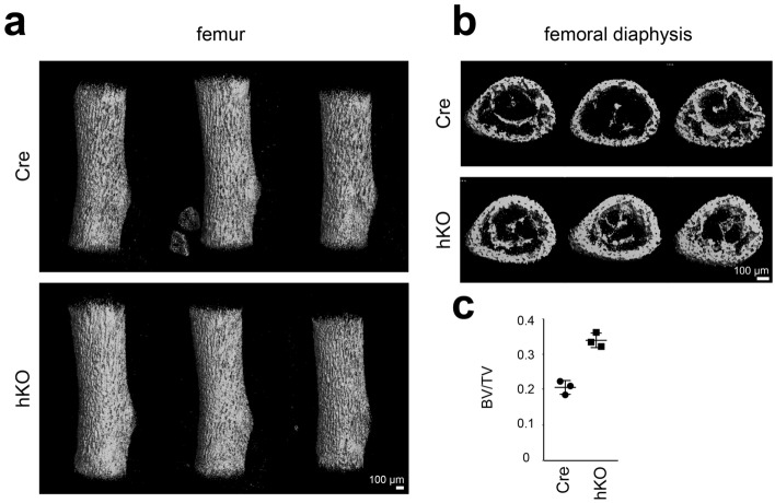
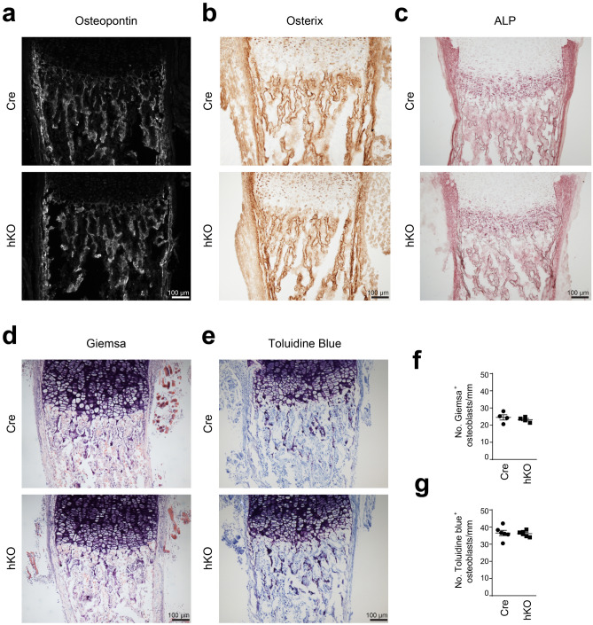
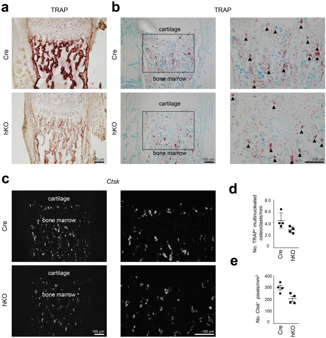
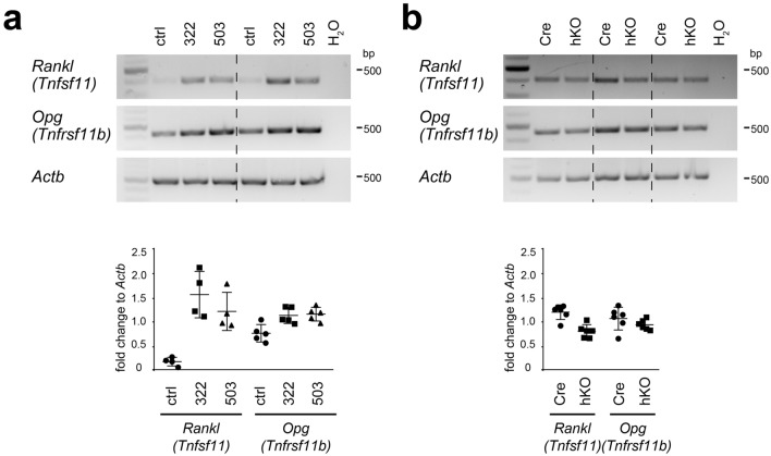
MicroRNAs (miRNAs) post-transcriptionally regulate cartilage and bone development and function, however, only few miRNAs have been described to play a role for cartilage to bone transition in vivo. Previously, we showed that cartilage-specific deletion of the Mirc24 cluster in newborn male mice leads to impaired growth plate cartilage development due to increased RAF/MEK/ERK signaling and affects the stability of the cartilage extracellular matrix on account of decreased SOX6 and SOX9 and increased MMP13 levels. Here, we studied how Mirc24 cluster inactivation in cartilage and osteoblasts leads to an increased bone density associated with defects in collagen remodeling in trabecular bone. No changes in osteoblast distribution were observed, whereas the number of osteoclasts was reduced and TRAP activity in osteoclasts decreased. Surprisingly, an increased level of cluster-encoded miR-322 or miR-503 raises Rankl gene expression and inactivation of the cluster in chondrocytes reduces Rankl expression. These results suggest that the Mirc24 cluster regulates Rankl expression in chondrocytes at the chondro-osseous border, where the cluster is mainly expressed to modulate osteoclast formation, bone remodeling and bone integrity.


Conflict of interest statement

The authors declare no competing interests.

Figures

Figure 1
Figure 1
Analysis of femora of Mirc24 cluster-deficient mice. μCT analysis of femora from newborn male Col2a1-Cre (Cre) and Col2a1-Cre-Mirc24tm1M/Y (hKO) mice. Three animals per genotype are shown. (a) Three dimensional reconstructions of the femora. (b) Cross-sections of the femoral diaphysis. (c) Relative bone volume (BV/TV). Individual and mean values and standard deviations are shown (graph). The figure was created with Adobe Illustrator CS6 (Version 16.0.3, https://adobe.com/products/illustrator).
Figure 2
Figure 2
Analysis of the abundance of fibrillar collagens in femoral sections of Mirc24 cluster-deficient mice. Detection was performed on plane matched sections of femora from newborn male Col2a1-Cre (Cre) and Col2a1-Cre-Mirc24tm1M/Y (hKO) mice. (a) Col1a1 mRNA distribution was analyzed by fluorescence in situ hybridization using a Col1a1 specific fluorescence probe. The abundance of (b) collagen I and (c) collagen II was studied by immunofluorescence. (d) In addition, to study bone remodeling the binding of collagen hybridizing peptide to unfolded collagen chains was analyzed by fluorescence microscopy. Note reduced intensity of the trabecular staining (arrowheads). Brightness was adjusted for visualization (ac) using Adobe Photoshop Elements 2020 (Version 18.0, https://www.adobe.com/products/photoshop-elements.html). Four animals per genotype were analyzed and representative images are shown. Additional images are shown in Supplementary Figs. S1 and S2 online. The figure was created using Adobe Illustrator CS6 (Version 16.0.3, https://adobe.com/products/illustrator).
Figure 3
Figure 3
Characterization of osteoblast organization in femoral sections from Mirc24 cluster-deficient mice. Osteoblast distribution in plane matched sections of femora from newborn male Col2a1-Cre (Cre) and Col2a1-Cre-Mirc24tm1M/Y (hKO) mice was analyzed by immunodetection of (a) osteopontin and (b) osterix. (c) In addition, alkaline phosphatase (ALP) activity was detected by histological staining. (d,e) To determine osteoblast numbers, paraffin sections were stained with Giemsa and Toluidine Blue. Brightness was adjusted using Adobe Photoshop Elements 2020 (Version 18.0, https://www.adobe.com/products/photoshop-elements.html) for visualization (d,e). At least three animals per genotype were analyzed and representative images are shown. Additional images are shown in Supplementary Figs. S3 and S4 online. (f,g) For quantification, the number of Giemsa+ and Toluidine Blue+ osteoblasts per mm trabecular bone were determined. Individual and mean values and standard deviations are shown (graph). The figure was created using Adobe Illustrator CS6 (Version 16.0.3, https://adobe.com/products/illustrator).
Figure 4
Figure 4
Characterization of osteoclast organization in femoral sections from Mirc24 cluster-deficient mice. (a) To identify osteoclasts, tartrate-resistant acid phosphatase (TRAP) activity stainings were performed on frozen sections of femora from newborn male Col2a1-Cre (Cre) and Col2a1-Cre-Mirc24tm1M/Y (hKO) mice. (b) To determine osteoclast numbers, TRAP activity (red) was detected in paraffin sections counterstained by Fast green (blue). Enlarged area is indicated by a box. Multinucleated osteoclasts are marked by arrowheads. (c) Ctsk mRNA distribution was analyzed by fluorescence in situ hybridization using a Ctsk specific fluorescence probe. Brightness was adjusted for visualization (b,c) using Adobe Photoshop Elements 2020 (Version 18.0, https://www.adobe.com/products/photoshop-elements.html). Four animals per genotype were analyzed and representative images are shown. Additional images are shown Supplementary Figs. S5 and S6 online. For quantification (d), the number of TRAP+ multinucleated osteoclasts per mm trabecular bone in (b) was determined and (e) the number of Ctsk+ particles per mm2 in (c) was calculated. Individual and mean values and standard deviations are shown (graph). The figure was created using Adobe Illustrator CS6 (Version 16.0.3, https://adobe.com/products/illustrator).
Figure 5
Figure 5
Determination of Rankl and Opg gene expression in miRNA mimic-transfected chondrocytes and in cultured chondrocytes from Mirc24 cluster-deficient mice. The gene expression of Rankl and Opg was analyzed by semi-quantitative PCR (a) in control (ctrl), miR-322 (322) or miR-503 (503) mimic-transfected primary epiphyseal chondrocytes (PECs) 2 days post transfection and (b) in PECs isolated from newborn male Col2a1-Cre (Cre) and Col2a1-Cre-Mirc24tm1M/Y (hKO) mice and cultured for 2 days. The control (H2O) contains no genomic DNA and Actb served as loading control. Rankl or Opg to Actb ratio was determined by ImageJ and individual and mean values and standard deviations are shown (graph). The size of NEB Quick-Load Purple 1 kb Plus DNA Ladder bands is shown. The figure was created using Adobe Illustrator CS6 (Version 16.0.3, https://adobe.com/products/illustrator).

Similar articles

Cited by

References

    1. Papaioannou G. miRNAs in bone development. Curr. Genomics. 2015;16:427–434. doi: 10.2174/1389202916666150817202425. - DOI - PMC - PubMed
    1. Zhou X, et al. Chondrocytes transdifferentiate into osteoblasts in endochondral bone during development, postnatal growth and fracture healing in mice. PLoS Genet. 2014;10:e1004820. doi: 10.1371/journal.pgen.1004820. - DOI - PMC - PubMed
    1. Yang L, Tsang KY, Tang HC, Chan D, Cheah KS. Hypertrophic chondrocytes can become osteoblasts and osteocytes in endochondral bone formation. Proc. Natl. Acad. Sci. U.S.A. 2014;111:12097–12102. doi: 10.1073/pnas.1302703111. - DOI - PMC - PubMed
    1. Long JT, et al. Hypertrophic chondrocytes serve as a reservoir for marrow-associated skeletal stem and progenitor cells, osteoblasts, and adipocytes during skeletal development. Elife. 2022;11:e76932. doi: 10.7554/eLife.76932. - DOI - PMC - PubMed
    1. Gibson G. Active role of chondrocyte apoptosis in endochondral ossification. Microsc. Res. Tech. 1998;43:191–204. doi: 10.1002/(SICI)1097-0029(19981015)43:2<191::AID-JEMT10>3.0.CO;2-T. - DOI - PubMed

Publication types