Review
. 2022 Aug;18(8):503-516.
doi: 10.1038/s41574-022-00688-1. Epub 2022 Jun 1.

Persistent coxsackievirus B infection and pathogenesis of type 1 diabetes mellitus

Affiliations
Review

Persistent coxsackievirus B infection and pathogenesis of type 1 diabetes mellitus

Magloire Pandoua Nekoua et al. Nat Rev Endocrinol. 2022 Aug.

Abstract

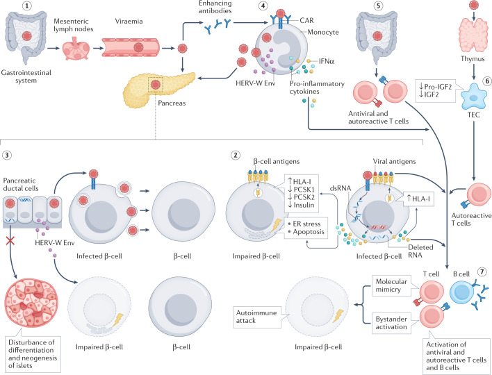
Enteroviruses are believed to trigger or accelerate islet autoimmunity in genetically susceptible individuals, thereby resulting in loss of functional insulin-producing β-cells and type 1 diabetes mellitus (T1DM). Although enteroviruses are primarily involved in acute and lytic infections in vitro and in vivo, they can also establish a persistent infection. Prospective epidemiological studies have strongly associated the persistence of enteroviruses, especially coxsackievirus B (CVB), with the appearance of islet autoantibodies and an increased risk of T1DM. CVB can persist in pancreatic ductal and β-cells, which leads to structural or functional alterations of these cells, and to a chronic inflammatory response that promotes recruitment and activation of pre-existing autoreactive T cells and β-cell autoimmune destruction. CVB persistence in other sites, such as the intestine, blood cells and thymus, has been described; these sites could serve as a reservoir for infection or reinfection of the pancreas, and this persistence could have a role in the disturbance of tolerance to β-cells. This Review addresses the involvement of persistent enterovirus infection in triggering islet autoimmunity and T1DM, as well as current strategies to control enterovirus infections for preventing or reducing the risk of T1DM onset.


Conflict of interest statement

The authors declare no competing interests.

Figures

Fig. 1
Fig. 1. The genome and capsid of enteroviruses.
The enteroviruses (viruses of the genus Enterovirus) are small (25–30 nm diameter) non-enveloped viruses with an icosahedral capsid that belong to the Picornaviridae family. a | The genome of enteroviruses (~7,400 bases) is a positive-sense single-stranded RNA genome, which contains a large open reading frame (ORF) flanked by a 5′-untranslated region (UTR) linked to the VPg (a viral non-structural protein also known as 3B) and 3′-UTR terminated with a poly(A) tail. A shorter ORF2 is located upstream from the main ORF,. The ORF encodes a polyprotein that is processed into four capsid proteins (VP1–VP4) (structural proteins) and seven other proteins, 2A, 2B, 2C, 3A, 3B, 3C and 3D (non-structural proteins), which are involved in viral replication. The ORF2 is translated into a single protein, ORF2p, which is involved in the infection of intestinal cells,. b | The icosahedral capsid consists of an arrangement of 60 protomers each consisting of four structural proteins (VP1, VP2, VP3 and VP4). VP4 is located on the internal side of the capsid according to studies based on X-ray diffraction carried out with virus particles frozen at −196°C.
Fig. 2
Fig. 2. Persistence of CVB in vitro and in vivo.
Coxsackievirus B (CVB) can persist in vitro in human and mouse pancreatic ductal and β-cells and thymic cells (primary cells and in cell lines) and lead to various structural or functional alterations of these cells. CVB persists in vivo in the gut mucosa, heart, thymus and pancreas of orally or intraperitoneally infected mice, weeks after the acute infectious period. 1.1B4, β-cell line derived from electrofusion of primary human β-cells with PANC-1; CAR, coxsackievirus and adenovirus receptor; GMCSF, granulocyte–macrophage colony-stimulating factor; INS-1, β-cell line derived from a rat insulinoma; LIF, leukaemia inhibitory factor; MTE, murine thymic epithelial cell line; NK, natural killer; PANC-1, human pancreatic ductal carcinoma cell line; PDX1, pancreatic duodenal homeobox factor 1; TEC, thymic epithelial cell.
Fig. 3
Fig. 3. Persistence of CVB and pathogenesis of T1DM.
(1) Coxsackievirus B (CVB) spreads through the gastrointestinal tract and possibly the oropharyngeal mucosa, and then to the pancreas via the lymphatics and the bloodstream. (2) CVB can persist in β-cells through 5′-UTR deletions of the viral RNA genome and the formation of double-stranded RNA (dsRNA), which activates host pathogen recognition receptors. This activation results in the expression of pro-inflammatory cytokines and upregulation of interferon response genes, resulting in production and release of type I interferons. These induce overexpression of HLA class I antigens on the β-cell surface (including neighbouring cells around infected cells), leading to enhanced presentation of β-cell and viral antigens. IFNα also causes endoplasmic reticulum (ER) stress, inhibition of PCSK1 and PCSK2, impaired insulin secretion and apoptosis in β-cells. CVB uses autophagosome-like vesicles, cellular protrusions or microvesicles to spread to β-cells by cell–cell transmission via membrane fusion. (3) CVB can also persist in pancreatic ductal cells, potentially spreading to β-cells, and can alter the differentiation of pancreatic ductal cells into insulin-producing cells. CVB4 activates the expression of human endogenous retrovirus-W envelope protein (HERV-W Env) in pancreatic ductal cells, which could have deleterious effects on β-cells. (4) CVB infection can be maintained in monocytes and macrophages via a mechanism involving enhancing antibodies, so that these cells behave as reservoirs for spreading of the virus to pancreatic cells. The persistent infection of immune cells could result in activation of HERV-W Env expression and a chronic inflammatory state, which can activate autoimmune T lymphocytes. (5) CVB persistence in the intestine might increase the number and activation of antiviral and autoreactive T lymphocytes. (6) CVB persistence in the thymus, especially in thymic epithelial cells (TECs), can disturb self-tolerance to β-cells, resulting in release of autoimmune T cells from the thymus. (7) CVB persistence results in the activation of antiviral T cells and might induce and/or aggravate autoimmune reactions against β-cells through various mechanisms, such as molecular mimicry and bystander activation. CAR, coxsackievirus and adenovirus receptor; IGF2, insulin-like growth factor 2; PCSK, pro-hormone convertase.

References

    1. Noble JA. Immunogenetics of type 1 diabetes: a comprehensive review. J. Autoimmun. 2015;64:101–112. doi: 10.1016/j.jaut.2015.07.014. - DOI - PubMed
    1. Ilonen J, Lempainen J, Veijola R. The heterogeneous pathogenesis of type 1 diabetes mellitus. Nat. Rev. Endocrinol. 2019;15:635–650. doi: 10.1038/s41574-019-0254-y. - DOI - PubMed
    1. Geravandi S, Liu H, Maedler K. Enteroviruses and T1D: is it the virus, the genes or both which cause T1D. Microorganisms. 2020;8:1017. doi: 10.3390/microorganisms8071017. - DOI - PMC - PubMed
    1. Sharp SA, et al. Development and standardization of an improved type 1 diabetes genetic risk score for use in newborn screening and incident diagnosis. Diabetes Care. 2019;42:200–207. doi: 10.2337/dc18-1785. - DOI - PMC - PubMed
    1. Qu HQ, et al. Improved genetic risk scoring algorithm for type 1 diabetes prediction. Pediatr. Diabetes. 2022;23:320–323. doi: 10.1111/pedi.13310. - DOI - PMC - PubMed

Publication types

MeSH terms