. 2022 Jun;606(7915):739-746.
doi: 10.1038/s41586-022-04760-8. Epub 2022 Jun 1.

Molecularly defined circuits for cardiovascular and cardiopulmonary control

Affiliations

Molecularly defined circuits for cardiovascular and cardiopulmonary control

Avin Veerakumar et al. Nature. 2022 Jun.

Abstract

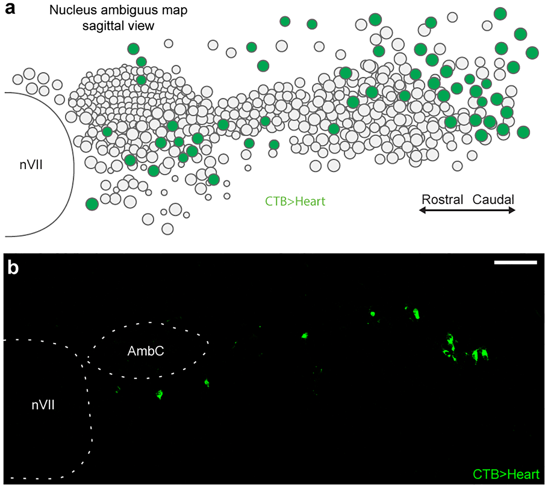
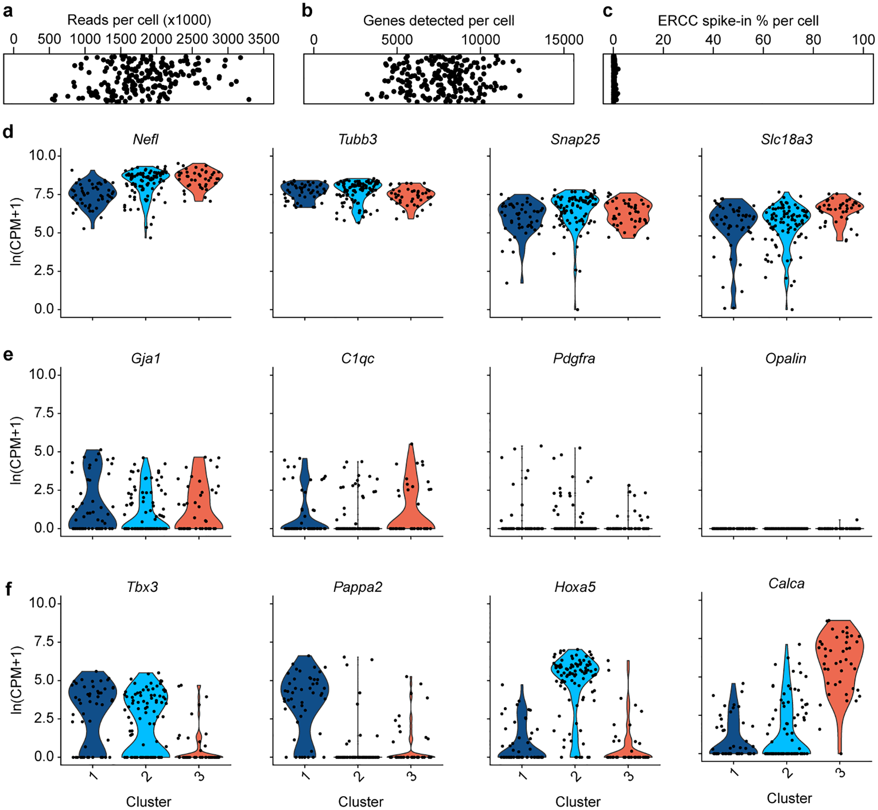
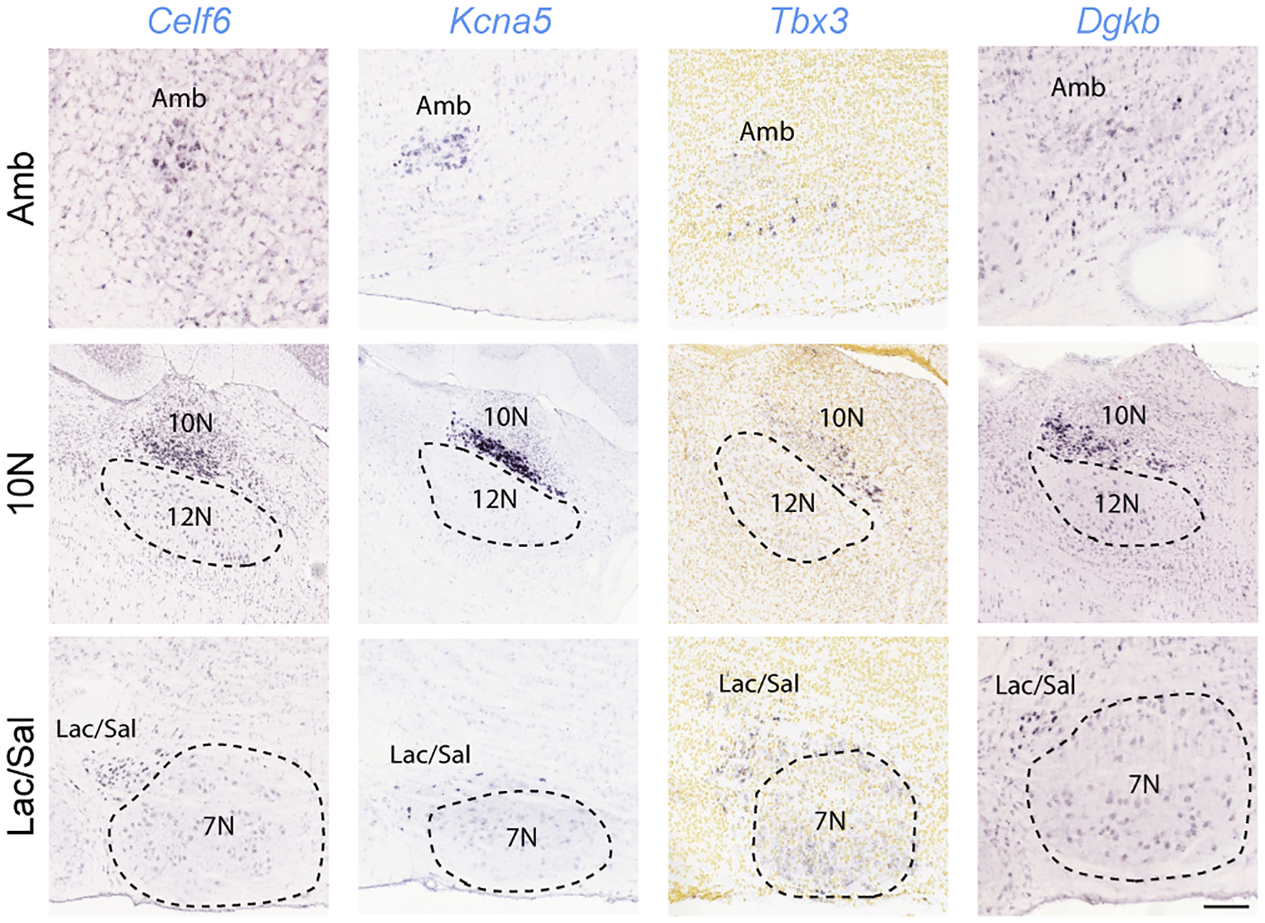
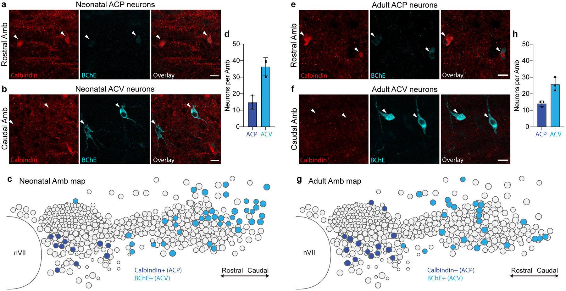
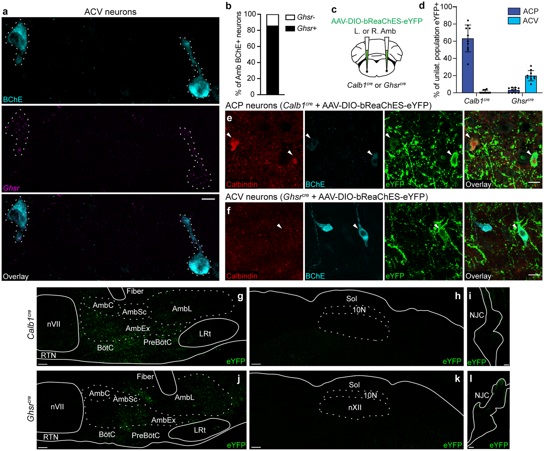
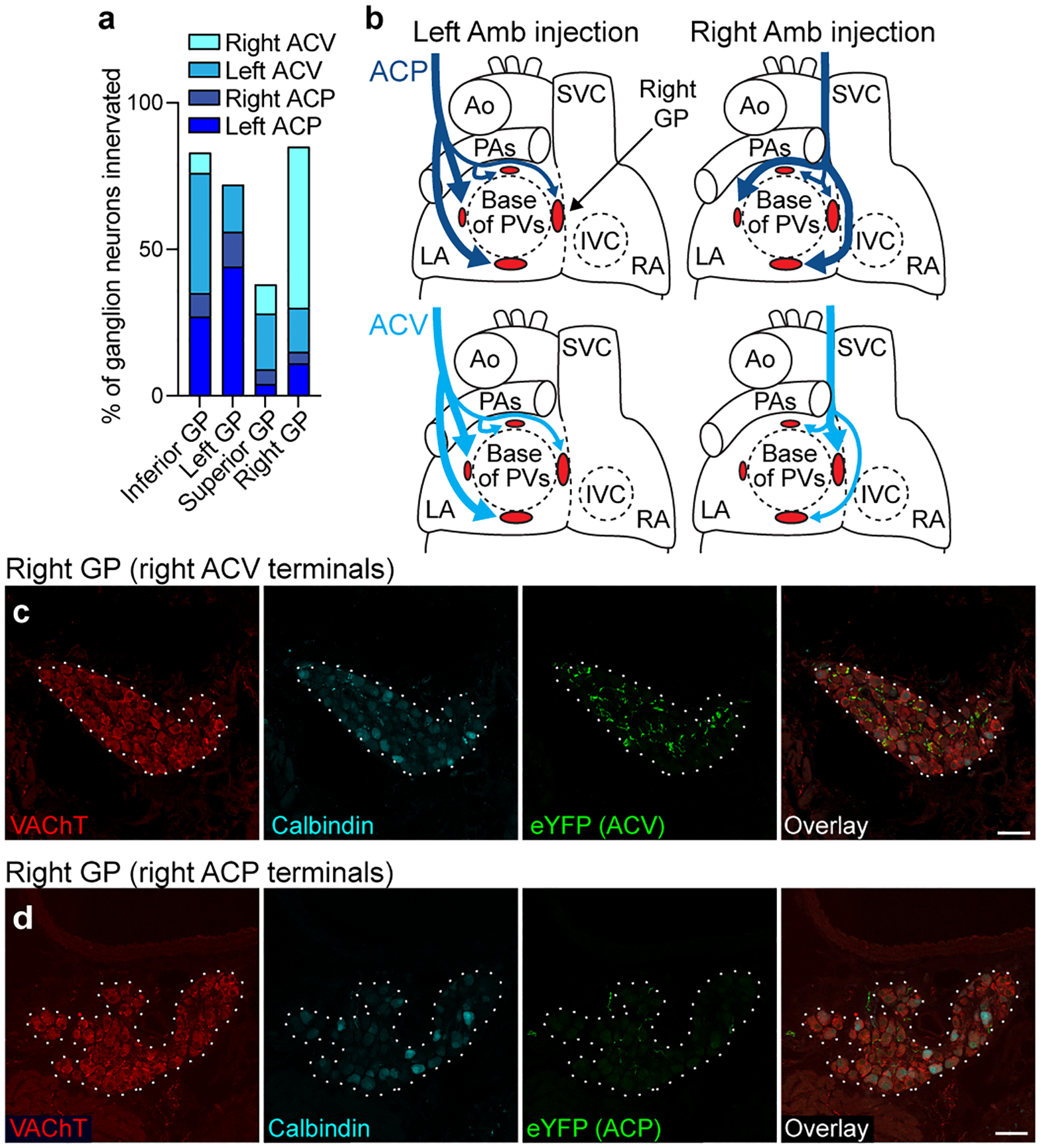
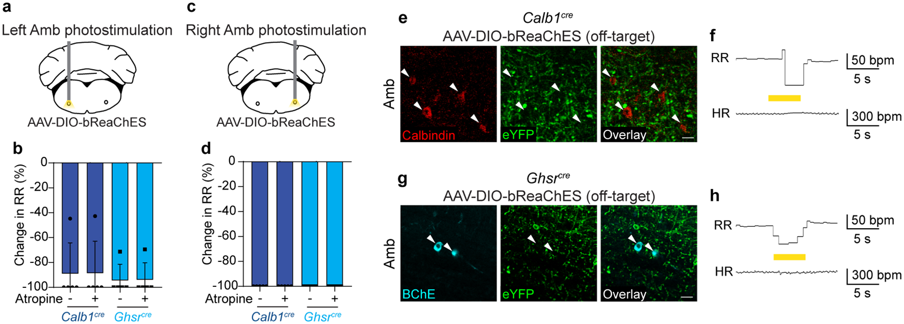
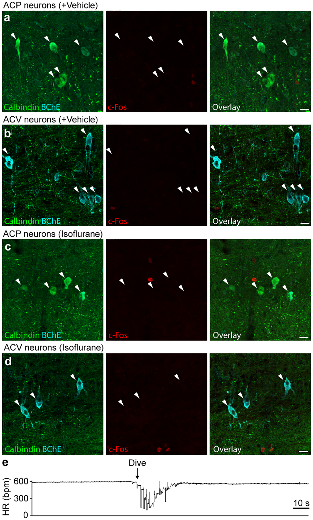
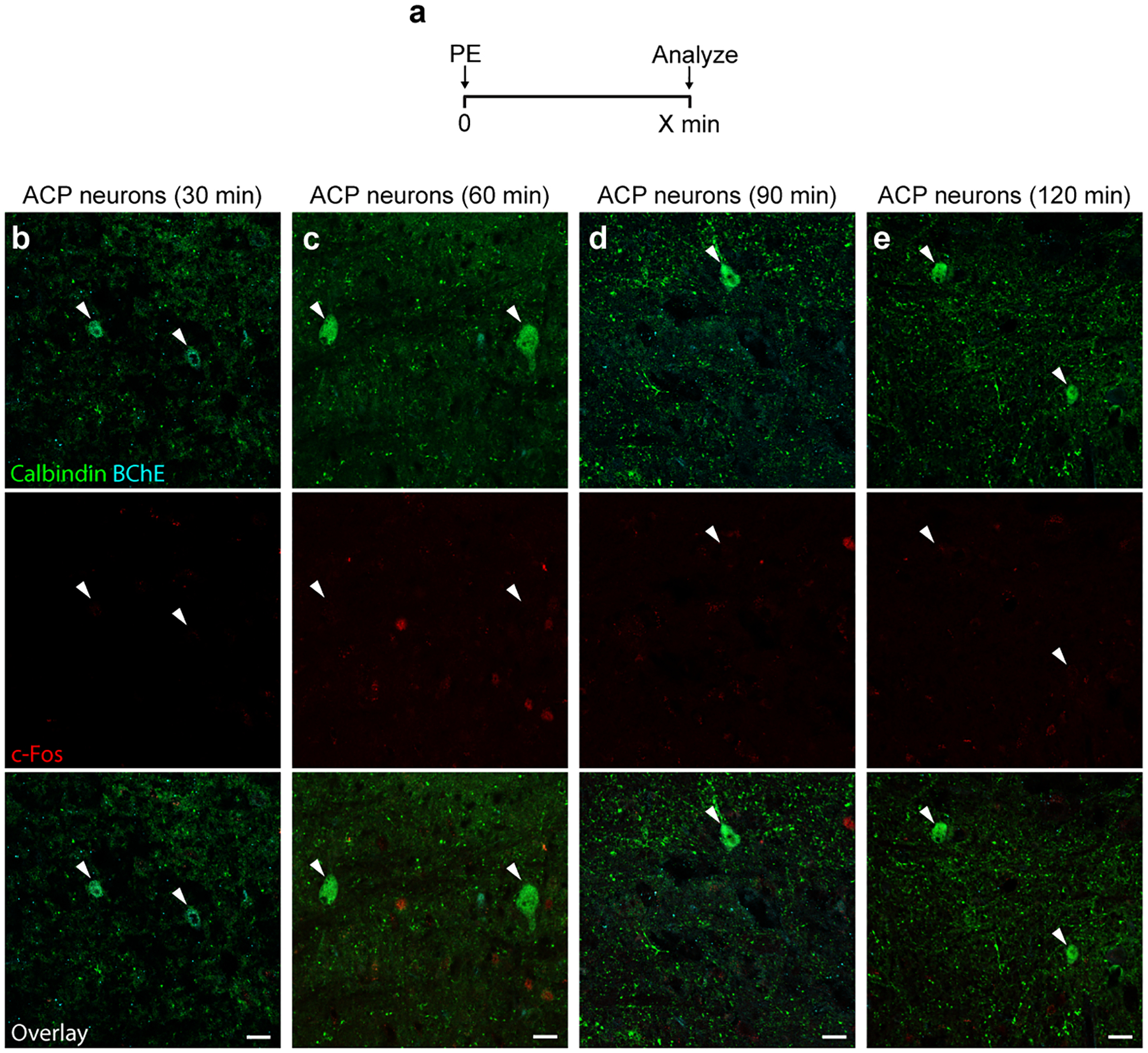
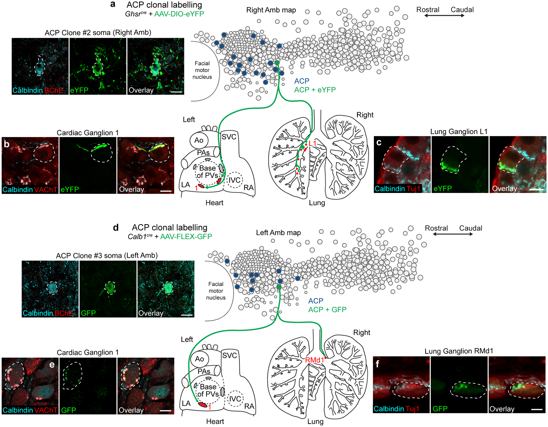
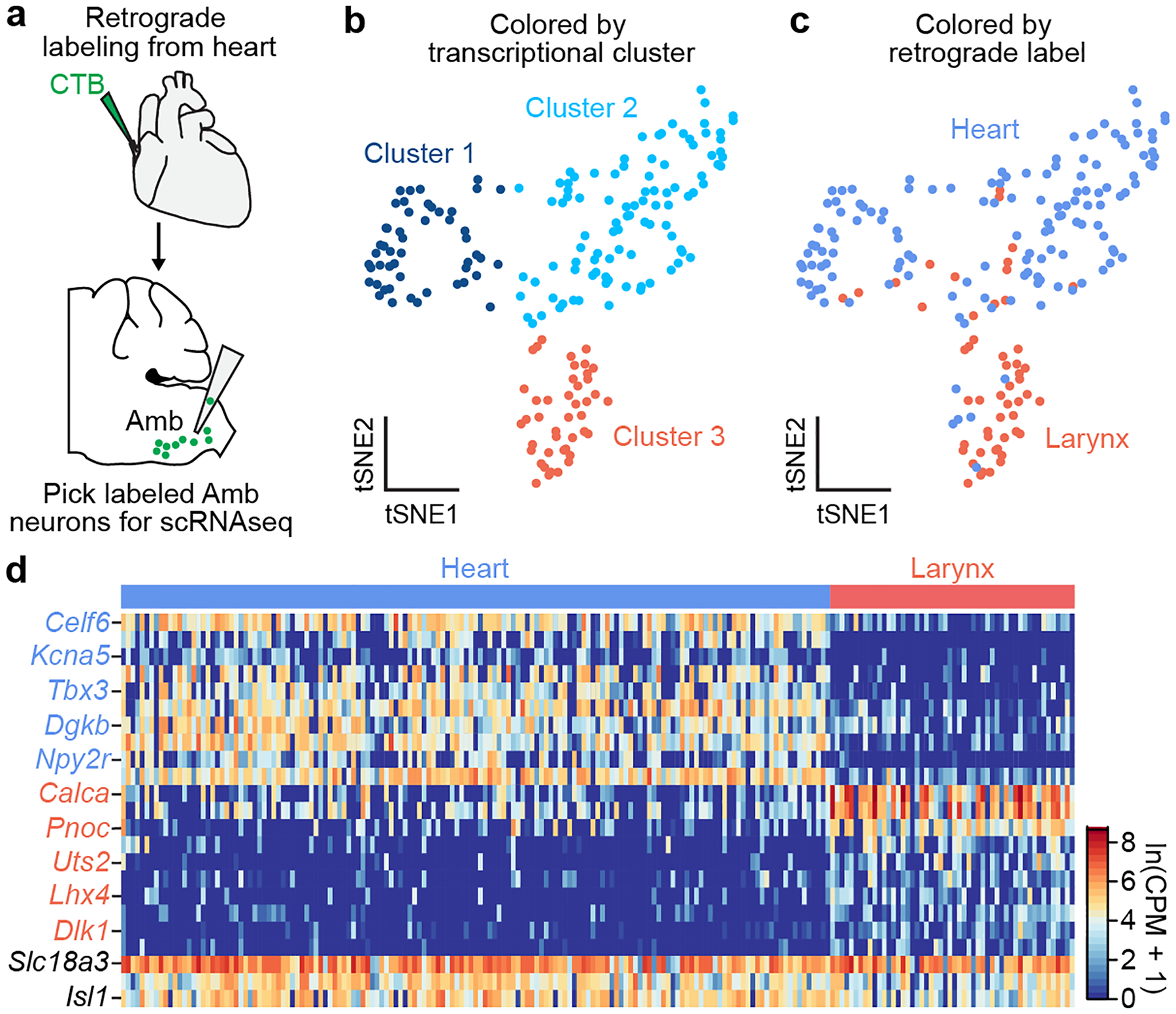
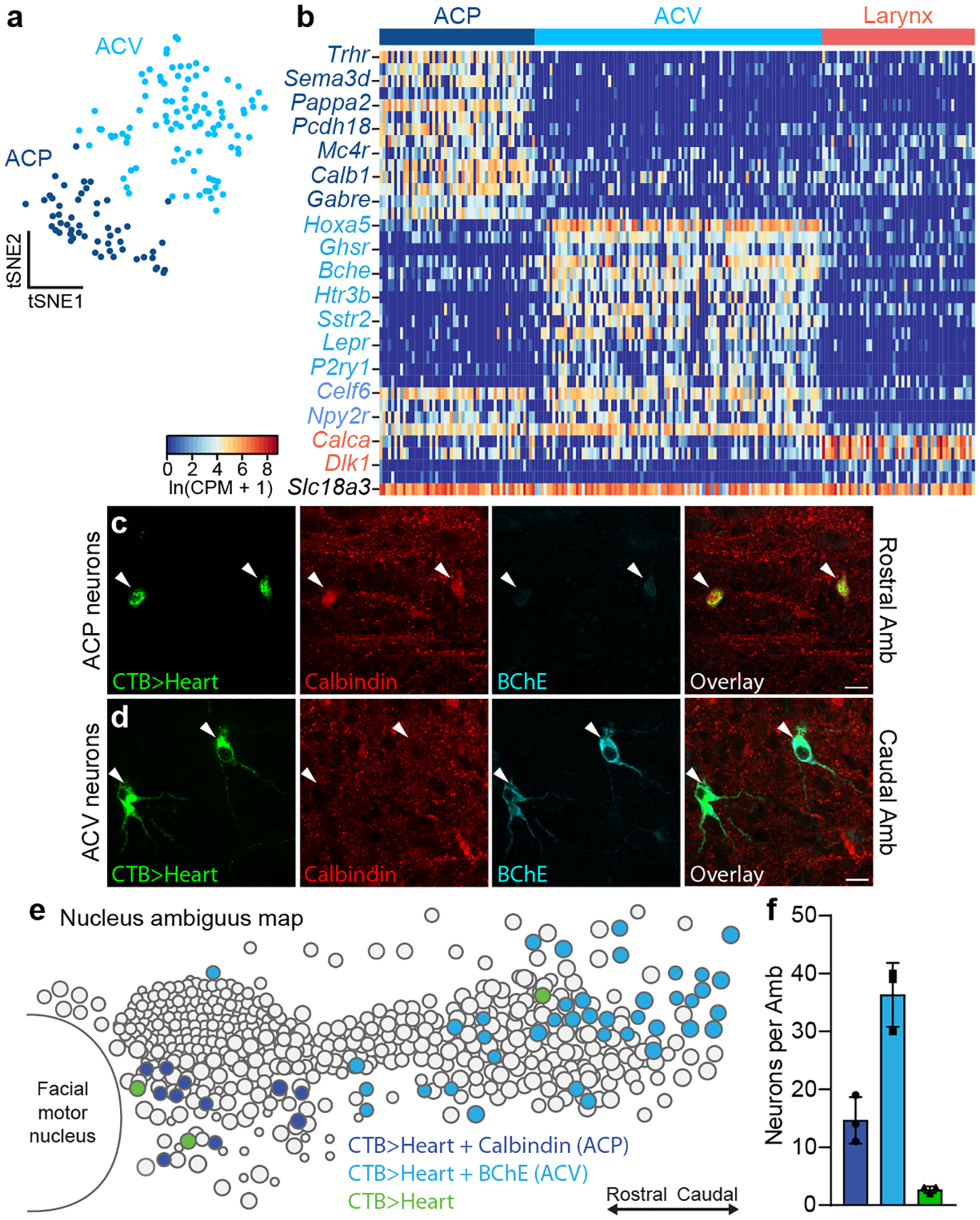
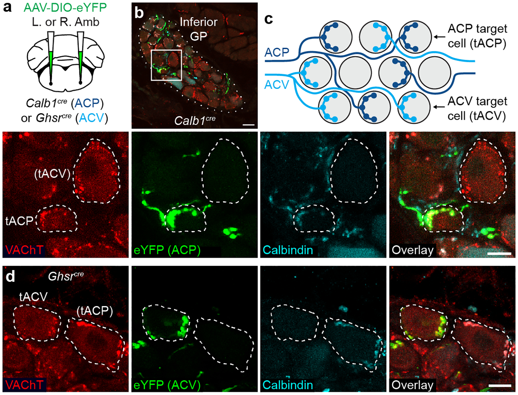
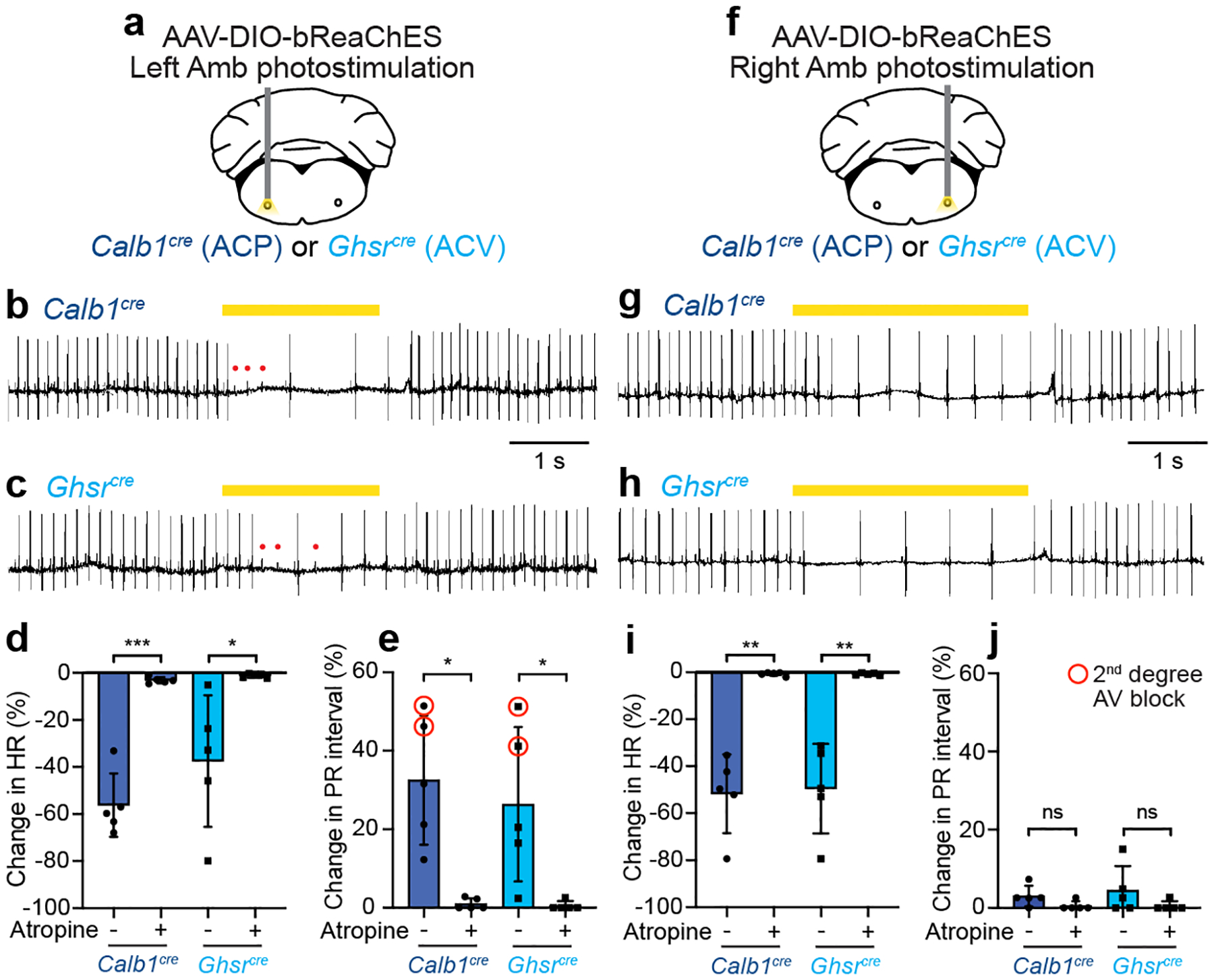
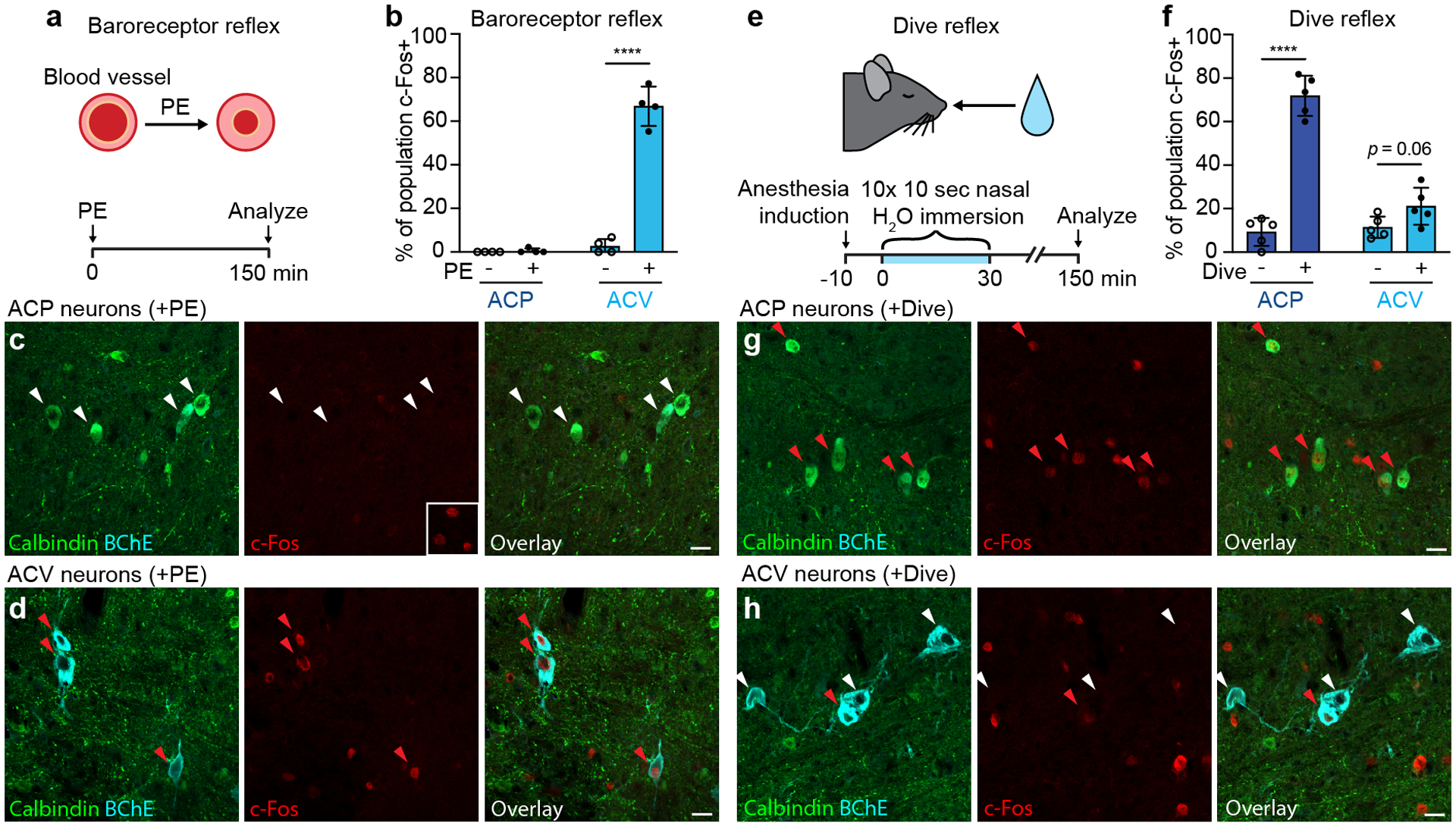
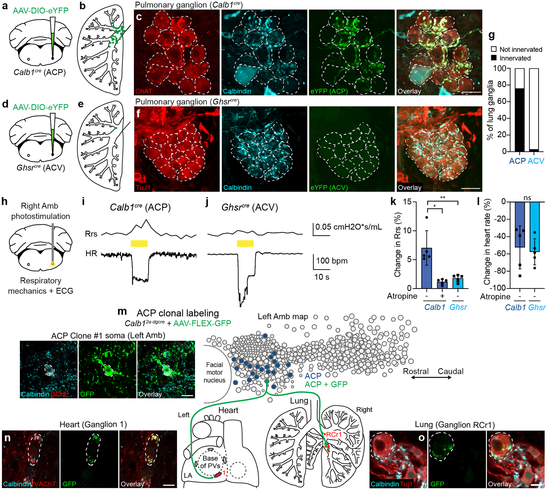
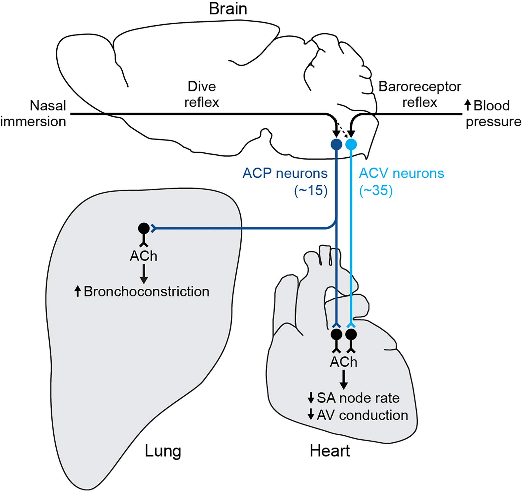
The sympathetic and parasympathetic nervous systems regulate the activities of internal organs1, but the molecular and functional diversity of their constituent neurons and circuits remains largely unknown. Here we use retrograde neuronal tracing, single-cell RNA sequencing, optogenetics and physiological experiments to dissect the cardiac parasympathetic control circuit in mice. We show that cardiac-innervating neurons in the brainstem nucleus ambiguus (Amb) are comprised of two molecularly, anatomically and functionally distinct subtypes. The first, which we call ambiguus cardiovascular (ACV) neurons (approximately 35 neurons per Amb), define the classical cardiac parasympathetic circuit. They selectively innervate a subset of cardiac parasympathetic ganglion neurons and mediate the baroreceptor reflex, slowing heart rate and atrioventricular node conduction in response to increased blood pressure. The other, ambiguus cardiopulmonary (ACP) neurons (approximately 15 neurons per Amb) innervate cardiac ganglion neurons intermingled with and functionally indistinguishable from those innervated by ACV neurons. ACP neurons also innervate most or all lung parasympathetic ganglion neurons-clonal labelling shows that individual ACP neurons innervate both organs. ACP neurons mediate the dive reflex, the simultaneous bradycardia and bronchoconstriction that follows water immersion. Thus, parasympathetic control of the heart is organized into two parallel circuits, one that selectively controls cardiac function (ACV circuit) and another that coordinates cardiac and pulmonary function (ACP circuit). This new understanding of cardiac control has implications for treating cardiac and pulmonary diseases and for elucidating the control and coordination circuits of other organs.


Conflict of interest statement

Competing Interests

The authors declare no competing interests.

Figures

Extended Data Figure 1.
Extended Data Figure 1.. Distribution of cardiac-innervating neurons in the nucleus ambiguus (Amb).
a, Sagittal schematic view displaying locations of Amb neurons (circles) within a postnatal day 2 Amb nucleus. Map is an overlay showing the locations of Amb neurons from all sagittal sections spanning a single Amb nucleus. Green fill circles, cardiac Amb neurons retrograde labeled by cholera toxin B (CTB) injection into pericardial space (CTB>Heart). Grey fill circles, other Amb neurons. Note cardiac Amb neurons localize primarily to “external formation” of Amb, surrounding the principal rostral-caudal column of Amb neurons. b, Representative sagittal section of Amb with AmbCardiac neurons labeled in green by retrograde labeling by CTB injection in heart (CTB>Heart). Retrograde labeled cells localized to the Amb external formation. AmbC, nucleus ambiguus compact formation. nVII, facial motor nucleus. Bar, 100 μm.
Extended Data Figure 2.
Extended Data Figure 2.. Quality control data for Amb single cell RNAseq.
a, Aligned reads per cell in combined AmbCardiac and AmbLaryngeal dataset. Cells with less than 500,000 reads were excluded from analysis. b, Genes detected per cell. c, Percent of reads per cell that aligned to ERCC spike-in RNA. d, Pan-neuronal genes Nefl, Tubb3, Snap25, and cholinergic gene Slc18a3 were highly expressed across all clusters from Fig. 1b. e, Glial genes were expressed at similarly low levels across the 3 clusters from Fig. 1b, indicating they did not contribute to clustering. Gja1; astrocyte marker, C1qc; microglia marker, Pdgfra; oligodendrocyte precursor cell marker, Opalin; oligodendrocyte marker. f, Examples of marker genes that significantly contributed to clustering of the 3 neuron types (Tbx3; AmbCardiac marker, Pappa2; ACP marker, Hoxa5; ACV marker, Calca; AmbLaryngeal marker).
Extended Data Figure 3.
Extended Data Figure 3.. AmbCardiac markers are expressed in other brainstem parasympathetic nuclei.
In situ hybridization data (from Allen Brain Atlas) showing expression (dark purple) of the indicated AmbCardiac-specific genes (Celf6, Kcna5, Tbx3, Dgkb, from Fig. 1) in parasympathetic neurons across the brainstem. Top row, Amb; middle row, dorsal motor nucleus of the vagus (10N); bottom row, lacrimal and salivatory nuclei (Lac/Sal). Note the genes are expressed in a subset of Amb neurons (top row panels), as predicted by scRNAseq (Fig. 1), but also in parasympathetic neurons of the dorsal motor nucleus of vagus (10N) that innervate thoracic and abdominal viscera (second row panels). Note absence of expression in the adjacent somatic motor neurons of the hypoglossal nucleus (12N). All four genes are also expressed in parasympathetic neurons of the facial nerve controlling the lacrimal and salivary glands (Lac/Sal) (third row panels) but had absent or lower expression in the adjacent facial motor nucleus (7N). Allen Brain Atlas images obtained from available postnatal day 56 sections (Celf6, Kcna5, Dgkb) and embryonic day 18.5 sections (Tbx3). Amb sections are sagittal except for Celf6 (coronal). 10N and Lac/Sal sections are sagittal. Bar of 200 μm applies to all sections.
Extended Data Figure 4.
Extended Data Figure 4.. Conservation of ACP and ACV markers across postnatal development.
a, Immunostaining of ACP neurons in the rostral Amb of a neonatal (postnatal day 2) mouse. ACP neurons stained positive for calbindin (red) and stained weakly positive for BChE (cyan). b, Immunostaining of neonatal ACV neurons in the caudal Amb of a postnatal day 2 mouse. ACV neurons stained positive for BChE and negative for calbindin. Bars, 20 μm. c, Map of ACP and ACV neurons in neonatal Amb. Sagittal schematic view showing overlay of soma of all neurons (circles) across all sections spanning a single neonatal (postnatal day 2) Amb that was stained for ACP marker calbindin (dark blue circles) and ACV marker BChE (light blue circles). d, Quantification of absolute numbers of ACP neurons (calbindin+, dark blue) and ACV neurons (BChE+, light blue) per Amb in neonatal mice (mean ± s.d., n = 3 mice). e, Immunostaining of ACP neurons in the rostral Amb of an adult (postnatal day 60) mouse. ACP neurons stained positive for calbindin (red) and stained weakly positive for BChE (cyan). f, Immunostaining of adult ACV neurons in the caudal Amb of a postnatal day 60 mouse. ACV neurons stained positive for BChE and negative for calbindin. Bars, 20 μm. g, Map of ACP and ACV neurons in adult Amb. Sagittal schematic view showing overlay of soma of all neurons (circles) across all sections spanning a single adult (postnatal day 60) Amb that was stained for calbindin (dark blue) and BChE (light blue). Note similarity in marker expression and cell type distribution in neonatal and adult Amb. h, Quantification of absolute numbers of ACP neurons (calbindin+, dark blue) and ACV neurons (BChE+, light blue) per Amb in adult mice (mean ± s.d., n = 3 mice).
Extended Data Figure 5.
Extended Data Figure 5.. Viral targeting of ACP or ACV neurons.
a, Combined immunostaining and smFISH showing overlap between BChE protein expression (cyan) and Ghsr mRNA expression (purple) in ACV neurons (dashed outlines). Bar, 100 μm. b, Quantification of panel a showing fraction of BChE+ ACV neurons that express Ghsr in Amb (n = 3 mice, 21 neurons total). c, Strategy for targeting ACP or ACV neurons. A Cre-dependent AAV encoding the opsin bReaChES fused to eYFP was delivered to the left or right Amb in Calb1cre mice (to target ACP neurons) or Ghsrcre mice (to target ACV neurons). d, Targeting specificity of AAV-DIO-bReaChES-eYFP vector used for left- and right-sided terminal mapping and optogenetics experiments in Figs. 3–4 (n = 10 Calb1cre, n = 10 Ghsrcre mice, n = 612 neurons total, mean ± s.d.). eYFP expression on the correct side of the brainstem was verified for all mice, and % of population eYFP+ was calculated for the unilateral (injected) Amb. When injected into Calb1Cre mice, ACP (calbindin+) neurons were specifically labeled. When injected into Ghsrcre mice, ACV (BChE+) neurons were specifically labeled, though note lower efficiency than the ACP neuron strategy. e, Immunostaining of Rostral Amb ACP neurons in Calb1cre mice injected with AAV-DIO-bReaChES-eYFP vector. Two calbindin+ neurons were eYFP-positive (arrowheads), indicating bReaChES-eYFP expression. f, Immunostaining of caudal Amb ACV neurons in Ghsrcre mice injected with AAV-DIO-bReaChES-eYFP vector. A BChE+ neuron was eYFP-positive (arrowhead), indicating bReaChES-eYFP expression. Bars, 20 μm. g, Distribution of eYFP expression after rostral Amb injection of AAV-DIO-bReaChES-eYFP in a Calb1cre mouse. Note eYFP expression in the Amb external formation (AmbEx) and in overlapping Bötzinger complex (BötC) and pre-Bötzinger complex (preBötC) breathing control regions, with sparing of Amb compact formation (AmbC, esophageal motor neurons), Amb semicompact formation (AmbSc, pharyngeal motor neurons), Amb loose formation (AmbL, laryngeal motor neurons), facial motor nucleus (nVII), retrotrapezoid nucleus (RTN), and lateral reticular nucleus (LRt). Bar, 100um. h, Sagittal brainstem section showing lack of eYFP expression in dorsal motor nucleus of vagus (10N) and nucleus of the solitary tract (Sol) after AAV-DIO-bReaChES-eYFP injection into Amb in Calb1cre mouse. Bar, 100um. i, Whole-mount immunostaining showing minimal eYFP expression in the nodose-jugular complex (NJC) after AAV-DIO-bReaChES-eYFP injection into Amb in Calb1cre mouse. Bar, 100 μm. j, Distribution of eYFP expression after caudal Amb injection of AAV-DIO-bReaChES-eYFP in a Ghsrcre mouse. Note eYFP expression in AmbEx, AmbL, and in overlapping BötC and preBötC, similar to Calb1cre mice in panel g but with a more caudal distribution. Bar, 100um. k, Sagittal brainstem section showing lack of eYFP expression in 10N and Sol after AAV-DIO-bReaChES-eYFP injection into Amb in Ghsrcre mouse. Bar, 100um. l, Whole-mount immunostaining showing lack of eYFP expression in the NJC after AAV-DIO-bReaChES-eYFP injection into Amb in Ghsrcre mouse. Bar, 100 μm.
Extended Data Figure 6.
Extended Data Figure 6.. Cardiac GP projection targets of ACP and ACV neurons.
a, Estimated proportions of ganglion neurons within each indicated ganglionated plexus (GP) that receive innervation from a given side and cell type, labeled as in Fig. 3 (n = 2067 neurons total, 2 mice per unilateral cell type). Remaining cells not innervated by ACP or ACV neurons are likely innervated by dorsal motor nucleus of vagus or possibly other ganglion neurons. b, Schematics (based on a) of left and right atria (LA and RA) of heart showing innervation of four cardiac GPs (red ovals) by left and right ACP (dark blue) and ACV (light blue) neurons. Thick arrows, dense innervation; thin arrows, sparse innervation. Note left and right ACP neurons innervate same set of GPs, whereas left and right ACV neurons innervated different sets of GPs. Ao, aorta; PA, pulmonary artery; PVs, pulmonary veins; SVC, superior vena cava; IVC, inferior vena cava. c, Immunostaining of the right cardiac GP (dotted outline) after right ACV fibers were labeled with eYFP. The right GP stained positive for vesicular acetylcholine transporter (VAChT), and many eYFP+ ACV fibers innervate ganglion neurons within the GP. d, Immunostaining of the right GP (dotted outline) after right ACP fibers were labeled with eYFP as in Fig. 3. In contrast to right ACV fibers, few eYFP+ fibers from right ACP neurons were found innervating right GP ganglion neurons. Bars, 50 μm.
Extended Data Figure 7.
Extended Data Figure 7.. Amb optogenetic stimulation in Calb1cre or Ghsrcre mice results in apnea mediated by non-cholinergic neurons.
Data are from same stimulation trials as Fig. 4. a, Schematic of optogenetic activation of left Amb ACP or ACV neurons in anesthetized Calb1cre or Ghsrcre mice. b, Before (−) atropine administration, optogenetic stimulation of left Amb Calb1 neurons (dark blue bars) or left Amb Ghsr neurons (light blue bars) resulted in apnea or reduction in respiratory rate (RR) (n = 5 mice per genotype). After (+) atropine administration, the apnea resulting from left Amb Calb1 or Ghsr neuron stimulation remained fully intact, indicating the effects are mediated by non-cholinergic neurons. c, Schematic of optogenetic activation of right Amb ACP or ACV neurons in anesthetized Calb1cre or Ghsrcre Mice. d, Before atropine administration, optogenetic stimulation of right Amb Calb1 neurons (dark blue bars) or right Amb Ghsr neurons (light blue bars) resulted in apnea (n = 5 mice per genotype). After atropine administration, the apnea resulting from right Amb Calb1 or Ghsr neuron stimulation remained fully intact, indicating the effects are mediated by non-cholinergic neurons. These respiratory effects are likely mediated by opsin expression in non-cholinergic interneurons of the pre-Bӧtzinger complex, an important breathing control region which overlaps significantly with Amb, contains Calb1- and Ghsr-expressing interneurons,, and where optogenetic stimulation of interneurons is known to result in apnea. e, Immunostaining of ACP neurons in Amb in a Calb1cre mouse injected with AAV-DIO-bReaChES-eYFP in Amb where injection failed to target ACP neurons (red), but still targeted nearby interneurons and fibers (green). Note lack of eYFP expression in ACP cell bodies (arrowheads). Bar, 25 μm. f, Respiratory rate (RR) and heart rate (HR) during optogenetic stimulation of interneurons surrounding ACP neurons in Calb1cre mouse from panel e. Note decrease in respiratory rate with optogenetic stimulation of interneurons (yellow bar, 40 Hz), with no changes in heart rate. bpm, breaths per minute (for RR) or beats per minute (for HR). g, Immunostaining of ACV neurons in Amb in Ghsrcre mouse injected with AAV-DIO-bReaChES-eYFP in Amb where injection failed to target ACV neurons (cyan), but still targeted nearby interneurons and fibers (green). Note lack of eYFP expression in ACV cell bodies (arrowheads). Bar, 25 μm. h, Respiratory rate (RR) and heart rate (HR) during optogenetic stimulation (yellow bar, 40 Hz) of interneurons surrounding ACV neurons in Ghsrcre mouse from panel g. Note decrease in respiratory rate with optogenetic stimulation of interneurons (yellow bar), with no changes in heart rate. bpm, breaths per minute (for RR) or beats per minute (for HR).
Extended Data Figure 8.
Extended Data Figure 8.. c-Fos negative control studies and heart rate response to dive reflex.
a, Immunostaining of ACP neurons in rostral Amb following vehicle injection (see Fig. 5). Note ACP neurons (calbindin+, white arrowheads) are c-Fos negative. b, Immunostaining of ACV neurons in caudal Amb following vehicle injection. Note ACV neurons (BChE+, white arrowheads) are c-Fos negative. c, Immunostaining of ACP neurons in rostral Amb following isoflurane anesthesia without nasal immersion. Note ACP neurons (calbindin+, white arrowheads) are c-Fos negative. d, Immunostaining of ACV neurons in caudal Amb following isoflurane anesthesia without nasal immersion. Note ACV neurons (BChE+, white arrowheads) are c-Fos negative. Bars, 20 μm. e, Example heart rate trace recorded by ECG during dive reflex activation for Fig. 5 experiments. Isoflurane-anesthetized mouse underwent nasal immersion (arrow, start of dive) for 10 s. Bradycardia and AV block were observed during nasal immersion, and heart rate returned to baseline following cessation of immersion.
Extended Data Figure 9.
Extended Data Figure 9.. ACP neurons are not activated early after phenylephrine injection.
a, Experimental time course paradigm. Phenylephrine (PE) (10 mg/kg, IP) was injected into awake mice and mice were sacrificed to perform immunostaining for c-Fos and the indicated AmbCardiac markers at the indicated time points following PE injection. b-e, ACP neurons (calbindin+, arrowheads) did not express c-Fos (red) at any of the time points (30 – 120 minutes) following PE injection. Bars, 20 μm.
Extended Data Figure 10.
Extended Data Figure 10.. Clonal analysis of ACP neurons.
a, ACP clonal labeling experiment in Ghsrcre mouse injected with AAV-DIO-eYFP. Left, immunostaining of the soma (dashed outline) in the right Amb of the single eYFP-labeled ACP neuron in this mouse (Clone #2); note it co-stained positive for calbindin (cyan) and negative for BChE (red), confirming ACP identity. Bar, 20 μm. Right, map of ACP neurons (dark blue fill circles) in right Amb (overlay of all sagittal sections of right Amb) showing location of the eYFP-labeled ACP clone (green fill circle). Terminals of the ACP clone (green fibers) were mapped in the heart (left) and lung (right), where it was found to innervate parasympathetic ganglia in both organs (red fill ovals/circles, targeted ganglia). LA, left atrium; RA, right atrium; Ao, aorta; PA, pulmonary artery; PVs, pulmonary veins; SVC, superior vena cava. b, Immunostaining of parasympathetic cardiac ganglion (Ganglion 1) showing a ganglion neuron (dashed outline) innervated by ACP Clone #2. Note innervated neuron stained positive for cholinergic marker VAChT (red) and receives innervation from a cholinergic (red), calbindin-positive (cyan) fiber labeled with eYFP (green). Bar, 10 μm. c, Immunostaining of lung parasympathetic ganglion (Ganglion L1) showing a ganglion neuron (dashed outline) innervated by ACP Clone #2. Note innervated neuron stained positive for neuronal marker Tuj1 (red) and receives innervation from a calbindin-positive (cyan), eYFP-positive (green) fiber. Bar, 10 μm. d, ACP clonal labeling experiment in Calb1cre mouse injected with limiting dose of AAV-FLEX-GFP. Left, immunostaining of the soma (dashed outline) in the L Amb of the single GFP-labeled ACP neuron in this mouse (Clone #3); note it co-stained positive for calbindin (cyan) and negative for BChE (red), confirming ACP identity. Bar, 20 μm. Right, map of ACP neurons (dark blue fill circles) in left Amb showing location of the GFP-labeled ACP clone (green fill circle, Clone #3). Terminals of the ACP clone (green fibers) were mapped in the heart (left) and lung (right), where it was found to innervate parasympathetic ganglia in both organs (red fill ovals/circles, targeted ganglia). e, Immunostaining of parasympathetic cardiac ganglion (Ganglion 1) showing a ganglion neuron (dashed outline) innervated by ACP Clone #3. Note innervated neuron stained positive for cholinergic marker VAChT (red) and receives innervation from a cholinergic (red), calbindin-positive (cyan) fiber labeled with GFP (green). Bar, 10 μm. f, Immunostaining of lung parasympathetic ganglion (Ganglion RMd1) showing a ganglion neuron (dashed outline) innervated by ACP Clone #3. Note innervated neuron stained positive for neuronal marker Tuj1 (red) and receives innervation from a calbindin-positive (cyan), GFP-positive (green) fiber. Bar, 10 μm.
Figure 1.
Figure 1.. scRNAseq of AmbCardiac neurons identifies a genetic signature of brainstem parasympathetic neurons.
a, Strategy for isolating AmbCardiac neurons. Fluorescent CTB was injected into the pericardial space, and 1–3 days later the retrograde-labeled cardiac-innervating neurons in the nucleus ambiguus (AmbCardiac neurons) were aspirated from acute physiological brainstem slices, then processed for scRNAseq. In separate mice (not shown), CTB was injected into the cricothyroid laryngeal muscle to similarly visualize, aspirate, and process control AmbLaryngeal neurons. b, t-distributed stochastic neighbor embedding (tSNE) plot comparing Amb neuron scRNA-seq expression profiles (dots). Note three transcriptionally distinct neuronal clusters defined by nearest neighbor analysis. c, tSNE plot of same Amb neurons as (b) but colored by their retrograde label origin. AmbLaryngeal neurons largely comprise cluster 3, whereas AmbCardiac neurons were largely split between clusters 1 and 2. d, Heat map showing log-transformed expression levels of selected genes differentially expressed between AmbCardiac and AmbLaryngeal neurons. Names in black, expected pan-Amb genes. Mouse brain image and those in Figs. 3, 4, 6, 7 and Extended Data Figs. 5 and 7 are reproduced from the Paxinos and Franklin atlas.
Figure 2.
Figure 2.. Two molecularly and anatomically distinct types of AmbCardiac neurons.
a, tSNE plot of AmbCardiac neuron expression profiles (dots). Nearest neighbor analysis defined two molecularly distinct clusters designated ACP (dark blue) and ACV (light blue) for reasons described later. b, Heat map showing expression of selected genes enriched in ACP neurons (dark blue gene names), ACV neurons (light blue), all AmbCardiac neurons (purple), all AmbLaryngeal neurons (“Larynx”, coral), and all Amb neurons (black). c, Immunostaining for ACP marker Calbindin and ACV marker BChE in rostral Amb. Rostral Amb neurons (arrowheads) labeled by intrapericardial CTB stained positive for Calbindin but expressed low levels of BChE, indicating they were ACP neurons. d, Immunostaining for ACP marker Calbindin and ACV marker BChE in caudal Amb of same sample as panel c. Caudal Amb neurons (arrowheads) labeled by intrapericardial CTB expressed high levels of BChE but stained negative for Calbindin, indicating they were ACV neurons. Bars, 20 μm. e, Map of ACP and ACV neurons in Amb. Sagittal schematic view showing soma of all neurons (circles) in a representative single postnatal day 2 Amb that was retrograde labeled with CTB from the heart (CTB>Heart) and stained for ACP marker Calbindin (dark blue circles) and ACV marker butyrylcholinesterase (BChE, light blue circles). A minority of retrograde-labeled cardiac neurons (green circles) did not stain for Calbindin or BChE. f, Quantification of absolute numbers of ACP neurons (dark blue), ACV neurons (light blue), and double negative cardiac neurons (green) per Amb (mean ± s.d., n = 3 mice).
Figure 3.
Figure 3.. Cardiac ganglion innervation patterns of ACP and ACV neurons.
a, Strategy for labeling ACP or ACV cardiac terminals by delivering AAV-DIO-eYFP encoding Cre-dependent eYFP reporter (green) to left (L) or right (R) Amb in Calb1cre (ACP driver) or Ghsrcre (ACV driver) mice. b, Immunostaining of ACP terminals (labeled with eYFP in Calb1cre mouse) in inferior pulmonary veins GP (dotted outline). Bar, 20 μm. Inset: close-up of boxed region showing two cardiac ganglion neurons (dashed outlines), one (tACP, target of ACP) with eYFP-positive ACP input that was also Calbindin+ (ACP marker) and vesicular acetylcholine transporter (VAChT)+. An adjacent cardiac ganglion neuron ((tACV), putative target of ACV) received eYFP-, Calbindin-, VAChT+ pre-ganglionic input (red puncta), indicating it was not innervated by an ACP, but likely by an ACV neuron. Bar, 10 μm. c, Schematic of typical ACP and ACV innervation pattern of individual cardiac ganglion neurons (grey circles) within a GP. ACP fibers (dark blue) provide all cholinergic input for tACP neurons. ACV fibers (light blue) provide all innervation for tACV neurons, which are intermingled with tACP neurons. d, Immunostaining of ACV terminals (labeled as above with eYFP in a Ghsrcre mouse) in two cardiac ganglion neurons (dashed outlines), one (tACV) with eYFP+, Calbindin-, VAChT+ ACV input. An adjacent cardiac ganglion neuron, (tACP), received eYFP-, Calbindin+, VAChT+ input, so was not innervated by an ACV but likely by an ACP neuron. Bar, 10 μm.
Figure 4.
Figure 4.. Cardiac effects of optogenetic activation of left and right ACP and ACV neurons.
a, Strategy for optogenetic activation of left ACP or ACV neurons. AAV-DIO-bReaChES was injected into left Amb of Calb1cre or Ghsrcre mice to express channelrhodopsin bReaChES in ACP (Calb1cre) or ACV neurons (Ghsrcre), which were photostimulated while recording ECG. b, ECG trace during optogenetic stimulation of left ACP neurons in a Calb1cre mouse. During stimulation (40 Hz, yellow bar), there was a rapid onset second-degree AV block (P waves (red dots) that failed to produce QRS complex (large inflection)). Also during stimulation, P-P interval was increased, indicating reduction in sinus rate. c, ECG trace during optogenetic stimulation of left ACV neurons in a Ghsrcre mouse. Note second-degree AV block and lengthening of P-P interval, as for left ACP stimulation. d, Quantification of effect on heart rate (HR, from P-P interval) in b, c (n = 5 mice per genotype) before (−) or after (+) muscarinic antagonist atropine (Calb1: p = 0.0009, Ghsr, p = 0.04). e, Quantification of effect on AV node conduction in b, c. Both cell types increased P-R interval (first-degree AV block) and caused second-degree AV block in some animals (red circled data points), and effects were abolished by atropine (Calb1: p = 0.01, Ghsr, p = 0.04). f-j, Strategy (f) and effects (g-j) of optogenetic activation of right ACP or ACV neurons (n = 5 mice per genotype), as for left ACP and ACV neurons in a-e (Calb1 HR: p = 0.002, Ghsr HR, p = 0.005, Calb1 PR: p = 0.1, Ghsr PR, p = 0.2). Data shown as mean ± S.D. *: p < 0.05, **: p < 0.01, ***: p < 0.001, ns: not significant by paired two-tailed t test.
Figure 5.
Figure 5.. Distinct ACP and ACV activation patterns in baroreceptor and dive reflexes.
a, Experimental design for baroreceptor reflex induction. Awake wild-type mice were administered α1 receptor agonist phenylephrine (PE), which causes peripheral blood vessel vasoconstriction, activating the baroreceptor reflex. After 150 minutes to allow c-Fos expression, mice were euthanized and activity of ACP and ACV neurons analyzed by c-Fos immunostaining. b, Fraction of ACP or ACV neurons positive for c-Fos following vehicle (- PE) or phenylephrine (+ PE) injections (n = 4 mice per condition, 418 total scored neurons). ACP: p = 0.4, ACV: p < 0.0001. c, Immunostaining for c-Fos in ACP neurons (Calbindin+, white arrowheads) in rostral Amb following baroreflex induction as above. Middle panel inset, positive immunostaining control showing c-Fos+ neurons in pre-Bӧtzinger complex of same brain section. d, Immunostaining for c-Fos in ACV neurons (BChE+, red arrowheads) in caudal Amb following baroreflex induction. e, Experimental design for dive reflex induction. Wild-type mice anesthetized with isoflurane received 10 nasal immersions (5–10 sec each) in thermoneutral water over 30 minutes, with ECG recorded to confirm dive reflex activation. Control mice were similarly anesthetized but did not receive immersions. ACP and ACV neurons were immunostained for c-Fos as in a-d. f, Fraction of ACP or ACV neurons that stained positive for c-Fos without (- Dive) or with dive reflex activation (+ Dive) (n = 5 mice per condition, 485 total scored neurons). ACP: p < 0.0001, ACV: p = 0.06. Note robust activation of ACP neurons but limited activation of ACV neurons. g, Immunostaining for c-Fos in ACP neurons (red arrowheads) following dive reflex induction. h, Immunostaining for c-Fos in ACV neurons following dive reflex induction. Note most ACV neurons are c-Fos- (white arrowheads), but rare ACV neurons are c-Fos+ (red arrowhead). Data shown as mean ± S.D. ****: p < 0.0001 by unpaired two-tailed t test. Bars, 20 μm.
Figure 6.
Figure 6.. ACP neurons innervate the lung and mediate bronchoconstriction.
a, Visualizing ACP lung terminals by injecting AAV-DIO-eYFP into Amb of Calb1cre mice. b, Locations in lung of ACP terminals. Green dots, overlay of all innervated ganglia from 5 mice. c, Immunostaining of innervated cholinergic pulmonary ganglion. Ganglion neurons (dashed outlines, expressing choline acetyltransferase (ChAT)) receive cholinergic innervation (red puncta) from Calbindin+, eYFP+ ACP neurons. Bar, 25μm. d, Visualizing ACV lung terminals in Ghsrcre mice. e, Locations in lung of ACV terminals. Green dots, overlay of all innervated ganglia from 5 mice. f, Immunostaining of representative pulmonary ganglion. Ganglion neurons (dashed outlines), expressing beta-tubulin III (TuJ1), receive Calbindin+ input (likely from ACP neurons) but no eYFP+ (ACV) input. Bar, 25μm. g, Percent of lung ganglia innervated by ACP or ACV neurons (n=33 (ACP) or 34 (ACV) ganglia, 5 mice each). h, Strategy for ACP or ACV optogenetic activation while measuring lung (respiratory mechanics) and heart (ECG) function. Right Amb of Calb1cre or Ghsrcre mice was targeted with AAV-DIO-bReaChES, then ACP or ACV neurons were activated via fiber optic. (i,j) Traces of total lung resistance (Rrs) and heart rate (HR) during optogenetic stimulation (yellow bar, 20 Hz) of ACP (i) or ACV (j) neurons. (k,l) Quantification of Rrs (k) and HR (l) changes measured simultaneously during optogenetic stimulation of ACP (Calb1) or ACV (Ghsr) neurons (n=5 mice each). Values are mean ±S.D. **, p=0.005; *, p=0.01; ns: not significant (p=0.7) by unpaired (Calb1 vs. Ghsr) or paired (- vs. +Atropine) two-tailed t test. m, ACP clonal labeling in Calb12a-dgcre mouse injected with limiting dose of AAV-FLEX-GFP. Left, immunostaining of soma in left Amb of the single GFP-labeled ACP neuron (Clone #1), dashed outline. Bar, 20μm. Right, location in left Amb of the GFP-labeled ACP clone (green circle). Blue circles, all other ACP neurons. Terminals of ACP clone (green fibers) were mapped in heart (left) and lung (right). and innervated parasympathetic ganglia in both organs (red ovals/circles, targeted ganglia). LA, left atrium; PVs, pulmonary veins. (n,o) Immunostaining of parasympathetic cardiac ganglion (n, Ganglion 1), showing ganglion neuron (dashed outline) receiving GFP+, Calbindin+, VAChT+ input from ACP Clone #1, and of lung parasympathetic ganglion (o, Ganglion RCr1), showing ganglion neuron receiving GFP+, Calbindin+ input from same clone. Bar, 10μm.
Figure 7.
Figure 7.. Parallel cardiovascular and cardiopulmonary control circuits.
ACV neurons (light blue circles, in caudal medulla) are central to the classical cardiovascular control circuit. Increased arterial blood pressure activates aortic arch baroreceptors, initiating baroreceptor reflex that activates ACV neurons. These in turn activate cholinergic cardiac ganglion neurons (black), releasing acetylcholine (ACh) that slows SA node rate and AV node conduction velocity to homeostatically maintain blood pressure. ACP neurons (dark blue circles, in rostral medulla) are central to the newly defined cardiopulmonary control circuit. Nasal water immersion activates sensory receptors in the nose, initiating the dive reflex that activates ACP neurons. These in turn activate cholinergic cardiac ganglion neurons (black) intermingled with ACV target neurons, releasing acetylcholine (ACh) that slows SA node rate and AV node conduction velocity like ACV target neurons. Single ACP neurons also project to the lung and together they provide the dominant or exclusive input to cholinergic ganglion neurons in the lung, causing simultaneous bronchoconstriction. Low level of ACV neuron activation during dive reflex suggests some ACV neurons can be additionally recruited during dive reflex, perhaps to enhance bradycardia during colder or deeper dives. In addition to these reflex pathways, ACV and ACP likely receive input from other reflex circuits and forebrain structures (not shown), and express receptors for distinct sets of circulating hormones, to regulate cardiovascular and cardiopulmonary physiology during other states.

Comment in

References

    1. Langley JN The autonomic nervous system. (W. Heffer & Sons, Ltd., 1921).
    1. Ernsberger U & Rohrer H Sympathetic tales: subdivisons of the autonomic nervous system and the impact of developmental studies. Neural Dev 13, 20 (2018). - PMC - PubMed
    1. Tao J et al. Highly selective brain-to-gut communication via genetically defined vagus neurons. Neuron (2021). - PMC - PubMed
    1. Palma JA & Benarroch EE Neural control of the heart: recent concepts and clinical correlations. Neurology 83, 261–271 (2014). - PubMed
    1. Appel ML, Berger RD, Saul JP, Smith JM & Cohen RJ Beat to beat variability in cardiovascular variables: noise or music? J. Am. Coll. Cardiol 14, 1139–1148 (1989). - PubMed