. 2022 Jun;606(7915):732-738.
doi: 10.1038/s41586-022-04782-2. Epub 2022 Jun 1.

Spatiotemporal dynamics of noradrenaline during learned behaviour

Affiliations

Spatiotemporal dynamics of noradrenaline during learned behaviour

Vincent Breton-Provencher et al. Nature. 2022 Jun.

Abstract

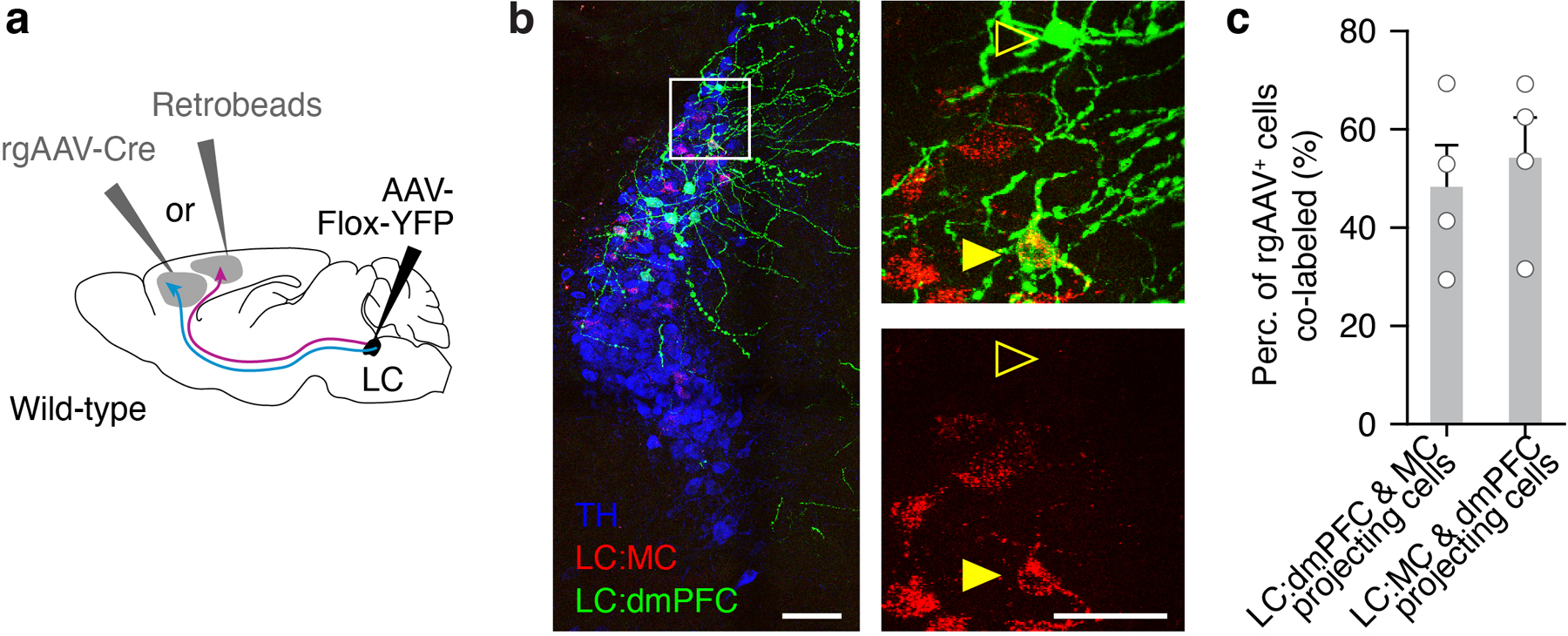
Noradrenaline released from the locus coeruleus (LC) is a ubiquitous neuromodulator1-4 that has been linked to multiple functions including arousal5-8, action and sensory gain9-11, and learning12-16. Whether and how activation of noradrenaline-expressing neurons in the LC (LC-NA) facilitates different components of specific behaviours is unknown. Here we show that LC-NA activity displays distinct spatiotemporal dynamics to enable two functions during learned behaviour: facilitating task execution and encoding reinforcement to improve performance accuracy. To examine these functions, we used a behavioural task in mice with graded auditory stimulus detection and task performance. Optogenetic inactivation of the LC demonstrated that LC-NA activity was causal for both task execution and optimization. Targeted recordings of LC-NA neurons using photo-tagging, two-photon micro-endoscopy and two-photon output monitoring showed that transient LC-NA activation preceded behavioural execution and followed reinforcement. These two components of phasic activity were heterogeneously represented in LC-NA cortical outputs, such that the behavioural response signal was higher in the motor cortex and facilitated task execution, whereas the negative reinforcement signal was widely distributed among cortical regions and improved response sensitivity on the subsequent trial. Modular targeting of LC outputs thus enables diverse functions, whereby some noradrenaline signals are segregated among targets, whereas others are broadly distributed.


Conflict of interest statement

Competing interests: The authors declare no competing interests.

Figures

Extended Data Fig.1.
Extended Data Fig.1.. Learning and execution of the go/no-go auditory detection task.
a, Probability of lever press (P(press)) for go or no-go trials as a function of number of sessions after both trial types were introduced. Each line represents a single mouse. b, Cumulative distribution of number of sessions to train mice. The dashed line indicates the mean for all mice. c, P(press) for different go/no-go tone intensities across sessions. d, P(press) for different go or no-go tone intensities (sGo or sNoGo respectively). Single dots correspond to the average performance for each tone intensity for either no-go (descending order) or go (ascending order) frequency. Single lines show unfitted single mouse data. FA: false alarms e, P(press) as a function of go/no-go tone intensity (circle) and their respective fitted data (solid line) for two example mice The fit was obtained using logistic regression for P(press) using sGo or sNoGo as regressors (see Methods). Beta weights for each regressor are indicated on the graph. Note the contrast between the intercept (β0) and slope (βgo) parameters of the logistic regression between mouse 1 and 2. f, Distribution of the different β parameters for all mice. β0 is the intercept and βgo and βnogo are the slopes resulting from the logistic regression of P(press) vs sNoGo, or sGo. *: P = 3.96*10−4 (β0), 0.0067 (βgo), and 3.96*10−4 (βnogo) calculated using two-tailed Wilcoxon test of median against zero with Bonferroni correction. g, d-prime for different tone intensities. Single lines show single mouse data. h, Top: an example session of mouse lever speed during hit or correct rejection trials aligned to tone onset. Bottom: corresponding lick rate for the same session. i, Example session of mouse lever speed sorted for different go/no-go tone intensities. j, Lever speed and reaction time as a function of go/no-go tone intensity. k, Example of optical fiber location with respect to the LC visualized with ArchT-tdTomato. Scale bar: 1 mm. l, Probability of pressing (P(press)) for different go or no-go tone intensities (sGo or sNoGo respectively) for 3 example sessions during laser on versus laser off trials. Each dot displays the average, and each solid line displays the results of the logistic regression for P(press) using sGo or sNoGo as regressors. m, Average false alarm, hit rate, and d-prime for laser off versus laser on trials for high and low stimulus intensity trials in control mice. n, P(press) at 0 dB intensity obtained by fitting the behavior with a logistic regression for control mice. o, Effect of LC-NE photoinhibition on P(early press) – or premature pressing during the delay period between the cue and the tone onset. p, Effect of LC-NE photoinhibition on reaction time. Values during laser on trials are subtracted from laser off trials. FA: False alarm. q, Session averages of pupil size traces aligned to the onset of laser illumination or control – laser off – trials. r, Average pupil size for a 4-second window during laser on or off trials. *: P = 0.016 using a two-tailed Wilcoxon test. s, Change in false alarm and hit rate at low tone intensities versus change in pupil size. Each dot represents the values for one mouse. P value for Pearson correlation = 0.41 and 0.75 for FA or hit versus pupil constriction respectively. n = 19 mice in a-d, f, g, 17 mice in j, 13 mice in l, m, and 7 mice in o-s. Data in a, d, e, g, j, p are mean ± 95% confidence intervals determined by bootstrapping. Data in c and q are mean ± s.e.m. Box and whisker plots indicate the median, the 25th and 75th percentile and the minimum to maximum values of the distribution (f).
Extended Data Fig.2.
Extended Data Fig.2.. Effect of LC-NE photoinhibition on behavior.
a, P(press) bias calculated by subtracting shuffled data from P(press) of trials following reward (blue – middle) and no reinforcement (gray – right). b, Change in beta weights obtained with logistic regression of P(Press) versus go/no-go tone intensity subtracted from the shuffled data. *: P = 0.02; ***: P = 3.9*10−4Go – post-punishment) and 6.3*10−40 – post-no reinf.) values calculated using two-tailed Wilcoxon test of data versus shuffled. c-e, Effect of tone intensity on performance bias on the subsequent trial. Difference in false alarm or hit rates and change in d-prime are shown following punishment (c), reward (d), and unreinforced trial (e). P values calculated with two-tailed Wilcoxon test of data versus shuffled (*) or one-way repeated measurement ANOVA of delta rate versus tone intensity (#) in c-e. *: P = 0.0096, 0.0065, and 0.028 (c, left to right); *: P = 0.012, 0.0074, 0.022, and 0.028 (d, left to right); *: P = 0.0005, 0.0014, 0.0074, 0.0021, 0.0096, 0.0002, 0.048, 0.018, 0.025, and 0.018 (e, left to right); #: P = 0.038 and 0.018 (d, false alarm and hit). f, Effect of photo-inhibiting LC-NE on the next trial’s P(press) bias. Data are displayed the same way as in a but for P(press) bias following LC-NE silenced trials. Left – post-reward and right - post-no reinforcement. g, Change in hit (left) or false alarm (right) rate following rewarded and non-reinforced trials with whole-trial LC-NE inhibition. h, Change in d-prime following rewarded and non-reinforced trials with whole-trial LC-NE inhibition. *: P = 0.031 using one-tailed Wilcoxon test (laser on vs. off). i, Change in intercept term (β0) and slope (βgo), calculated as in (b), with or without LC-NE photoinhibition on the previous trial. Each line represents a beta weight from one mouse for laser on/off trials. *: P = 0.031 using one-tailed Wilcoxon test (laser on vs. off). j, Change in false alarm and hit rate following rewarded trials with LC-NE inhibition during the reinforcement epoch. k, Change in d-prime following punished or rewarded trials with LC-NE inhibition during the reinforcement epoch. l, Change in false alarm and hit rate following false alarm trials as a function of days from the first go/no-go training session. m, Change in false alarm and hit rate following rewarded trials as a function of days from the first Go/No-go training session. Data were binned by 5 sessions in l, m. n, o, Effect of LC-NE photoinhibition during all reward epochs during the go-only stage of learning for 3 mice compared with the data of LC-NE intact mice. Reaction time (n) and P(press) (o) are plotted across training sessions for control mice and mice receiving LC-NE photoinhibition. n = 18 mice in a-e and l-o. n = 5 mice in f-k. Data are mean ± s.e.m. in a-f, l-o
Extended Data Fig. 3:
Extended Data Fig. 3:. Effect of unexpected reward on correct rejection trials.
a, P(press) for trials following correct rejection trials (grey) or correct rejection trials with a surprising reward (purple). b, Effect of an unexpected reward on a correct rejection trial on false alarm rate, hit rate, and d-prime on the subsequent trial. *: P = 0.011 using the normal approximation to binomial test for rewarded versus unrewarded post-correct rejection data. c, Effect of LC-NE photoinhibition on correct rejection trials with a surprising reward on false alarm rate, hit rate, and d-prime. P = 0.031, 0.563, and 0.031 for false alarm, hit and d-prime using one-tailed Wilcoxon test (laser on vs. off). Data are from 5 mice. d, Spike raster plot aligned to timing of tone for example unit on correct rejection trials (left), and correct rejection with reward trials (right). Session averaged firing rate is shown at the bottom e, Comparison of session average firing rate of a single unit on false alarm, reward, correct rejection, and correct rejection with reward trials. f, Comparison of spike rate during correct rejection with reward and false alarm trials for 3 units. Data are from concatenating 7658 and 128 control and surprise trials respectively from 5 mice in a, b. n = 5 mice in c, and 3 units in f. Data are mean ± s.e.m in b, f.
Extended Data Fig.4.
Extended Data Fig.4.. Spiking activity of photo-tagged LC-NE neurons during the task.
a, Example of two recording sites during the go/no-go task. Dbh-Cre mice were injected with Flox-YFP-ChR2 virus; the 16-channel optrode was coated with DiI to mark the recording location. Scale bar: 0.5 mm. b, The waveform of the photo-tagged units recorded for this study. The non-laser-evoked – or spontaneous – waveform is compared to laser-evoked waveform for each unit. c, Scatter plot of average firing rate and spike duration for all photo-tagged units in comparison with 141 non-identified units obtained during the same sessions. d-f, Average photo-evoked spike latency (d), jitter (e), and photo-evoked vs. spontaneous waveform correlation (f) for photo-tagged units. Each dot represents a unit and the corresponding mean ± s.e.m. is shown on the left side of each graph. g, Spike raster plot aligned to the timing of lever press for false alarm and hit trials. Session averaged firing rate is shown at the bottom. Top panel – Timing of tone, lever press, and reinforcement. For the recordings shown in this panel there was a delay of 250 ms between press and reinforcement. h, i, Mean firing rate of LC-NE photo-tagged units aligned to tone onset (h) or light cue (i) for hit, miss, false alarm, or correct rejection trials. j, Mean firing rate of LC-NE photo-tagged units aligned to lever press during false alarm and hit. The population average (solid line) and the corresponding s.e.m. (shaded area) are shown at the bottom. k, l, Raster plots of spike time-stamps and the underlying average firing rate for two example neurons plotted for all 4 go-tone intensities. m, n, Mean firing activity for a 200-ms window before press or a 100-ms window after reinforcement for go (m) or no-go trials (n). P values were calculated using two-tailed Wilcoxon signed-rank test (vs. baseline) with Bonferroni correction. In order of tone intensity, P = 2.0*10−4, 1.1*10−6, 3.1*10−8, and 2.1*10−8 for pre-press (m) and P = 0.013, 9.7*10−4, 0.0028, and 0.038 for post reward (m); P = 2.5*10−7, 3.9*10−5, 2.5*10−5, and 2.3*10−4 for pre-press (n) and P = 4.4*10−5, 6.8*10−5, 0.0053, and 0.0037 for post-punishment (n). o, Average LC-NE response as a function of animal’s exposure to the behavior, measured with the number of expert sessions (or sessions with 4 tones). Each dot is the average response of all LC units for a given session. P value for Pearson correlation = 0.151, 0.068 and 0.8205 for pre-press, post-reward or post-punishment versus number of sessions with 4 tones. p, 3 example units showing heterogeneous encoding of press, reward, and punishment by single LC-NE neurons. Top panel shows spike raster plots aligned to time of press for three individual neurons on false alarm and hit trials; bottom panel shows the average firing rate. q, Percentage of responsive units during pre-press (43/45), post-reward (16/27), and post-punishment (10/10). Different shades of gray indicate units responding with high phasic bursts (absolute firing rate above 5 Hz) and units that are significantly responsive but with a lower response (< 5Hz). r, Trial-to-trial spiking variability, measured with Fano factor, versus average response rate for pre-press, post-reward, and post-punishment. n = 45 units acquired over 15 sessions in 9 mice in b-f, h, i, o, for calculation of press activity in m, n, q, r. n = 27 units acquired over 15 sessions 9 mice, used for calculation of reinforcement activity in m, n, q, r. n = 18 units acquired over 6 sessions in 5 mice in j. Box plot parameters as in Extended data Fig. 1.
Extended Data Fig.5.
Extended Data Fig.5.. Recording LC-NE calcium activity with two-photon micro-endoscopy.
a, Example coronal slice stained with DAPI showing the location of the micro-endoscope (GRIN lens) with respect to the LC. Scale bar: 1 mm. b, Example of GCaMP6m ΔF/F signals in two LC-NE neurons recorded simultaneously. Arrow highlights the most decorrelated calcium transients in each cell. c, Raster plot aligned to timing of press during hit or false alarm trials. Pairs of columns represent two simultaneously recorded cells (LC-NE+ 1 vs. 2 or 3 vs. 4) recorded from two mice (session 1 vs. 2). Session averages for these two pairs of LC-NE cells are shown in Figure 4c. d, Population averages for same ROIs as in Figure 4e; aligned to tone for hit, miss, correct rejection, and false alarm trials. Black dashed lines delineate the three clusters (see Figure 4). e Timing of calcium spike versus average time from first lick for calcium imaging animals. f-n, Data from example LC-NE ROIs tracked over several sessions. Example ROI from reward (f-h), punishment (i-k), and press (l-n) cluster (see Figure 4e-g) tracked for 3 sessions over 7 days for false alarm and hit trials. For each ROI, we show raster plots of hit and false alarm trials aligned to timing of press (f, i, l), corresponding session averages (g, j, m), and the within-session (WS) and between-session (BS) correlation coefficient (h, k, n) from day 0 separated for false alarm and hit trials. o, WS and BS correlation coefficient from day 0 separated for false alarm (top) and hit trials (bottom). P = 0.243 and 0.864 using 2-way ANOVA assessing the effect of days from first recording over correlation coefficient. p, Signal drift index. P= 0.753 using 2-way ANOVA assessing the effect of days from first recording over signal drift index. P= 0.753 using 2-way ANOVA assessing the effect of days from first recording over signal drift index. n = 65 ROIs from 3 mice in d. n = 128 ROIs from 11 mice (GRIN and axonal data included) in e. n = 9 ROIs from 2 mice tracked over 10+ days in o, p. Data are mean ± s.e.m. in o,p.
Extended Data Fig.6.
Extended Data Fig.6.. Modeling behavioral correlates of LC-NE activity using a multiple linear regression model.
a, The timing of light cue, tone onset, lever press, reward, and punishment were used as regressors to predict the ΔF/F signal of LC-NE neurons. b, Each regressor was convolved by rectangular functions evenly spaced in time to produce the predictor matrix. c, By using Lasso regression to weight each of the 79 predictors in predicting LC-NE neuron ΔF/F signal, we obtained a set of beta weight functions. This graph shows the grouped average of beta weight for each of the 5 regressors aligned to the timing of lever press (n = 142 LC-NE cells). d, Cumulative distribution of the explained variance (E.V.) obtained using 5-fold cross-validation of our modeled ΔF/F. We predicted 41.7, 44.7, and 45.4% of the E.V. for the LC, LC:dmPFC, and LC:MC conditions respectively. As a comparison, we show the E.V. obtained from a model where trial orders were shuffled. e, Comparison of the real versus modeled ΔF/F for 4 trials taken in 3 example ROIs. f, Scatter plots of the partial model – model with one regressor removed – versus the full model E.V. (obtained with 5-fold cross validation). n = 65 (3 mice), 34 (4 mice), and 43 (4 mice) LC, LC:dmPFC, and LC:MC ROIs respectively in d and f.
Extended Data Fig.7.
Extended Data Fig.7.. Retrograde tracings of LC-NE neurons projecting to dmPFC or MC
a, Schematic of experimental design for tracing experiments. We injected rgAAV-Cre and retrobeads into dmPFC or MC and quantified co-labeled TH+ neurons in the LC. b, Representative image of TH+ LC neurons (blue) with neurons projecting to dmPFC labeled with YFP and neurons projecting to MC labeled with red retrobeads. Arrows indicate example neurons, one labeled with just YFP (outlined arrow), another with both YFP and retrobeads (filled arrow). Scale bars: 100 μm left panel, 50 μm right panels. c, Quantification of the percent of YFP+ cells co-labeled with retrobeads when rgAAV-Cre was injected in dmPFC and retrobeads were injected in MC (group 1; n = 4 mice) and when rgAAV-Cre was injected in MC and retrobeads were injected in dmPFC (group 2; n = 4 mice). Data are mean ± s.e.m. in c.
Extended Data Fig.8.
Extended Data Fig.8.. LC-NE axonal imaging correlates highly with cortical NE release.
a, Raster plots aligned to the timing of lever press during hit or false alarm trials for LC-NE:dmPFC and LC-NE:MC axons. b, Corresponding session average (shaded areas indicate s.e.m.) for the two examples shown in a. c, Population activity of all LC:dmPFC and LC:MC axons for hit, miss, correct rejection, and false alarm trials, aligned to time of tone. d, Example ROI of two axons recorded simultaneously. and the distance between them. Scale bar: 50 μm. e, Raster plots aligned to the timing of press during hit trials for the two axons shown in d. f, Trial by trial correlation versus distance between axons for all simultaneously recorded axons in the LC-NE:MC and LC-NE:dmPFC conditions. P value for Pearson correlation = 0.537. Note the similar trial-by-trial correlation between the two conditions. g, Example ROI of two segments from the same axon from the LC-NE:MC condition. Scale bar: 50 μm. h, Session average during hit (left) or false alarm (right) for the two axonal segments in g, compared with the signal from the entire axon. i, Comparison of the correlation between the signal from an axonal segment and the signal from the entire visible part of the axon. Note the high correlation for both conditions indicating that within-axon Ca2+ dynamics are low. j, Strategy and schematic for sparse labeling and imaging of GRABNE2m in the cortex. k, GRABNE2m ΔF/F signal for a full 450 × 450 μm field of view in the MC. Dashed lines indicate timing of lever press for hit or false alarm trials. l,m, Average GRABNE2m signal on hit, miss, correct rejection, and false alarm trials, aligned to time of tone in l and the timing of press in m. Solid lines and shaded areas display mean ± s.e.m. n, Normalized cross-correlation (xcorr) of axonal ΔF/F versus average GRABNE2m ΔF/F as a function of lag between the two signals during false alarm (FA) and hit trials. o, Pearson r correlation for axonal ΔF/F versus average GRABNE2m ΔF/F during false alarm and hit trials. 33/44 LC:MC axons were significantly correlated with GRABNE2m signal (P < 0.05, two-tailed, from Pearson’s correlation). n = 43 LC:MC and 34 LC:dmPFC axons from 4 mice each in c. n = 71 axonal pairs in f. n = 8 LC:MC and 7 LC:dmPFC axons from 4 mice each in i. n = average GRABNE2m signal from 4 mice in l,m,n,o. n = 43 LC:MC axons in m,o. Data are mean ± s.e.m. in b, l, m, n. Box plot parameters as in Extended data Fig. 1.
Extended Data Fig. 9.
Extended Data Fig. 9.. MC is involved in the behavioral response.
a, Muscimol (GABAA receptor agonist) or saline (control) were locally injected in the MC of both hemispheres. b, 90 to 120 minutes after injection, we tested the mouse performance on the go/no-go auditory detection task. c, Coronal slices at the level of MC showing the extent of our local injection with fluorescein, a fluorophore with a similar molecular weight than muscimol. Scale bars: 1 mm. d, Probability of pressing (P(press)) for different go or no-go tone intensities (sGo or sNoGo respectively) for an example mouse. Each dot displays the average, and each solid line displays the results of the logistic regression for P(press) using sGo or sNoGo as regressors. e, Change in P(press) following muscimol injections in MC from saline injected controls. Data are mean ± 95% confidence intervals determined by bootstrapping. P values were calculated using two-tailed Wilcoxon signed-rank test (vs. baseline). f, Change in average false alarm, hit rate, and d-prime following muscimol injection in MC. P = 0.031 (false alarm), 0.016 (hit), and 0.016 (d-prime) using one-sided Wilcoxon test of saline versus muscimol condition. g, Coronal sections at the LC, MC – forelimb, and dmPFC levels showing Jaws-tdTomato in LC and fiber location above MC and dmPFC. Scale bars = 1 mm. h, Example axonal expression of Jaws-tdTomato in the dmPFC. Scale bar = 20 μm. i, Effect of LC-NE photoinhibition on the change in d-prime following different trial types. Delta d-prime was calculated by subtracting the average d-prime measured after a certain reinforcement to the global d-prime measured by shuffling trial sequences. n = 6 mice in e,f. n = 7 and 5 mice for LC-NE:dmPFC and MC photoinhibition respectively in i.
Extended Data Fig.10
Extended Data Fig.10. Summary of spatiotemporal dynamics of LC-NE in learned behavior.
a, In a sensory-motor task, LC-NE neurons are transiently activated during the execution (lever press) and following a positive or negative reinforcement. The execution activity scales up while the reward response scales down with sensory evidence. Negative reinforcement produces the largest LC-NE response during the task regardless of sensory evidence. b, Temporal (top) and spatial (bottom) dynamics of LC-NE during learned behavior. LC-NE signals to cortical outputs are targeted modularly to motor cortex during press and distributed focally or broadly following reward or punishment respectively. These distinct spatiotemporal dynamics facilitate task execution (lever movement) and serial response bias.
Fig. 1.
Fig. 1.. LC-NE activity facilitates behavioral responses to low stimulus evidence.
a, Behavioral apparatus for head-fixed go/no-go auditory detection task. b, Summary of the trial sequence and its trial outcomes. FA: false alarm and CR: correct rejection. c, Probability of lever press (P(press)) for different go or no-go tone intensities (sgo or snogo respectively). Single dots correspond to the average performance for each of 4 tone intensities for either no-go (descending order) or go (ascending order) frequency. Single lines correspond to the fitted P(press) using logistic regression for sgo or snogo (see Methods). d, Methods for photoinhibition of LC-NE activity. e, Example trial sequence showing the trial type, lever presses, and “laser on” trials. Top panel shows the timing of photoinhibition with respect to task epochs. f, P(Press) for different go/no-go tone intensities for trials with laser on or off in a mouse during one example session. g, Average false alarm, hit rate, and d-prime for laser off versus laser on trials for high and low stimulus intensity trials. h, P(press) at 0 dB intensity obtained by fitting the behavior with a logistic regression. P values in g and h calculated using two-tailed Wilcoxon test. i, Change in P(press) for different go/no-go tone intensities for trials where laser was turned on for LC-ArchT-tdTomato mice (green), and for LC-tdTomato controls (grey). P value calculated using 2-way ANOVA. n = 19 mice in c, 7 LC-ArchT mice in g-i, and 13 LC-tdTomato mice in i. Data are means ± 95% confidence intervals determined by bootstrapping (c) and means ± s.e.m. (i).
Fig. 2.
Fig. 2.. LC-NE activity promotes serial response bias.
a, Serial response bias was calculated as the change in P(press) on the subsequent trial following either punishment, reward, or no reinforcement. P(press) for trials following punishment (red), reward (blue), and no reinforcement (gray) are shown in comparison to P(press) following a shuffled order for one mouse. b, P(press) bias calculated by subtracting shuffled data from P(press) of trials following punishment. c, Change in false alarm, hit rate, and D-prime following punishment, reward, and no reinforcement. P values calculated using two-tailed Wilcoxon test of data versus shuffled. d, Timing of LC-NE photo-inhibition during full trial or reinforcement-only inactivation experiments. e, Effect of LC-NE full trial inactivation on the next trial’s P(press) bias following a punishment. Data are displayed the same way as in b. P values calculated using one-tailed Mann-Whitney U-test comparing with control bias (b) for Sgo intensities > 5dB. f, g, Effect of LC-NE whole-trial (f) or punishment-only (g) photoinhibition on the change in FA and hit rate, calculated as in c following punishment trials. P values in f and g calculated using one-tailed Wilcoxon test. Data comparable to d-g for other trial types are shown in Extended Data Fig. 2g-k. n = 18 mice (b,c), 6 mice (e) and 5 mice (f,g). Data are mean ± s.e.m (b,e) and mean ± 95% confidence intervals determined by bootstrapping (c).
Fig. 3.
Fig. 3.. Transient LC-NE neuronal activity is linked with execution and reinforcement surprise.
a, Recording the spiking activity of LC-NE neurons using photo-tagging b, Units included for analysis spiked reliably after the onset of laser illumination, and their light-elicited waveform matched non-light-evoked spikes (blue versus black lines in inset). c, Mean firing rate of LC-NE photo-tagged units aligned to lever press during false alarm and hit trials. The population average (solid line) and the corresponding s.e.m. (shaded area) are shown at the bottom. d, Average firing rate activity during a 300 ms window after tone. FA: false alarm, CR: Correct rejection. P values calculated using a two-tailed Wilcoxon signed-rank test with Bonferroni correction. e, Pre-trial tonic activity calculated over a 1 s window before the light cue, preceding any of the 4 trial types. Post-trial tonic activity calculated over a 2 s window, 3 s after the tone. P values calculated using a Kruskal-Wallis test. f, g, Spike rate of LC-NE neurons during hit or false alarm trials averaged before press (f) and after reinforcement (g) for all tone intensities. P calculated using two-tailed Mann-Whitney U-test. h, Average firing rate as a function of tone intensity for three LC-NE neurons. Single dots represent average for each tone intensity. Solid lines were obtained with least-square linear regression. Shaded areas indicate the 95% confidence interval of the regression. P-values test the significance of the correlation. i, Slope of the spike rate versus no-go (left) or go (right) tone intensities for different behavioral epochs. Slope of baseline activity is shown as control. P calculated using two-tailed Mann-Whitney U-test with Bonferroni correction. n = 45 units, 9 mice (d-f, calculation of pre-press activity in i), and 27 units, 5 mice (calculation of reinforcement activity in g, i). Box and whisker plots indicate the median, the 25th and 75th percentile and the minimum to maximum values of the distribution (d-g, i).
Fig. 4.
Fig. 4.. LC-NE activity before press and after reward is modular, while LC-NE activity after punishment is global.
a, Calcium activity of LC-NE neurons was imaged by implanting a microendoscope above the LC of Flox-GCaMP6m injected Dbh-Cre mice. b, Example 2-photon image of GCaMP6m and mCherry signals obtained through the implanted microendoscope. Scale bar: 50 μm. c, Session average ΔF/F traces aligned to the timing of press for false alarm and hit trials. Pairs of columns represent two simultaneously recorded cells (LC-NE+ 1 vs. 2 or 3 vs. 4) recorded from two mice (session 1 vs. 2). Data are mean ± s.e.m.. d, Signal correlation obtained by calculating the Pearson correlation of the average signal of all pairs of LC-NE neurons recorded during false alarm trials, hit trials or a baseline period taken during the inter-trial interval (ITI). Kruskal-Wallis test for comparing the 3 distributions. e, Left – multiple regression linear model to predict the behavioral correlates of LC-NE neurons during the task (see Extended Data Fig. 6). Right – Average calcium activity for all 65 LC-NE neurons recorded separated by each cluster. f, Average activity aligned to lever press for each cluster. g, Area under the curve (AUC) of the normalized ΔF/F curve during press and after reward or punishment for each cluster. P values calculated using one-way ANOVA. n = 197 pairs, 3 mice (d). n = 26, 15 and 24 LC-NE cells (3 mice) for press, reward, and punishment clusters respectively (e-g). Box plot parameters as in Figure 3.
Fig. 5.
Fig. 5.. LC-NE cortical outputs are modular.
a, Experimental strategy to record LC-NE axonal activity in the cortex. b, 2-photon image in the MC or dmPFC of LC-NE+ axons expressing GCaMP7b. Scale bar: 50 μm. c, Example ROIs and calcium traces of LC-NE axonal segments in fields of view shown in b. d, Comparison of the average LC-NE axonal activity in dmPFC or MC aligned to the timing of lever press during false alarm and hit trials. e, Area under the curve of the normalized DF/F signal during press and after reward or punishment for LC-NE:dmPFC or LC-NE:MC axonal segments. f, Left – The fraction of explained variance (EV) for each axon was sorted into 3 clusters as in Figure 4e. Right – Average calcium activity for all LC-NE axons recorded. Each cluster is separated by a dashed line within LC-NE:dmPFC (top) or LC-NE:MC (bottom) groups. g, Comparison of linear model contribution to LC-NE cortical axons for press, reward, and punishment predictors. h, Fraction of LC-NE axons from each cluster in LC:dmPFC and LC:MC. i, Experimental strategy to silence LC-NE axonal activity using photoinhibition. j, Average change in false alarm, hit, and d-prime for trials where laser was turned on for LC:dmPFC (top) or LC:MC (bottom). k, Change in P(Press) for different go/no-go tone intensities for trials where laser was turned on for LC:dmPFC (top) or LC:MC (bottom). n = 34 LC:dmPFC and 43 LC-NE:MC axons, 4 mice each (d-h), and 7 and 5 mice for LC:dmPFC and LC:MC photoinhibition (j,k). P values calculated with Mann-Whitney U test using Bonferroni correction for multiple comparison (e, g), normal approximation to binomial test (h), and hierarchical bootstrapping (j, k). Box plot parameters as in Figure 3 (e,g). Data are mean ± s.e.m. (d) and mean ± 95% confidence intervals determined by bootstrapping (j,k).

Similar articles

Cited by

References

    1. Poe GR et al. Locus coeruleus: a new look at the blue spot. Nature Reviews Neuroscience 21, 644–659, doi:10.1038/s41583-020-0360-9 (2020). - DOI - PMC - PubMed
    1. Aston-Jones G & Cohen JD An Integrative Theory of Locus Coeruleus-Norepinephrine Function: Adaptive Gain and Optimal Performance. Annual Review of Neuroscience 28, 403–450, doi:10.1146/annurev.neuro.28.061604.135709 (2005). - DOI - PubMed
    1. Sara SJ The locus coeruleus and noradrenergic modulation of cognition. Nature Reviews Neuroscience 10, 211–223, doi:10.1038/nrn2573 (2009). - DOI - PubMed
    1. Breton-Provencher V, Drummond GT & Sur M Locus Coeruleus Norepinephrine in Learned Behavior: Anatomical Modularity and Spatiotemporal Integration in Targets. Frontiers in Neural Circuits 15, doi:10.3389/fncir.2021.638007 (2021). - DOI - PMC - PubMed
    1. Jones BE Arousal and sleep circuits. Neuropsychopharmacology 45, 6–20, doi:10.1038/s41386-019-0444-2 (2020). - DOI - PMC - PubMed

Methods references

    1. Dana H et al. High-performance calcium sensors for imaging activity in neuronal populations and microcompartments. Nature Methods 16, 649–657, doi:10.1038/s41592-019-0435-6 (2019). - DOI - PubMed
    1. Feng J et al. A Genetically Encoded Fluorescent Sensor for Rapid and Specific In Vivo Detection of Norepinephrine. Neuron 102, 745–761.e748, doi:10.1016/j.neuron.2019.02.037 (2019). - DOI - PMC - PubMed
    1. Carlén M What constitutes the prefrontal cortex? Science 358, 478, doi:10.1126/science.aan8868 (2017). - DOI - PubMed
    1. Barthas F & Kwan AC Secondary Motor Cortex: Where ‘Sensory’ Meets ‘Motor’ in the Rodent Frontal Cortex. Trends in Neurosciences 40, 181–193, doi:10.1016/j.tins.2016.11.006 (2017). - DOI - PMC - PubMed
    1. Slotnick B A SIMPLE 2-TRANSISTOR TOUCH OR LICK DETECTOR CIRCUIT. Journal of the Experimental Analysis of Behavior 91, 253–255, doi:10.1901/jeab.2009.91-253 (2009). - DOI - PMC - PubMed