

doi: 10.1186/s12891-022-05443-1.
Comparison of electromagnetic and optical navigation assisted Endo-TLIF in the treatment of lumbar spondylolisthesis
Affiliations
- PMID: 35650587
- PMCID: PMC9158260
- DOI: 10.1186/s12891-022-05443-1
Item in Clipboard
Comparison of electromagnetic and optical navigation assisted Endo-TLIF in the treatment of lumbar spondylolisthesis
BMC Musculoskelet Disord.
.
Erratum in
-
Correction: Comparison of electromagnetic and optical navigation assisted Endo-TLIF in the treatment of lumbar spondylolisthesis.BMC Musculoskelet Disord. 2022 Jul 19;23(1):686. doi: 10.1186/s12891-022-05649-3. BMC Musculoskelet Disord. 2022. PMID: 35854288 Free PMC article. No abstract available.
Abstract
Uniportal full endoscopic posterolateral transforaminal lumbar interbody fusion (Endo-TLIF) with percutaneous pedicle screw fixation is a promising, minimally invasive method for the treatment of lumbar spondylolisthesis. However, repeated radiation exposure from X-rays and the steep learning curve remain to be improved.
Keywords: Electromagnetic navigation; Endo-TLIF; Optical navigation.
© 2022. The Author(s).
Conflict of interest statement
The authors declare that they have no Conflicts of Interest.
Figures
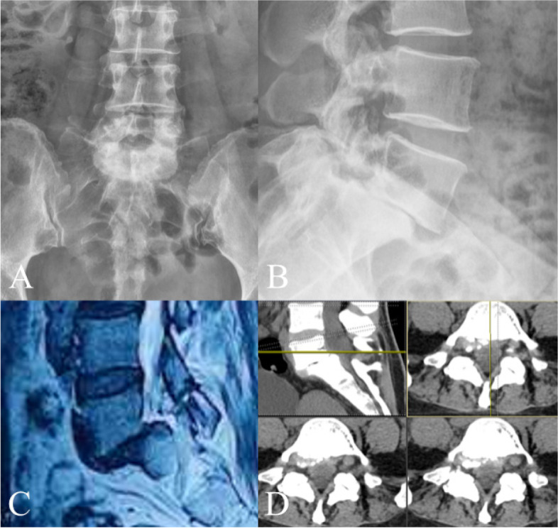
Representative case of a patient before Endo-TLIF. A and B: the fluoroscopic AP (a) and lateral (b) views. C and D: CT scan and MRI of the case with lumbar spondylolisthesis
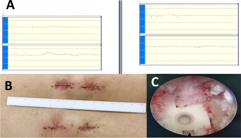
A The result of transcranial electrical stimulation-induced motor-evoked potentials (MEPs) and electromyography (EMG) monitoring during the operation. B The surgical incision. C An interbody fusion cage was observed under endoscope
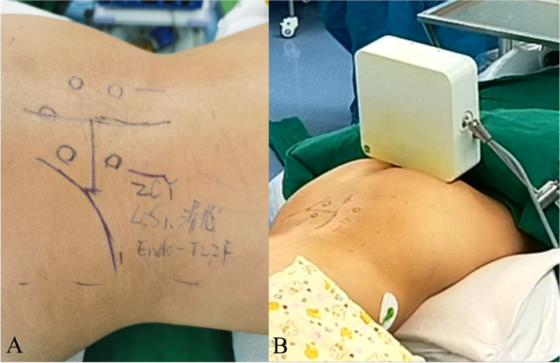
The incision plan. A According to C-arm positioning, bilateral iliac crest and pedicles were marked. B Navigation sensor frame is fixed beside operating bed

Preparation of Electromagnetic navigation. A After the field generator and a patient tracker equipped with signal coils was fixed on the ilium. B and C Anteroposterior and lateral fluoroscopic views of the lumbar segment were taken. D The software made surface matching on the respective vertebral body with the preoperative CT data in the electromagnetic coordinate system

A Access Tracker was successfully registered. B and E Guide wires insertion. In the navigation system, the procedure and track of Access Tracker is real-time visible in all spatial planes so the operator can make a quick adjustment as needed. C and D Determine the position of the guide wire and the endoscopic working channel with anteroposterior and lateral fluoroscopic views
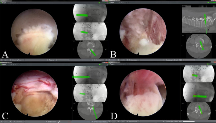
Operation field under endoscope, green indicates safe operation. A The Laminotomy was achieved via the circular saw assisted by electromagnetic navigation under the view of the endoscope. B The discectomy was achieved assisted by electromagnetic navigation under the view of the endoscope. Cartilage endplate was exposed. C The dural sac was exposed, and pulsation of the dural sac improved. D The model cage was implanted to the center of the intervertebral space in appropriate depth, with the location confirmed by electromagnetic navigation

A The circular saw was registered to be connected to the electromagnetic navigation system. B The pedicle screws were installed to replace four guide wires. C and D The position of screws and cage were verified under C-arm fluoroscopy
A case of Endo-TLIF assisted by optical navigation navigation. A and B Intraoperative C-arm image of the guide wires. B and C The position of screws and cage were verified under C-arm fluoroscopy

The pedicle screws were installed assisted by optical navigation
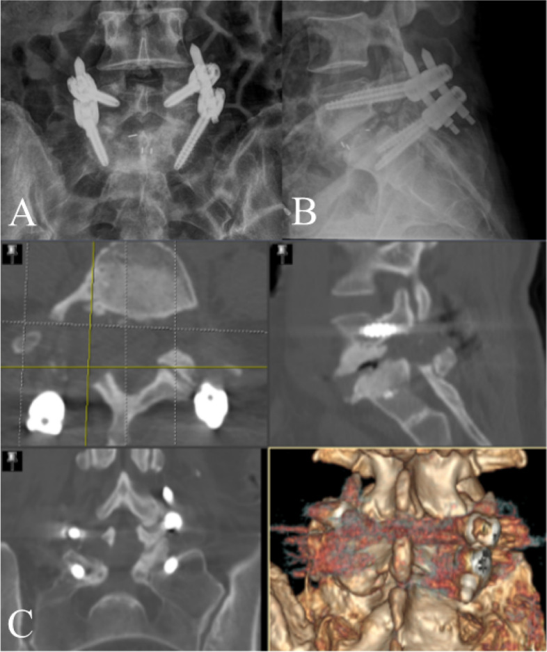
Postoperative radiographs (A and B) and CT scan (C)
References
-
- Glassman SD, Carreon LY, Ghogawala Z, Foley KT, McGirt MJ, Asher AL. Benefit of Transforaminal lumbar Interbody fusion vs Posterolateral spinal fusion in lumbar spine disorders: a propensity-matched analysis from the National Neurosurgical Quality and outcomes database registry. Neurosurgery. 2016;79(3):397–405. doi: 10.1227/NEU.0000000000001118. - DOI - PubMed
-
- Millimaggi DF, Norcia VD, Luzzi S, Alfiero T, Galzio RJ, Ricci A. Minimally invasive Transforaminal lumbar Interbody fusion with percutaneous bilateral pedicle screw fixation for lumbosacral spine degenerative diseases. A retrospective database of 40 consecutive cases and literature review. Turk Neurosurg. 2018;28(3):454–461. - PubMed
MeSH terms
LinkOut - more resources
Full Text Sources
