. 2022 Sep;55(9):447-452.
doi: 10.5483/BMBRep.2022.55.9.061.

MiR-30a-5p and miR-153-3p regulate LPS-induced neuroinflammatory response and neuronal apoptosis by targeting NeuroD1

Affiliations

MiR-30a-5p and miR-153-3p regulate LPS-induced neuroinflammatory response and neuronal apoptosis by targeting NeuroD1

Hye-Rim Choi et al. BMB Rep. 2022 Sep.

Abstract

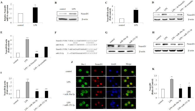
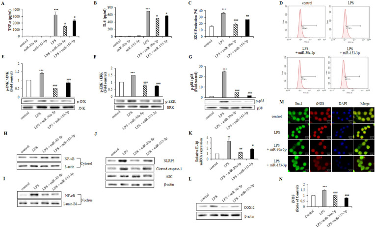
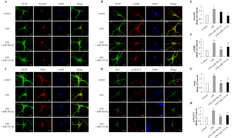
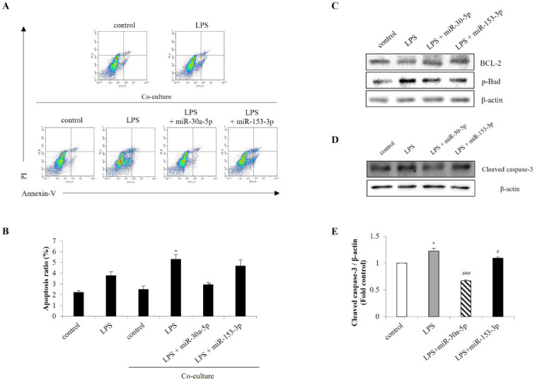
Neurogenic differentiation 1 (NeuroD1) is an essential transcription factor for neuronal differentiation, maturation, and survival, and is associated with inflammation in lipopolysaccharide (LPS)- induced glial cells; however, the concrete mechanisms are still ambiguous. Therefore, we investigated whether NeuroD1-targeting miRNAs affect inflammation and neuronal apoptosis, as well as the underlying mechanism. First, we confirmed that miR-30a-5p and miR-153-3p, which target NeuroD1, reduced NeuroD1 expression in microglia and astrocytes. In LPS-induced microglia, miR-30a-5p and miR-153-3p suppressed pro-inflammatory cytokines, reactive oxygen species, the phosphorylation of c-Jun N-terminal kinase, extracellular-signal-regulated kinase (ERK), and p38, and the expression of cyclooxygenase and inducible nitric oxide synthase (iNOS) via the NF-κB pathway. Moreover, miR-30a-5p and miR-153-3p inhibited the expression of NOD-like receptor pyrin domain containing 3 (NLRP3) inflammasomes, NLRP3, cleaved caspase-1, and IL-1β, which are involved in the innate immune response. In LPS-induced astrocytes, miR-30a-5p and miR-153-3p reduced ERK phosphorylation and iNOS expression via the STAT-3 pathway. Notably, miR-30a-5p exerted greater anti-inflammatory effects than miR-153-3p. Together, these results indicate that miR-30a-5p and miR-153-3p inhibit MAPK/NF-κB pathway in microglia as well as ERK/STAT-3 pathway in astrocytes to reduce LPS-induced neuronal apoptosis. This study highlights the importance of NeuroD1 in microglia and astrocytes neuroinflammation and suggests that it can be regulated by miR-30a-5p and miR-153-3p. [BMB Reports 2022; 55(9): 447-452].


Conflict of interest statement

CONFLICTS OF INTEREST

The authors have no conflicting interests.

Figures

Fig. 1
Fig. 1
Expression of NeuroD1 by LPS is targeted by miR-30a-5p and miR-153-3p. BV-2 cells were treated with LPS (1 μg/ml) for 1 h. After stimulation, the expression of NeuroD1 was assessed by (A) RT-qPCR and (B, C) western blot. (D, E) BV-2 cells were transfected with NeuroD1 shRNA (1 μg) for 24 h followed by treatment with LPS (1 μg/ml) for 1 h. The protein was extracted and the expression of NeuroD1 was detected by western blot. (F) MiR-30a-5p and miR-153-3p binding site were predicted in the NeuroD1 mRNA. (G) NeuroD1 was knocked down by miR-30a-5p and miR-153-3p (50 nM) in microglia cells. Knockdown efficiency was determined by protein levels of NeuroD1. (H, I) BV-2 cells were transfected with miR-30a-5p and miR-153-3p followed by LPS (1 μg/ml) treatment for 1 h. (J, K) Primary microglia were treated with LPS (1 μg/ml) for 1 h, and immunofluorescence was conducted to detect NeuroD1 using a Iba-1 as a microglia marker (Scale bars: 15 μm). Data are presented as the means ± S.D. Values of *P < 0.05, **P < 0.01, ***P < 0.001 versus control; #P < 0.05, ##P < 0.01, ###P < 0.001 versus LPS group.
Fig. 2
Fig. 2
MiR-30a-5p and miR-153-3p regulate inflammatory factors through MAPK pathway. BV-2 cells were transfected with miR-30a-5p and miR-153-3p (50 nM) for 24 h followed by treatment with LPS (1 μg/ml) for 1 h. (A, B) Cell-free supernatant were collected and TNF-α and IL-6 were evaluated using ELISA. (C, D) LPS-induced ROS was measured through flow cytometry with H2DCF-DA (5 μM) for 1 h. (E-G) The protein was extracted, and the expression of protein level was detected by western blot to measure inflammatory factors p-JNK, p-ERK and p-p38. (H, I) Translocation of NF-κB was detected by western blot. BV-2 cells were lysed to cytosol and nucleic extracts. β-actin and Lamin-B1 were used as an internal control. (J) BV-2 cells were lysed to whole lysates and NLRP3 inflammasome factors, NLRP3, cleaved caspase-1 and ASC. (K) The IL-1β mRNA expression was analyzed by RT-qPCR. (L) Western blot analysis was performed to measure the expression of COX-2 in BV-2 cells. (M, N) Primary microglia cells were transfected with miR-30a-5p and miR-153-3p for 24 h followed by treated with LPS (1 μg/ml) for 1 h. The expression of iNOS was detected by immunofluorescence analysis (Scale bars: 15 μm). Data are presented as the means ± S.D. Values of **P < 0.01, ***P < 0.001 versus control; #P < 0.05, ##P < 0.01, ###P < 0.001 versus LPS group.
Fig. 3
Fig. 3
Inflammatory mediators are decreased by miR-30a-5p and miR-153-3p in astrocytes. Primary astrocytes were transfected with miR-30a-5p and miR-153-3p (50 nM) for 24 h followed by treatment with LPS (1 μg/ml) for 24 h. Immunofluorescence was conducted with (A, E) NeuroD1, (B, F) p-ERK, (C, G) iNOS and (D, H) p-STAT-3 with GFAP as an astrocytes marker (Scale bars: 15 μm). Data are presented as the means ± S.D. Values of **P < 0.01, ***P < 0.001 versus control; ##P < 0.01, ###P < 0.001 versus LPS group.
Fig. 4
Fig. 4
MiR-30a-5p and miR-153-3p in microglia cells alleviate neuronal apoptosis. BV-2 cells were transfected with miR-30a-5p and miR-153-3p (50 nM) for 24 h. Then BV-2 cells and SH-SY5Y cells were co-cultured followed by treated with LPS (1 μg/ml) for 1 h in BV-2 cells. (A, B) The Annexin-V/PI images were detected by flow cytometry. (C) Western blot analysis was performed to measure the expression of BCL-2 and p-Bad in SH-SY5Y co-cultured with BV-2 cells. (D, E) Primary mixed cells were transfected with miR-30a-5p and miR-153-3p for 24 h followed by LPS (1 μg/ml) for 1 h. Cells were harvested, and the expression protein levels of cleaved caspase-3 was analyzed by western blot. Data are presented as the means ± S.D. Values of *P < 0.05 versus control; #P < 0.05, ###P < 0.001 versus LPS group.

Similar articles

Cited by

References

    1. Ransohoff RM, Brown MA. Innate immunity in the central nervous system. J Clin Invest. 2012;122:1164–1171. doi: 10.1172/JCI58644. - DOI - PMC - PubMed
    1. Reemst K, Noctor SC, Lucassen PJ, Hol EM. The indispensable roles of microglia and astrocytes during brain development. Front Hum Neurosci. 2016;10:566. doi: 10.3389/fnhum.2016.00566. - DOI - PMC - PubMed
    1. Zhang P, Lokuta KM, Turner DE, Liu B. Synergistic dopaminergic neurotoxicity of manganese and lipopolysaccharide: differential involvement of microglia and astroglia. J Neurochem. 2010;112:434–443. doi: 10.1111/j.1471-4159.2009.06477.x. - DOI - PMC - PubMed
    1. Jha MK, Lee WH, Suk K. Functional polarization of neuroglia: implications in neuroinflammation and neurological disorders. Biochem Pharmacol. 2016;103:1–16. doi: 10.1016/j.bcp.2015.11.003. - DOI - PubMed
    1. Wang S, Colonna M. Microglia in Alzheimer's disease: a target for immunotherapy. J Leukoc Biol. 2019;106:219–227. doi: 10.1002/JLB.MR0818-319R. - DOI - PubMed

MeSH terms