. 2022 May 16:14:911220.
doi: 10.3389/fnagi.2022.911220. eCollection 2022.

Multi-Modal Neuroimaging Neural Network-Based Feature Detection for Diagnosis of Alzheimer's Disease

Affiliations

Multi-Modal Neuroimaging Neural Network-Based Feature Detection for Diagnosis of Alzheimer's Disease

Xianglian Meng et al. Front Aging Neurosci. .

Abstract

Alzheimer's disease (AD) is a neurodegenerative brain disease, and it is challenging to mine features that distinguish AD and healthy control (HC) from multiple datasets. Brain network modeling technology in AD using single-modal images often lacks supplementary information regarding multi-source resolution and has poor spatiotemporal sensitivity. In this study, we proposed a novel multi-modal LassoNet framework with a neural network for AD-related feature detection and classification. Specifically, data including two modalities of resting-state functional magnetic resonance imaging (rs-fMRI) and diffusion tensor imaging (DTI) were adopted for predicting pathological brain areas related to AD. The results of 10 repeated experiments and validation experiments in three groups prove that our proposed framework outperforms well in classification performance, generalization, and reproducibility. Also, we found discriminative brain regions, such as Hippocampus, Frontal_Inf_Orb_L, Parietal_Sup_L, Putamen_L, Fusiform_R, etc. These discoveries provide a novel method for AD research, and the experimental study demonstrates that the framework will further improve our understanding of the mechanisms underlying the development of AD.

Keywords: LassoNet; diffusion tensor imaging; feature detection; multi-modal; resting state functional magnetic resonance imaging.


Conflict of interest statement

The authors declare that the research was conducted in the absence of any commercial or financial relationships that could be construed as a potential conflict of interest.

Figures
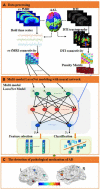
FIGURE 1
FIGURE 1
An illustration of the proposed multi-modal framework for AD. (A) Data processing. The fMRI and DTI images were preprocessed, and then the regions of interest were extracted as fMRI and DTI features through the AAL template, and the corresponding brain networks of fMRI and DTI were obtained, respectively. Then, computed the inverse proportional function of the structural brain network as a penalty matrix. (B) Multi-modal LassoNet Modeling with a neural network. We constructed a multi-modal network framework for feature selection and classification based on the LassoNet model. It consisted of residual connection and an arbitrary feed-forward neural network. The input to the network was the fMRI feature information. The penalty matrix was introduced to the residual connection to sparse features. (C) The detection of the pathological mechanism of AD. We visualized brain regions for selected features to analyze the affected discriminative brain regions.
FIGURE 2
FIGURE 2
The relationship between the accuracies and λ.
FIGURE 3
FIGURE 3
The prediction accuracy was obtained through 10 experiments for five methods in three groups. (A) Prediction accuracy of AD-HC group. (B) Prediction accuracy of AD-EMCI group. (C) Prediction accuracy of EMCI-HC group.
FIGURE 4
FIGURE 4
The ROC curve of the five methods in three groups. (A) Prediction accuracy of AD-HC group. (B) Prediction accuracy of AD-EMCI group. (C) Prediction accuracy of EMCI-HC group.
FIGURE 5
FIGURE 5
Visualization of discriminative brain regions. (A) AD-HC, (B) AD-EMCI, and (C) EMCI-HC.

References

    1. Abbasi W. A., Abbas S. A., Andleeb S. (2021). PANDA: Predicting the change in proteins binding affinity upon mutations by finding a signal in primary structures. J. Bioinform. Comput. Biol. 19:2150015. 10.1142/S0219720021500153 - DOI - PubMed
    1. Aderghal K., Afdel K., Benois-Pineau J., Catheline G. (2020). Improving Alzheimer’s stage categorization with Convolutional Neural Network using transfer learning and different magnetic resonance imaging modalities. Heliyon 6:e05652. 10.1016/j.heliyon.2020.e05652 - DOI - PMC - PubMed
    1. Chen Z., Zeng W., Yang Z., Yu L., Fu C. W., Qu H. (2019). LassoNet: deep Lasso-Selection of 3D Point Clouds. IEEE Trans. Vis. Comput. Graph. 2019:2934332. 10.1109/tvcg.2019.2934332 - DOI - PubMed
    1. Choo I. H., Lee D. Y., Oh J. S., Lee J. S., Lee D. S., Song I. C., et al. (2010). Posterior cingulate cortex atrophy and regional cingulum disruption in mild cognitive impairment and Alzheimer’s disease. Neurobiol. Aging 31 772–779. 10.1016/j.neurobiolaging.2008.06.015 - DOI - PubMed
    1. Cogswell P. M., Wiste H. J., Senjem M. L., Gunter J. L., Weigand S. D., Schwarz C. G., et al. (2021). Associations of quantitative susceptibility mapping with Alzheimer’s disease clinical and imaging markers. Neuroimage 224:117433. 10.1016/j.neuroimage.2020.117433 - DOI - PMC - PubMed

LinkOut - more resources