

BMP-4, TGF-β and Smad3 as Modulators of Synovial Fluid Cells Viability
- PMID: 35652012
- PMCID: PMC9142237
- DOI: 10.1055/s-0041-1724076
BMP-4, TGF-β and Smad3 as Modulators of Synovial Fluid Cells Viability
Abstract
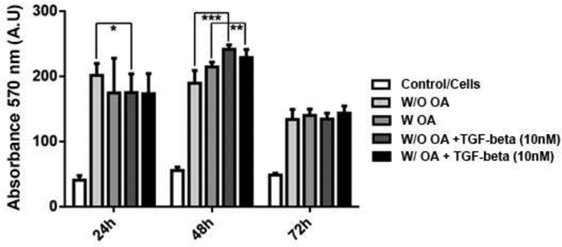
Objective Our goal was to evaluate the modulation of the synovial fluid cells (SFC) from patients with and without osteoarthritis (OA) by bone morphogenetic protein 4 (BMP-4), Smad-3 and transforming growth factor beta (TGF-β). Methods Synovial fluid was collected from patients submitted to knee arthroscopy or replacement and were centrifuged to isolate cells from the fluid. Cells were cultured for 21 days and characterized as mesenchymal stem cells (MSCs) according to the criteria of the International Society of Cell Therapy. Then, we performed an [3-(4,5-dimethylthiazol-2-yl)-2,5-diphenyltetrazolium bromide] assay (MTT) assay after exposing cells with and without OA to TGF-β, Smad3 and BMP-4 pathway inhibitors and to different concentrations of BMP4. Results Exposure to the TGF-β, Smad3 and BMP-4 inhibitors modifies the mitochondrial activity of the SFCs. The activity of the SFCs is modified by influences of increasing concentrations of BMP4, but there is no difference in cellular activity between patients with and without OA. Conclusion TGF-β, Smad3 and BMP-4 modulate the activity of SFCs from patients with and without knee OA.
Keywords: TGF-β pathway; mesenchymal stem cells; osteoarthritis; synovial fluid cells.
Sociedade Brasileira de Ortopedia e Traumatologia. This is an open access article published by Thieme under the terms of the Creative Commons Attribution-NonDerivative-NonCommercial License, permitting copying and reproduction so long as the original work is given appropriate credit. Contents may not be used for commecial purposes, or adapted, remixed, transformed or built upon. ( https://creativecommons.org/licenses/by-nc-nd/4.0/ ).
Conflict of interest statement
Conflitos de Interesse Os autores declaram não haver conflitos de interesse.
Figures


Similar articles
-
Osteoarthritic Synovial Fluid Modulates Cell Phenotype and Metabolic Behavior In Vitro.Stem Cells Int. 2019 Jan 15;2019:8169172. doi: 10.1155/2019/8169172. eCollection 2019. Stem Cells Int. 2019. PMID: 30766606 Free PMC article.
-
Transforming growth factor-β(1) represses bone morphogenetic protein-mediated Smad signaling in pulmonary artery smooth muscle cells via Smad3.Am J Respir Cell Mol Biol. 2013 Dec;49(6):1135-45. doi: 10.1165/rcmb.2012-0470OC. Am J Respir Cell Mol Biol. 2013. PMID: 23937428 Free PMC article.
-
Chondrogenic potential of human synovial mesenchymal stem cells in alginate.Osteoarthritis Cartilage. 2007 Oct;15(10):1178-89. doi: 10.1016/j.joca.2007.03.015. Epub 2007 May 14. Osteoarthritis Cartilage. 2007. PMID: 17502159
-
Role of the TGF-β/BMP-7/Smad pathways in renal diseases.Clin Sci (Lond). 2013 Feb;124(4):243-54. doi: 10.1042/CS20120252. Clin Sci (Lond). 2013. PMID: 23126427 Review.
-
TGF-β and BMP signaling in osteoblast differentiation and bone formation.Int J Biol Sci. 2012;8(2):272-88. doi: 10.7150/ijbs.2929. Epub 2012 Jan 21. Int J Biol Sci. 2012. PMID: 22298955 Free PMC article. Review.
References
-
- Johnson K, Zhu S, Tremblay M S.A stem cell-based approach to cartilage repair Science 2012336(6082):717–721. - PubMed
-
- Barry F, Murphy M. Mesenchymal stem cells in joint disease and repair. Nat Rev Rheumatol. 2013;9(10):584–594. - PubMed
-
- Pers Y M, Ruiz M, Noël D, Jorgensen C. Mesenchymal stem cells for the management of inflammation in osteoarthritis: state of the art and perspectives. Osteoarthritis Cartilage. 2015;23(11):2027–2035. - PubMed
-
- Jones E A, Crawford A, English A. Synovial fluid mesenchymal stem cells in health and early osteoarthritis: detection and functional evaluation at the single-cell level. Arthritis Rheum. 2008;58(06):1731–1740. - PubMed
LinkOut - more resources
Full Text Sources