

Histomorphometry of Bone Microarchitecture in Rats Treated with Vitamin D and Bisphosphonate in the Management of Osteoporosis
- PMID: 35652013
- PMCID: PMC9142217
- DOI: 10.1055/s-0041-1741023
Histomorphometry of Bone Microarchitecture in Rats Treated with Vitamin D and Bisphosphonate in the Management of Osteoporosis
Abstract
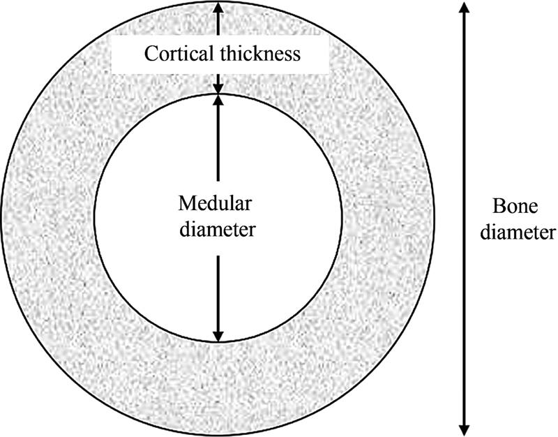
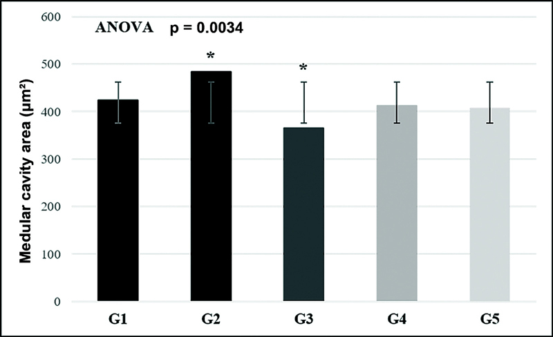
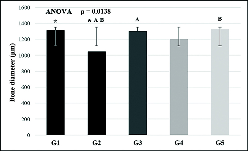
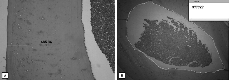
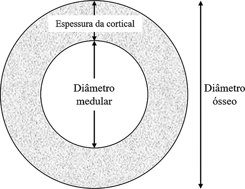
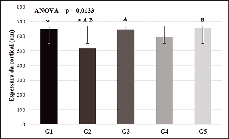
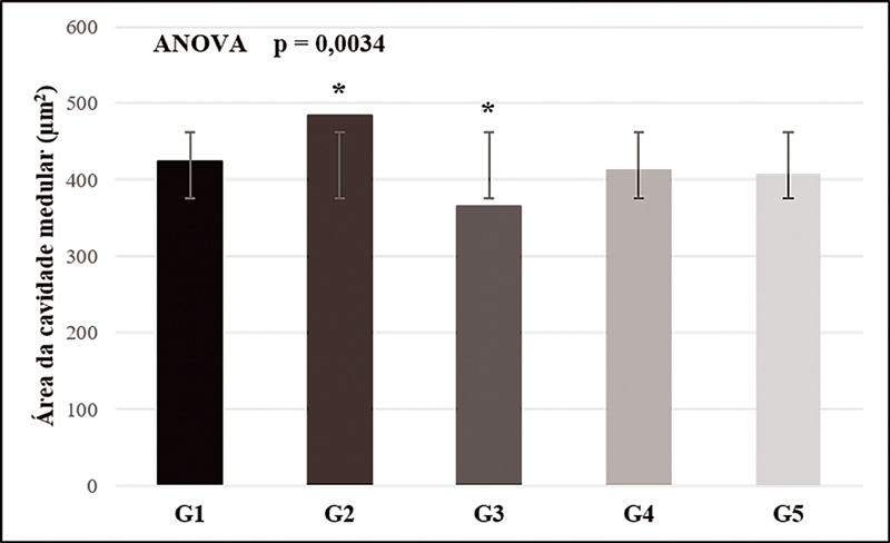
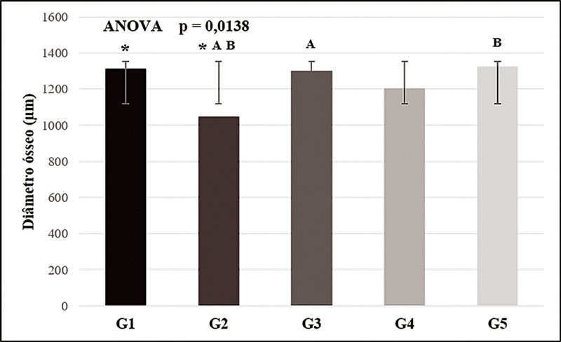
Objective To verify how the combined administration of alendronate (ALN) and vitamin D3 (VD) acts on the bone microarchitecture in rats with glucocorticoid-induced osteoporosis. Methods The experiment used 32 90-day-old female Wistar rats weighing between 300 and 400g. The induction of osteoporosis consisted of intramuscular administration of dexamethasone at a dose of 7.5 mg/kg of body weight once a week for 5 weeks, except for the animals in the control group. The animals were separated into the following groups: G1 (control group without osteoporosis), G2 (control group with osteoporosis without treatment), G3 (group with osteoporosis treated with ALN 0.2 mg/kg), G4 (group with osteoporosis treated with VD 10,000UI/500μL), and G5 (group with osteoporosis treated with ALN + VD). The right femurs of the rats were fixed in 10% buffered formaldehyde, decalcified, and processed for inclusion in paraffin. Histological sections were stained with hematoxylin-eosin for histomorphometric analysis. Cortical thickness and medullary cavity were measured in cross-sections. Results There was a statistical difference ( p < 0.05) between groups G3 and G5 compared with the positive control group (G2), both related to the measurement of cortical thickness and to the total diameter of the bone. In the evaluation of the spinal area, only the G3 group has shown to be statistically different from the G2 group. Conclusion Concomitant treatment with daily ALN and weekly VD is effective in preventing glucocorticoid-induced bone loss. However, there was no difference between the therapy tested and treatment with ALN alone.
Keywords: alendronate; femur; menopause; rats; vitamin D.
Sociedade Brasileira de Ortopedia e Traumatologia. This is an open access article published by Thieme under the terms of the Creative Commons Attribution-NonDerivative-NonCommercial License, permitting copying and reproduction so long as the original work is given appropriate credit. Contents may not be used for commecial purposes, or adapted, remixed, transformed or built upon. ( https://creativecommons.org/licenses/by-nc-nd/4.0/ ).
Conflict of interest statement
Conflito de Interesses Os autores declaram não haver conflito de interesses.
Figures


Similar articles
-
Biometric, histomorphometric, and biochemical profile in atorvastatin calcium treatment of female rats with dexamethasone-induced osteoporosis.Rev Bras Ortop. 2018 Jul 27;53(5):607-613. doi: 10.1016/j.rboe.2018.07.007. eCollection 2018 Sep-Oct. Rev Bras Ortop. 2018. PMID: 30245999 Free PMC article.
-
Individual and combined effects of exercise and alendronate on bone mass and strength in ovariectomized rats.Bone. 2007 Aug;41(2):290-6. doi: 10.1016/j.bone.2007.04.179. Epub 2007 Apr 24. Bone. 2007. PMID: 17544352
-
Can the alendronate dosage be altered when combined with high-frequency loading in osteoporosis treatment?Osteoporos Int. 2017 Apr;28(4):1287-1293. doi: 10.1007/s00198-016-3859-1. Epub 2016 Dec 5. Osteoporos Int. 2017. PMID: 27921147
-
Effects of combined treatment with eldecalcitol and alendronate on bone mass, mechanical properties, and bone histomorphometry in ovariectomized rats: a comparison with alfacalcidol and alendronate.Bone. 2013 Jan;52(1):181-8. doi: 10.1016/j.bone.2012.09.031. Epub 2012 Oct 2. Bone. 2013. PMID: 23041510
-
Sequential administration of alendronate and strontium ranelate: histomorphometry and bone biomechanics in ovariectomized animals.Acta Odontol Latinoam. 2016 Sep;29(2):168-177. Acta Odontol Latinoam. 2016. PMID: 27731487 English.
Cited by
-
The Relationship between Bone Health Parameters, Vitamin D and Iron Status, and Dietary Calcium Intake in Young Males.Nutrients. 2024 Jan 9;16(2):215. doi: 10.3390/nu16020215. Nutrients. 2024. PMID: 38257108 Free PMC article.
References
-
- Radominski S C, Bernardo W, Paula A P.Diretrizes brasileiras para o diagnóstico e tratamento da osteoporose em mulheres na pós-menopausa Rev Bras Reumatol 201757(S 2):S452–S466.
-
- Si L, Winzenberg T M, Chen M, Jiang Q, Palmer A J. Residual lifetime and 10 year absolute risks of osteoporotic fractures in Chinese men and women. Curr Med Res Opin. 2015;31(06):1149–1156. - PubMed
-
- NIH Consensus Development Panel on Osteoporosis Prevention, Diagnosis, and Therapy . Osteoporosis prevention, diagnosis, and therapy. JAMA. 2001;285(06):785–795. - PubMed
-
- Lazaretti-Castro M, Eis S R, Marques Neto J F. A prevenção da osteoporose levada a sério: uma necessidade nacional. Arq Bras Endocrinol Metabol. 2008;52(04):712–713. - PubMed
LinkOut - more resources
Full Text Sources