

Isolated distal pisiform dislocation: Case Report
- PMID: 35652023
- PMCID: PMC9142225
- DOI: 10.1055/s-0040-1722589
Isolated distal pisiform dislocation: Case Report
Abstract
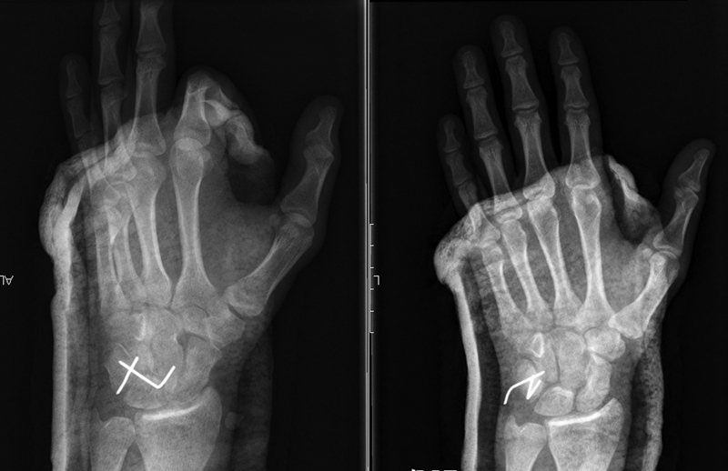
Isolated pisiform dislocation is a rare lesion with few cases described in the literature. This type of lesion is typically observed in young males and can be easily overlooked at first assessment. Isolated proximal dislocation is more common due to the action of the flexor carpi ulnaris (FCU) muscle. We present the case of a 19-year-old male patient with isolated distal pisiform dislocation after wrist trauma. He underwent open reduction and internal fixation with Kirschner wires with excellent functional outcomes. Although there is no consensual therapeutic method, closed reduction is a first-line treatment for acute presentations. Pisiform open reduction or excision may be performed alternatively or after a failed closed reduction.
Keywords: carpal bones; joint dislocations; pisiform; wrist injuries.
Sociedade Brasileira de Ortopedia e Traumatologia. This is an open access article published by Thieme under the terms of the Creative Commons Attribution-NonDerivative-NonCommercial License, permitting copying and reproduction so long as the original work is given appropriate credit. Contents may not be used for commecial purposes, or adapted, remixed, transformed or built upon. ( https://creativecommons.org/licenses/by-nc-nd/4.0/ ).
Conflict of interest statement
Conflito de Interesses Os autores declaram não haver conflito de interesses. O estudo foi realizado em conformidade com a Declaração da Associação Médica Mundial de Helsinque sobre Princípios Éticos para Pesquisa Médica Envolvendo Seres Humanos.
Figures


References
-
- Kwon O S, Choi S P, Won H Y. Acute isolated pisiform dislocation: A case report. J Korean Orthop Assoc. 2007;42(05):688–691.
-
- Minami M, Yamazaki J, Ishii S. Isolated dislocation of the pisiform: a case report and review of the literature. J Hand Surg Am. 1984;9A(01):125–127. - PubMed
-
- Schädel-Höpfner M, Junge A, Böhringer G. [Dislocation of the pisiform bone. A review of the literature] Handchir Mikrochir Plast Chir. 2002;34(03):168–172. - PubMed
-
- Sharara K H, Farrar M. Isolated dislocation of the pisiform bone. J Hand Surg Br. 1993;18(02):195–196. - PubMed
-
- Campbell E, Magi E. A dislocated pisiform: Case report. Can J Plast Surg. 1999;7(02):57–58.
LinkOut - more resources
Full Text Sources