

Case Report: Multimodal Imaging in a Rare Case of Morning Glory Disc Anomaly Complicated With Choroidal Ossification
- PMID: 35652078
- PMCID: PMC9149162
- DOI: 10.3389/fmed.2022.826860
Case Report: Multimodal Imaging in a Rare Case of Morning Glory Disc Anomaly Complicated With Choroidal Ossification
Abstract
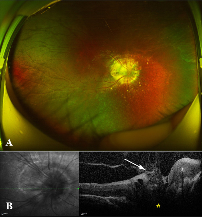
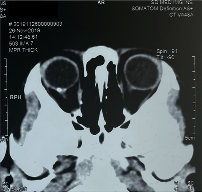
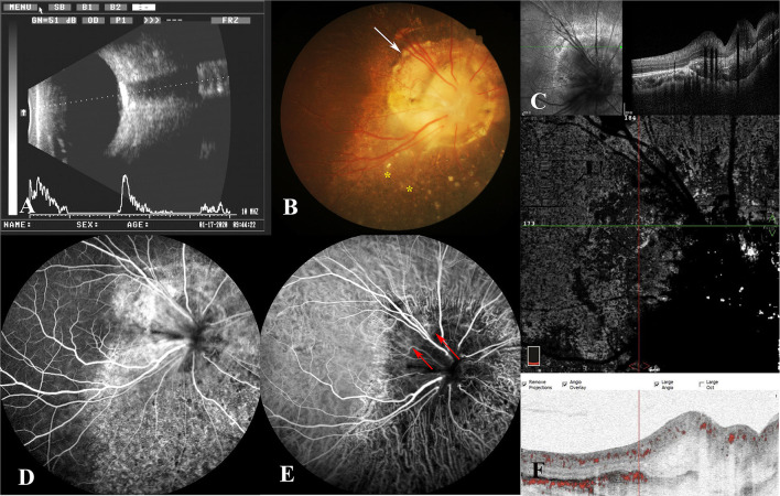
Purpose: The authors described a 7-year-old boy who was diagnosed with morning glory disc (MGD) anomaly in the right eye via fundus examination. However, during the head CT examination, a hyperdense choroidal lesion was discovered around the optic disc. Comprehensive investigations revealed that the lesion's characteristics were extremely consistent with choroidal osteoma (CO), so the patient was diagnosed with MGD with CO on his second visit. However, in the subsequent follow-up, the author discovered pigmentary alterations in the retinal pigment epithelium (RPE) in the patient's right eye. Finally, the diagnosis was corrected to MGD with choroidal ossification following a thorough etiological analysis. Meanwhile, the characteristics of choroidal ossification were described in detail through multimodal imaging in this article.
Methods: Retrospective review of a case note.
Conclusions: Similar to CO, choroidal ossification is the consequence of structured osseous tissue formation regulated by osteoblasts and osteoclasts. It consists of bone trabecular and vascular components and is difficult to be distinguished from CO on imaging examinations. In contrast to the congenital prevalence of CO, there are often incentives for the occurrence of choroidal ossification. These inducements will eventually mediate the inflammation in the eye, resulting in the activation of many cytokines and the production of choroidal ossification. Around one-third of patients with MGD will experience retinal detachment, and in certain cases, the subretinal fluid will be absorbed spontaneously, resulting in alterations to the RPE. These processes can activate inflammatory factors in the eye, bringing about a cascade of abnormalities, including the development of CO. Therefore, the proper diagnosis of disease should not be made exclusively on the basis of the imaging findings. A thorough analysis of the epidemiology and etiology is crucial.
Keywords: choroidal ossification; choroidal osteoma; etiology analysis; morning glory disk anomaly; multimodal imaging.
Copyright © 2022 Wang, Wang, Wang and Bi.
Conflict of interest statement
The authors declare that the research was conducted in the absence of any commercial or financial relationships that could be construed as a potential conflict of interest.
Figures
Similar articles
-
Postoperative follow-up of a case of atypical morning glory syndrome associated with persistent fetal vasculature.BMC Ophthalmol. 2019 Jul 16;19(1):150. doi: 10.1186/s12886-019-1154-6. BMC Ophthalmol. 2019. PMID: 31311513 Free PMC article. Review.
-
Case Report: Fibroglial Retinal Tissue in Contractile Morning Glory Disc Anomaly.Case Rep Ophthalmol. 2021 Jun 11;12(2):525-530. doi: 10.1159/000510958. eCollection 2021 May-Aug. Case Rep Ophthalmol. 2021. PMID: 34248586 Free PMC article.
-
Management of Retinal Detachment Associated with Morning Glory Disc Syndrome.Case Rep Ophthalmol. 2021 May 25;12(2):457-463. doi: 10.1159/000516205. eCollection 2021 May-Aug. Case Rep Ophthalmol. 2021. PMID: 34177542 Free PMC article.
-
Morning glory disc anomaly with an ipsilateral enlargement of the optic nerve pathway.Eur J Paediatr Neurol. 2017 Sep;21(5):787-791. doi: 10.1016/j.ejpn.2017.04.1334. Epub 2017 May 19. Eur J Paediatr Neurol. 2017. PMID: 28666648
-
[Congenital abnormalities of the optic disc].J Fr Ophtalmol. 2019 Sep;42(7):778-789. doi: 10.1016/j.jfo.2018.09.011. Epub 2019 Mar 29. J Fr Ophtalmol. 2019. PMID: 30935696 Review. French.
References
-
- Munteanu M, Munteanu G, Giuri S, Zolog I, Motoc AG. Ossification of the choroid: three clinical cases and literature review of the pathogenesis of intraocular ossification. Rom J Morphol Embryol. (2013) 54(3 Suppl):871–7. - PubMed
Publication types
LinkOut - more resources
Full Text Sources