. 2022 May 16:10:812244.
doi: 10.3389/fcell.2022.812244. eCollection 2022.

Validation of Effective Extracellular Vesicles Isolation Methods Adapted to Field Studies in Malaria Endemic Regions

Affiliations

Validation of Effective Extracellular Vesicles Isolation Methods Adapted to Field Studies in Malaria Endemic Regions

Matteo Zoia et al. Front Cell Dev Biol. .

Abstract

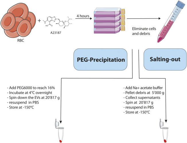
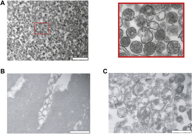
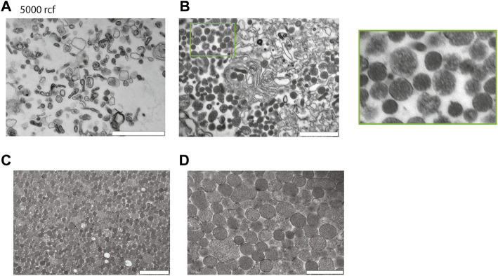
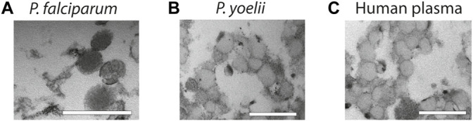
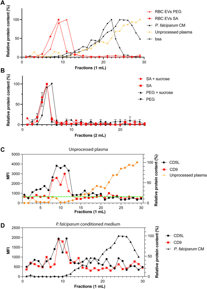
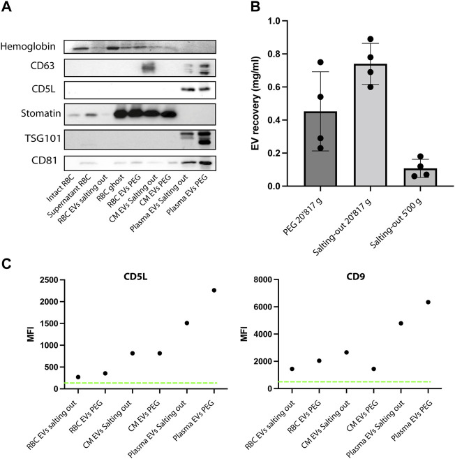
Malaria affects the poorer regions of the world and is of tremendous health and economic burden for developing countries. Extracellular vesicles (EVs) are small vesicles released by almost any cells in the human body, including malaria infected red blood cells. Recent evidence shows that EVs might contribute to the pathogenesis of malaria. In addition, EVs hold considerable value in biomarker discovery. However, there are still significant gaps in our understanding of EV biology. So far most of our knowledge about EVs in malaria comes from in vitro work. More field studies are required to gain insight into their contribution to the disease and pathogenesis under physiological conditions. However, to perform research on EVs in low-income regions might be challenging due to the lack of appropriate equipment to isolate EVs. Therefore, there is a need to develop and validate EV extraction protocols applicable to poorly equipped laboratories. We established and validated two protocols for EV isolation from cell culture supernatants, rodent and human plasma. We compared polyethylene glycol (PEG) and salting out (SA) with sodium acetate for precipitation of EVs. We then characterized the EVs by Transmission Electron Microscopy (TEM), Western Blot, Size-exclusion chromatography (SEC), bead-based flow cytometry and protein quantification. Both protocols resulted in efficient purification of EVs without the need of expensive material or ultracentrifugation. Furthermore, the procedure is easily scalable to work with large and small sample volumes. Here, we propose that both of our approaches can be used in resource limited countries, therefore further helping to close the gap in knowledge of EVs during malaria.

Keywords: Plasmodium falciparum; extracellular vesicles; malaria; microvesicles; polyethylene glycol precipitation; salting-out.


Conflict of interest statement

The authors declare that the research was conducted in the absence of any commercial or financial relationships that could be construed as a potential conflict of interest.

Figures

FIGURE 1
FIGURE 1
Schematic representation and description of the main steps involved in the 2 methodologies used for the isolation of EVs. Created with BioRender.com.
FIGURE 2
FIGURE 2
Characterization of red blood cell derived EVs isolated by PEG precipitation. (A) Transmission electron microscopy (TEM) visualization of RBC derived EVs isolated from A23487 treated RBCs by PEG precipitation and collected by centrifugation at 20′817 g. Representative TEM image shows individual EVs and a few clumps of varying sizes and intact lipid bilayers. The image on the right is a zoom in of the first panel. The scale bar is 1 μm. (B), (C) Analysis of hypo-osmotic lysed RBCs by TEM. The cellular debris were pelleted by centrifugation and the pellet was resuspended in PBS for visualization by TEM. The scale bar is 1 μm.
FIGURE 3
FIGURE 3
Characterization of red blood cell derived EVs isolated by salting out precipitation. (A) Healthy Human RBCs EVs extracted via Salting-out precipitation and pelleted at 5′000 g. The images were taken at 24′ × 500 magnification by TEM. Scale bar = 1 μm. (B) Healthy Human RBCs EVs extracted via Salting-out precipitation at 20′817 g, imaging has been taken at × 24500 magnification by TEM. Scale bar = 1 μm. (C) EVs were precipitated by Salting out and debris were pelleted at 5′000 g, the EVs were then collected by centrifugation at 20′817 g from the resulting supernatant. Scale bar = 1 μm. (D) Scale bar = 500 nm.
FIGURE 4
FIGURE 4
Characterization of EVs from biofluids. (A) EVs derived from in vitro P. falciparum infected RBC cultures reveal vesicular structures of 100–200 nm. Scale bar = 500 nm. (B) EVs isolated from plasma of P. yoelii infected Balb/c mice. Scale bar = 500 nm. (C) EVs isolated from healthy human plasma. Scale bar = 500 nm. All the EV preparations were analyzed by TEM.
FIGURE 5
FIGURE 5
Only one population of EVs are identified by size-exclusion chromatography. (A) Standard curves containing the elution profiles of EVs purified by ultracentrifugation, BSA and human serum. Points represent the mean ± s.d. of two independent analyses. (B) Elution profile of purified RBC EVs isolated by PEG or salting out precipitation as measured by absorbance at 280 nm. The dashed lines represent EVs further purified by sucrose gradient. Points represent the mean ± s.d. of three experiments. (C) MFI values of CD9 and CD5L in SEC fractions. Total of 30 fractions of 1 ml were collected. (D) MFI values of CD9 and CD5L in SEC fractions of P. falciparum conditioned medium.
FIGURE 6
FIGURE 6
EVs express specific markers. (A) Western Blot analysis of RBC EV markers, stomatin, GAPDH and Hemoglobin. In total, 10 μg of protein were loaded on a SDS-PAGE gel. (B) A comparison of EV recovery yield between PEG and salting out precipitation as measured by protein content. The mean ± s.d. of four independent analyses is shown. (C) Representative experiment of purified EVs from RBCs, plasma and conditioned medium. The presence of EV markers (CD9 and CD5L), was assessed by bead-based flow cytometry assay. The Mean Fluorescence Intensity (MFI) was calculated after measuring 50,000 events (one representative of three experiments).

Similar articles

Cited by

References

    1. Allan D., Billah M. M., Finean J. B., Michell R. H. (1976). Release of Diacylglycerol-Enriched Vesicles from Erythrocytes with Increased Intracellular [Ca2+]. Nature 261, 58–60. 10.1038/261058a0 - DOI - PubMed
    1. Allan D., Thomas P., Limbrick A. R. (1980). The Isolation and Characterization of 60 Nm Vesicles ('nanovesicles') Produced during Ionophore A23187-Induced Budding of Human Erythrocytes. Biochem. J. 188, 881–887. 10.1042/bj1880881 - DOI - PMC - PubMed
    1. Andrea Hernandez-Castaneda M., Mbagwu S., Babatunde K. A., Walch M., Filgueira L., Mantel P. Y. (2018). Evaluation of Extracellular Vesicle Function during Malaria Infection. J. Vis. Exp. 2018, 57067. 10.3791/57067 - DOI - PMC - PubMed
    1. Antwi-Baffour S., Malibha-Pinchbeck M., Stratton D., Jorfi S., Lange S., Inal J. (2020). Plasma mEV Levels in Ghanain Malaria Patients with Low Parasitaemia Are Higher Than Those of Healthy Controls, Raising the Potential for Parasite Markers in mEVs as Diagnostic Targets. J. Extracell Vesicles 9, 1697124. 10.1080/20013078.2019.1697124 - DOI - PMC - PubMed
    1. Babatunde K. A., Mbagwu S., Hernández-Castañeda M. A., Adapa S. R., Walch M., Filgueira L., et al. (2018). Malaria Infected Red Blood Cells Release Small Regulatory RNAs through Extracellular Vesicles. Sci. Rep. 8, 884. 10.1038/s41598-018-19149-9 - DOI - PMC - PubMed