Review
. 2022 Apr 8;4(2):fcac083.
doi: 10.1093/braincomms/fcac083. eCollection 2022.

Emerging insights into synapse dysregulation in Alzheimer's disease

Affiliations
Review

Emerging insights into synapse dysregulation in Alzheimer's disease

Raquel Martínez-Serra et al. Brain Commun. .

Abstract

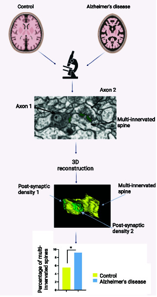
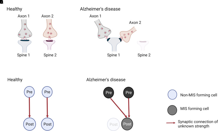
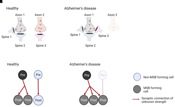
Alzheimer's disease is the leading cause of dementia and a growing worldwide problem, with its incidence expected to increase in the coming years. Since synapse loss is a major pathology and is correlated with symptoms in Alzheimer's disease, synapse dysfunction and loss may underlie pathophysiology. In this context, this review focuses on emerging insights into synaptic changes at the ultrastructural level. The three-dimensional electron microscopy technique unequivocally detects all types of synapses, including multi-synapses, which are indicators of synaptic connectivity between neurons. In recent years it has become feasible to perform sophisticated three-dimensional electron microscopy analyses on post-mortem human Alzheimer's disease brain as tissue preservation and electron microscopy techniques have improved. This ultrastructural analysis found that synapse loss does not always precede neuronal loss, as long believed. For instance, in the transentorhinal cortex and area CA1 of the hippocampus, synapse loss does not precede neuronal loss. However, in the entorhinal cortex, synapse loss precedes neuronal loss. Moreover, the ultrastructural analysis provides details about synapse morphology. For example, changes in excitatory synapses' post-synaptic densities, with fragmented postsynaptic densities increasing at the expense of perforated synapses, are seen in Alzheimer's disease brain. Further, multi-synapses also appear to be altered in Alzheimer's disease by doubling the abundance of multi-innervated spines in the transentorhinal cortex of Alzheimer's disease brain. Collectively, these recent ultrastructural analyses highlight distinct synaptic phenotypes in different Alzheimer's disease brain regions and broaden the understanding of synapse alterations, which may unravel some new therapeutic targets.

Keywords: Alzheimer’s disease; multi-innervated spine; multi-spine bouton; synapses; three-dimensional electron microscopy.


Figures

Graphical Abstract
Graphical Abstract
Figure 1
Figure 1
Identification of synapses in EM image obtained by FIB/SEM on the transentorhinal cortex from post-mortem human brain (control). Excitatory synapses (arrows) on dendritic spines (ds) are shown. Presynaptic elements contain numerous and visible vesicles. The arrows point at the asymmetric PSDs. Scale bar: 500 nm.
Figure 2
Figure 2
Identification of synapses in EM serial images obtained by FIB/SEM on the transentorhinal cortex from post-mortem human brain (control). (A–C) A sequence of serial images showing a multi-innervated dendritic spine (ds) with an excitatory A–C and an inhibitory synapse B indicated by arrows. (D–F) A sequence of serial images to illustrate a multi-synaptic bouton establishing two excitatory synapses with two dendritic spines (arrows). Scale bar in F 500 nm.
Figure 3
Figure 3
Examples of 3D reconstructed axonal boutons establishing synapses with dendritic spines. (AC) 3D reconstructions of a multi-spine bouton after axis rotation of the axon. The MSB includes one axonal bouton, three post-synaptic densities (PSD) on A–B and three dendritic spines (Sp) on C. (DF) 3D reconstructions of a multi-innervated spine after axis rotation of the dendritic spine. The MIS consists of one dendritic spine, two post-synaptic densities (PSD) on D–E, and two axonal boutons (Ax) on F. Scale bar (in F) indicates 1 μm in A–F.
Figure 4
Figure 4
Model for generation of multi-innervated dendritic spines (MIS) in Alzheimer’s disease brains and impact on synaptic connectivity. (A and B) Two synapses in a healthy brain and Alzheimer’s disease brain are shown. (A) In a healthy brain, one synapse is formed between axon 1 and spine 1, while the other synapse is made between axon 2 and spine 2. (B) In Alzheimer’s disease, spine 2 has degenerated, and its presynaptic input from axon 2 established a new connection with spine 1, generating an MIS. As a consequence, synapse density is maintained, but MIS number is increased. (C and D) Illustration of the difference in synaptic connectivity and resulting information flow as a consequence of dendritic spine loss and MIS generation in Alzheimer’s disease. It is less likely that the higher MIS number increases connectivity between two neurons (scenario not shown), as two axonal branches from one pre-synaptic neuron would have to connect to one spine. - Created with BioRender.
Figure 5
Figure 5
Model for generation of multi-spine boutons in Alzheimer’s disease brains and impact on synaptic connectivity. (A and B) Three synapses in a healthy and Alzheimer’s disease brain are shown. (A) An MSB is formed between axon 1 and spines 1 and 2, and a single synapse between axon 2 and spine 3 is shown for a healthy brain. (B) In Alzheimer’s disease, the presynaptic input from axon 2 has degenerated, and spine 3 from the single synapse established a new connection with the existing MSB. As a consequence, synapse density and MSB number are maintained, but MSB’s complexity is increased. (C and D) Illustration of the difference in synaptic connectivity and resulting information flow as a consequence of dendritic spine loss and MIS generation in Alzheimer’s disease. Note that in a healthy brain, the vast majority of most MSBs are formed between one presynaptic neuron and two post-synaptic neurons. Thus, the higher MSB complexity in Alzheimer’s disease brain is unlikely to include spines from the same dendrite, which would not lead to connecting of previously unconnected neurons (scenario not shown). Created with BioRender.

References

    1. DeKosky ST, Scheff SW. Synapse loss in frontal cortex biopsies in Alzheimer’s disease: Correlation with cognitive severity. Ann Neurol 1990;27(5):457–464. - PubMed
    1. Scheff SW, DeKosky ST, Price DA. Quantitative assessment of cortical synaptic density in Alzheimer’s disease. Neurobiol Aging 1990;11(1):29–37. - PubMed
    1. Shankar GM, Walsh DM. Alzheimer’s disease: synaptic dysfunction and Aβ. Mol Neurodegener 2009;4(1):48. - PMC - PubMed
    1. DeKosky ST, Scheff SW, Styren SD. Structural correlates of cognition in dementia: Quantification and assessment of synapse change. Neurodegeneration 1996;5(4):417–421. - PubMed
    1. Terry RD, Masliah E, Salmon DP, et al. . Physical basis of cognitive alterations in alzheimer’s disease: Synapse loss is the major correlate of cognitive impairment. Ann Neurol 1991;30(4):572–580. - PubMed

LinkOut - more resources