. 2022 Jul;9(21):e2202102.
doi: 10.1002/advs.202202102. Epub 2022 Jun 2.

Magnesium-Encapsulated Injectable Hydrogel and 3D-Engineered Polycaprolactone Conduit Facilitate Peripheral Nerve Regeneration

Affiliations

Magnesium-Encapsulated Injectable Hydrogel and 3D-Engineered Polycaprolactone Conduit Facilitate Peripheral Nerve Regeneration

Zhi Yao et al. Adv Sci (Weinh). 2022 Jul.

Abstract

Peripheral nerve injury is a challenging orthopedic condition that can be treated by autograft transplantation, a gold standard treatment in the current clinical setting. Nevertheless, limited availability of autografts and potential morbidities in donors hampers its widespread application. Bioactive scaffold-based tissue engineering is a promising strategy to promote nerve regeneration. Additionally, magnesium (Mg) ions enhance nerve regeneration; however, an effectively controlled delivery vehicle is necessary to optimize their in vivo therapeutic effects. Herein, a bisphosphonate-based injectable hydrogel exhibiting sustained Mg2+ delivery for peripheral nerve regeneration is developed. It is observed that Mg2+ promoted neurite outgrowth in a concentration-dependent manner by activating the PI3K/Akt signaling pathway and Sema5b. Moreover, implantation of polycaprolactone (PCL) conduits filled with Mg2+ -releasing hydrogel in 10 mm nerve defects in rats significantly enhanced axon regeneration and remyelination at 12 weeks post-operation compared to the controls (blank conduits or conduits filled with Mg2+ -absent hydrogel). Functional recovery analysis reveals enhanced reinnervation in the animals treated with the Mg2+ -releasing hydrogel compared to that in the control groups. In summary, the Mg2+ -releasing hydrogel combined with the 3D-engineered PCL conduit promotes peripheral nerve regeneration and functional recovery. Thus, a new strategy to facilitate the repair of challenging peripheral nerve injuries is proposed.

Keywords: hydrogel; magnesium; peripheral nerve regeneration.


Conflict of interest statement

The authors declare no conflict of interest.

Figures
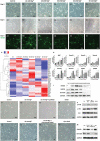
Figure 1
Figure 1
Mg2+ promotes neurite outgrowth in a concentration‐dependent manner by activating the PI3K/Akt signaling pathway and upregulating Sema5b expression. A–F) Primary culture of DRG neurons treated with varying Mg2+ concentrations (Ctrl, 10, 20, 30, 40, and 50 mM). G) Gene expression heatmap of axon guidance molecules and neurotrophic factors (n = 3). H) mRNA expression levels of NGF, Sema3b, Sema3e, Sema4c, Sema5b, NetrinG1, EFNA2, EFNB3 (mean ± standard deviation; one‐way ANOVA and Tukey's post hoc tests, *P < 0.05; **P < 0.01 compared to the mRNA levels in the Ctrl group; n = 3 per group). I) Protein expression levels of NF200, GAP43, Sema5b, and GAPDH assessed by western blotting. J) Primary culture of DRG neurons treated with additional Mg2+ ions, LY294002, or DMSO. K) Protein expression levels of pPI3K, Akt, and p‐Akt in four groups (Ctrl, Mg2+, Mg2+ with LY294002, and DMSO). L) Primary culture of DRG neurons with additional Mg2+, Lipo: Sema5b‐siRNA or Lipo: Blank; M) Western blot results of GAP43, Sema5b expression in four groups. DRG, dorsal root ganglia; DMSO, dimethyl sulfoxide; Ctrl, control.
Figure 2
Figure 2
Fabrication, characterization, mechanical properties, and scanning electron microscopic observation of HA‐pamidronate‐magnesium hydrogel. A) Fabrication of the HA‐Pam‐Mg hydrogels. B) Good injectability and self‐molding properties of the HA‐Pam‐Mg hydrogels. C) In vitro degradation of the HA‐Pam‐Mg hydrogels encapsulating different concentrations of Mg2+ (25, 50, 75, and 100 mM). D) Accumulated Mg2+ release within 14 days of HA‐Pam‐Mg hydrogels. E) Time sweep at a constant shear strain of 0.1%. F) Frequency sweep from 10–0.1 Hz at a constant shear strain of 0.1%. G) Strain sweep from 0.01% to the crossover point of G′ and G″ at a constant shear frequency of 1 Hz. H) Shear‐thinning test. I: Stress relaxation test. J–M) SEM characterization of the HA‐Pam‐Mg hydrogels. N) EDS analysis of the existing elements within the hydrogel; O) SEM characterization of MeHA hydrogels for comparison. HA, hyaluronic acid; Pam, pamidronate; Mg, magnesium; SEM, scanning electron microscopy; MeHA, methacrylated HA; EDS, energy dispersive X‐ray spectroscopy.
Figure 3
Figure 3
Fabrication, mechanical analysis, and scanning electron microscopy of 3D‐engineered PCL nerve conduits. A) Schematic diagram of the 3D printing process of the PCL nerve conduits. B) PCL nerve conduit with a diameter of 2 mm and a length of 12 mm. C) Mechanical analysis of PCL nerve conduits. D) Strain–stress curve. E–H) SEM analysis of the PCL conduits. PCL, polycaprolactone; SEM, Scanning Electron Microscopy.
Figure 4
Figure 4
Functional recovery analysis via catwalk gait and electrophysiological assessment at 12 weeks post nerve grafts implantation. A) Footprints and gaits were automatically captured by the catwalk gait analysis system. B) Percentage print area of RH/LH. C) Percentage maximum intensity of RH/LH. D) Percentage swing duration of RH/LH. E) Percentage swing speed of RH/LH (n = 5). F1–I1) NAP amplitude of the autograft, PCL nerve conduit, HA‐Pam‐Mg hydrogel, and MeHA hydrogel groups. F2–I2) CAMP amplitude of the four groups. J) NAP amplitude in mV. K) CAMP amplitude in mV. Data are expressed as mean ± standard deviation. One‐way ANOVA and Tukey's post‐hoc test is performed for statistical analysis. *P < 0.05; **P < 0.01. HA, hyaluronic acid; Pam, pamidronate; Mg magnesium; PCL, polycaprolactone; NAP, nerve action potential; CAMP, compound muscle action potential.
Figure 5
Figure 5
Immunofluorescence analysis of early nerve regeneration. A) Full‐length longitudinal sections of nerve samples displaying newly regenerated axons. A1–4) Autograft group, PCL nerve conduits group, HA‐Pam‐Mg hydrogel group, and MeHA hydrogel group; B–E) NF200/S100 staining of regenerated axons. Timepoint: week 2. HA, hyaluronic acid; Pam, pamidronate; Mg magnesium; PCL, polycaprolactone; MeHA, methacrylated HA.
Figure 6
Figure 6
H&E staining and immunohistochemical analysis of the regenerated nerves. A1–D1) H&E staining in the autograft, PCL nerve conduit, HA‐Pam‐Mg hydrogel, and MeHA hydrogel group samples. A2–D2) Immunohistochemical analysis of NF200 staining in all four groups. A3–D3) Immunohistochemical analysis of S100 staining in all four groups. E) Diameter of the regenerated nerves (µm; n = 4). F) Percentage of NF200‐positive area (n = 4). G: percentage of S100‐positive area (n = 4). Data are expressed as mean ± standard deviation. Statistical analysis is evaluated by one‐way ANOVA and Tukey's post‐hoc test. *P < 0.05; **P < 0.01. H&E, hematoxylin and eosin; HA, hyaluronic acid; Pam, pamidronate; Mg magnesium; PCL, polycaprolactone.
Figure 7
Figure 7
Remyelination of regenerated nerves. A) Toluidine blue staining of transverse sections of nerve grafts harvested 12 weeks post‐surgery. B) TEM analysis of the remyelinated axons. C) Number of the myelinated axons (n = 5). D) Diameter of the myelinated axons (µm; n = 5). E) Thickness of the new myelin sheath (µm; n = 5). F) G‐ratio (n = 5). Data are expressed as the mean ± standard deviation. One‐way ANOVA followed by Tukey's post‐hoc test is performed to analyze statistical significance. *P < 0.05; **P < 0.01. TEM, Transmission Electron Microscopy.
Figure 8
Figure 8
Masson's trichome staining of transverse sections of the triceps surae muscles. A–D) Triceps surae muscle samples harvested 12 weeks post‐operation from the autograft, PCL nerve conduit, HA‐Pam‐Mg hydrogel, and MeHA hydrogel groups. E) Wet weight muscle ratio (%). F) CSA of muscle fibers (µm2). G) CSA of collagen fibers (µm2). H) Percentage area of collagen fibers (%; n = 4). Data are expressed as mean ± standard deviation. One‐way ANOVA followed by Tukey's post‐hoc test is performed to analyze statistical significance. *P < 0.05; **P < 0.01. CSA, cross‐sectional area; HA, hyaluronic acid; Pam, pamidronate; Mg magnesium; PCL, polycaprolactone.
Figure 9
Figure 9
Schematic illustration of animal surgery. A) Surgical procedures performed on SD rats belonging to four groups, that is, the autograft, PCL nerve conduit, HA‐Pam‐Mg hydrogel, and MeHA hydrogel groups. B) Repair of a nerve defect using autografts. C) Repair of a nerve defect using PCL nerve conduits. D,E) Surgical procedures in the HA‐Pam‐Mg hydrogel and MeHA hydrogel groups, wherein first one nerve stump is sutured, followed by injection of the hydrogels into the nerve conduit cavities. HA, hyaluronic acid; Pam, pamidronate; Mg, magnesium; SEM, scanning electron microscopy; MeHA, methacrylated HA.

Similar articles

Cited by

References

    1. Millesi H., Acta Neurochir Suppl 2007, 100, 37 - PubMed
    1. He F. L., Qiu S., Zou J. L., Gu F. B., Yao Z., Tu Z. H., Wang Y. Y., Liu X. L., Zhou L. H., Zhu Q. T., J. Neurosurg. 2020, 134, 1599. - PubMed
    1. Yao Z., Yan L. W., Qiu S., He F. L., Gu F. B., Liu X. L., Qi J., Zhu Q. T., Biomed Res. Int. 2019, 2019, 3845780. - PMC - PubMed
    1. Yi S., Xu L., Gu X., Exp. Neurol. 2019, 319, 112761. - PubMed
    1. Zhu W. Y., Guo J., Yang W. F., Tao Z. Y., Lan X., Wang L., Xu J., Qin L., Su Y. X., J Orthop Translat 2022, 33, 153. - PMC - PubMed

LinkOut - more resources