Review
. 2022 Aug;9(22):e2201734.
doi: 10.1002/advs.202201734. Epub 2022 Jun 2.

Immunogenic Cell Death Activates the Tumor Immune Microenvironment to Boost the Immunotherapy Efficiency

Affiliations
Review

Immunogenic Cell Death Activates the Tumor Immune Microenvironment to Boost the Immunotherapy Efficiency

Zhilin Li et al. Adv Sci (Weinh). 2022 Aug.

Abstract

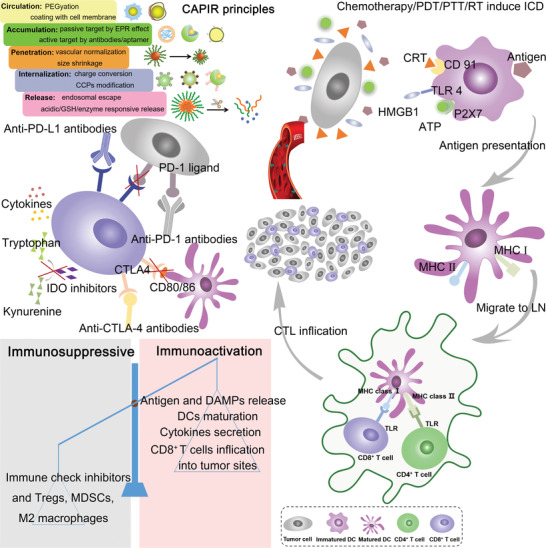
Tumor immunotherapy is only effective in a fraction of patients due to a low response rate and severe side effects, and these challenges of immunotherapy in clinics can be addressed through induction of immunogenic cell death (ICD). ICD is elicited from many antitumor therapies to release danger associated molecular patterns (DAMPs) and tumor-associated antigens to facilitate maturation of dendritic cells (DCs) and infiltration of cytotoxic T lymphocytes (CTLs). The process can reverse the tumor immunosuppressive microenvironment to improve the sensitivity of immunotherapy. Nanostructure-based drug delivery systems (NDDSs) are explored to induce ICD by incorporating therapeutic molecules for chemotherapy, photosensitizers (PSs) for photodynamic therapy (PDT), photothermal conversion agents for photothermal therapy (PTT), and radiosensitizers for radiotherapy (RT). These NDDSs can release loaded agents at a right dose in the right place at the right time, resulting in greater effectiveness and lower toxicity. Immunotherapeutic agents can also be combined with these NDDSs to achieve the synergic antitumor effect in a multi-modality therapeutic approach. In this review, NDDSs are harnessed to load multiple agents to induce ICD by chemotherapy, PDT, PTT, and RT in combination of immunotherapy to promote the therapeutic effect and reduce side effects associated with cancer treatment.

Keywords: antitumor; drug delivery system; immunogenic cell death; immunotherapy; nanomedicines; synergic therapy.


Conflict of interest statement

The authors declare no conflict of interest.

Figures

Scheme 1
Scheme 1
Schematic illustration of “CAPIR” principles for engineering smart NDDSs and the process of chemotherapy/PDT/PTT and RT‐based ICD combined with immunotherapy for cancer therapy. After a rationally designed NDDSs delivers therapeutic agents to tumor cells, the released tumor‐associated antigens can be phagocytosed by DCs and DAMPs (CRT, ATP, and HMGB1) in situ to promote DCs maturation and activate immune responses that reverse the tumor immune microenvironment from immunosuppression to immunoactivation. Furthermore, immunotherapeutic agents such as ICIs can block the PD‐1/PD‐L1 or CTLA‐4/CD28 axis to prevent the immune escape; in turn, the ICD‐inducing immunoresponsive tumor microenvironment can boost the ICIs therapeutic effect.
Figure 1
Figure 1
a) Schematic illustration of dual pH and MMP‐2 responsive micelles (sAMcP) to load both PTX and anti‐PD‐1 antibodies. b) TEM images of sAMcP after treatment at pH 7.4, pH 6.5, and pH 6.5 in the presence of 1 × 10⁻⁸ m MMP‐2 to shed the PEG layer and responsively release anti‐PD‐1 antibodies in the tumor microenvironment. c) Charge switchable property of sAMcP at pH 7.4, 6.5, and 6.5 in the presence of 10 × 10−9 m MMP‐2. d) Confocal images of CRT released on tumor cells incubated with sAMcP under different pH values. Antitumor effects on B16F10 tumor‐bearing mice: e) tumor growth curves and f) survival rates. Reproduced with permission.[ 86 ] Copyright 2020, Wiley‐VCH.
Figure 2
Figure 2
a) Illustration of nanoscale coordination polymeric core‐shell particles loaded with DHA and OxPt to induce ICD for cancer treatment. DHA and OXA were responsively released in the presence of abundant GSH in a tumor physiological condition. b) ROS generation ability in CT26 cells after OxPt/DHA treatment. c) Flow cytometry analysis of CRT exposure on the surface of tumor cells. Treatment with OxPt/DHA + anti‐PD‐L1 antibodies (αPD‐L1) remarkably enhanced d) DCs (CD11c+) and e) macrophages (F4/80+) infiltration into tumors for antigens presentation, and facilitated migration of f) CD8+ T cells (CD3ε + CD8+) in tumors for adaptive immune responses. Reproduced with permission.[ 90 ] Copyright 2019, Springer Nature.
Figure 3
Figure 3
Chemotherapy combined with cytokines and adjuvants. a–c) Schematic illustration of the preparation process to engineer an erythrocyte membrane‐coated nanogel (NR) for co‐delivery of PTX and IL‐2. Chemical structures of a) chitosan derivatives of amphoteric methacrylamide N‐carboxyethyl chitosan (CECm) and b) positively charged methacrylamide N‐(2‐hydroxy)propyl‐3‐trimethylammonium chitosan chloride (HTCCm). Immune cells in the tumor microenvironment after different treatments, d) reducing the ratio of Tregs and increasing infiltration of immune effector cells such as e) mature DCs, f) NK cells. Reproduced with permission.[ 93 ] Copyright 2017, American Chemical Society. g) Preparation of cross‐linked lipid‐polymer NPs between anionic thiolated hyaluronic acid (HA‐SH) and cationic maleimide‐modified unilamellar liposomes via charge‐mediated complexation to load CpG. Reproduced with permission.[ 96 ] Copyright 2017, American Chemical Society.
Figure 4
Figure 4
a) IR780 (green) was connected with hydrophilic APP (red) by short methionine fragments (blue) and MMP‐2 responsive cleaved linkers (PLGLAG: purple). b) Schematic illustration of the self‐assembly process to construct IP780‐M‐APP NPs. c) TEM images of IP780‐M‐APP NPs and smaller particles after incubation with MMP‐2. After intravenous injection, d) PDT increased the number of CD3+CD8+ T cells, e) while down‐regulated the ratio of Tregs. IR780‐M‐APP NPs killed both f) primary and g) distant tumors. Reproduced with permission from.[ 129 ] Copyright 2020, Elsevier.
Figure 5
Figure 5
a) Schematic illustration of constructing a supramolecular prodrug nanoplatform from HA‐CD and adamantine‐conjugated heterodimers of JQ1 and Ppa (AD‐SS‐JQ1, AD‐SS‐Ppa) via the host–guest interaction. Schematic diagram of b) the glucose metabolism in tumor cells, JQ1 down‐regulated the expression of c) HK2, d) LDHA, and e) PD‐L1. The synergic therapy promoted f) DCs maturation and g) increased the population of effective memory T lymphocytes, demonstrating effective inhibition of h) abscopal tumor growth and i) lung metastasis. Reproduced under the terms of the Creative Commons CC‐BY license.[ 136 ] Copyright 2021, The Authors. Published by Wiley‐VCH.
Figure 6
Figure 6
a) Schematic illustration of enzyme activatable prodrug vesicles (EAPVs) for PDT combined with an IDO inhibitor (NLG919). b) Confocal images analysis for revealing the penetration depth of EAPVs on CT26 multicellular spheroids. c) NLG919 release profiles after incubation with 10 mm GSH and 2.5 µg mL−1 MMP‐2. d) The ratio of kynurenine to tryptophan on CT26 after different treatments, indicating NLG 919 effectively inhibited the activity of IDO. EAPVs induced ICD, leading to e) an increase in the amount of CD8+ T cells and f) a decrease in the population of Tregs in tumor sites. Reproduced with permission.[ 140 ] Copyright 2019, American Chemical Society.
Figure 7
Figure 7
a) Preparation of BP‐based nanocomposites (BPCP) from BP nanosheets incubated with bPEI‐PEG and CpG. b) Infrared thermal images of 4T1 subcutaneous tumor mice after treatment with PBS, BP, and BPCP. c) Temperature rise curves of 4T1 cells incubated with BP nanosheets after NIR laser irradiation. BPCP‐based PTT induced ICD, evidenced with secretion of d) extracellular HMGB1 and e) ATP via ELISA. Serum cytokines levels in mice after different treatments: f) TNF‐α, g) IFN‐γ, and h) IL‐2 (a: PBS; b: laser; c: CpG; d: BP + laser; e: BPCP + laser). Reproduced with permission.[ 149 ] Copyright 2020, Elsevier.
Figure 8
Figure 8
a) Controllable Au NPs aggregation on fluidic liposomes to mediate NIR‐II responsive PTT. b) After irradiation, tumor cells released DAMPs including CRT, HMGB1, and ATP. c) The CRT at different depths of the tumor (0, 3, 6, 9 mm) to demonstrate the great penetration ability of NIR‐II PTT. PTT‐induced ICD to elicit the innate and adaptive immune response, which promoted d) DCs maturation and e) increase the number of CD8+ T cells. Reproduced with permission.[ 151 ] Copyright 2019, American Chemical Society. f) Schematic illustration of the preparation process of the AM@DLMSN@CuS/R848 nanohybrid. Cetyltrimethylammonium bromide (CTAB) and deferred acid (DFX) co‐assembled in an aqueous solution and hydrolyzed by tetraethyl orthosilicate (TEOS) to obtain DLMSNs. g) Confocal fluorescence semi‐quantitative results of HSP 70 in different groups. h) Photographs of primary and metastatic tumors after different treatments two times. Reproduced with permission.[ 155 ] Copyright 2020, American Chemical Society.
Figure 9
Figure 9
a,b) Schematic illustration of SPNs coated with membranes from 4T1 cells and DCs (SPNE) for PTT‐mediated immunotherapy. c) 4T1 cells were treated with DOX to expose CRT on the membrane surface and DCs were stimulated with resiquimod to provide T cell binding moieties (MHCI, CD80/86). d) The tumor temperature changes under 1064 nm laser irradiation after 24 h administration of SPNE. SPNU were controls without coating cell membranes. e) PTT up‐regulated the expression of HMGB1 in the tumor cytoplasm to induce ICD. f,g) Flow cytometric plots of mature DCs and cytotoxic T cells in lymph nodes after PTT. h) SPNE activated central memory T cells for the long‐term antitumor immunity. Reproduced with permission.[ 158 ] Copyright 2021, Wiley‐VCH.
Figure 10
Figure 10
a) Schematic illustration of IMD@Hf‐DBP‐αCD47 activated tumor microenvironment to inhibit tumor growth. b) Chemical structures of Hf‐DBP and trimethylsilyl trifluoroacetate (TMS‐TFA)‐modified Hf‐DBP. c) The loading efficiency of anti‐CD47 antibodies in Hf‐DBP and TFA‐modified Hf‐DBP. d,e) Macrophage repolarization ratios after X‐ray irradiation by flow cytometry. f) Primary and g) distant tumor volumes after different treatments. Reproduced with permission from.[ 172 ] Copyright 2020, American Chemical Society.
Figure 11
Figure 11
a) Schematic illustration of PLGA NPs for co‐delivery of catalase and imiquimod for RT. Catalase relieved the hypoxia in the tumor microenvironment to intensify the RT effect, and R837, a toll‐like receptor 7 agonist, acted as an adjuvant. b) Hypoxia positive area in tumor slices. c) Oxygen generation by PLGA‐R837 or PLGA‐R837@Cat in a H2O2 solution (0.1 × 10⁻³ m). d) The percentage of M2 macrophages (CD206+) in CD11b+F4/80+ cells after different treatments. Antitumor effect of PLGA‐R837@Cat based‐RT in combination of an anti‐CTLA‐4 antibody (αCTLA4) in 4T1 breast tumors: survival curves of e) the 4T1 tumor metastasis mice; f) spread and growth of fluc‐4T1 cancer cells in different treatment groups. Reproduced with permission.[ 176b ] Copyright 2019, Wiley‐VCH.
Figure 12
Figure 12
a) Schematic diagram for the assembly process of Cu2+ and 5′‐guanosine monophosphate disodium salt to construct Cu‐based nanoscale coordination polymers (Cu‐NCPs), and formation of a supramolecular network from Cu‐NCPs. b) The mechanism of mixed‐valence (Cu+ and Cu2+) to generate •OH and deplete GSH. (c) Cu‐NCPs converted GSH to GSSG in vitro. d) •OH generation at different radiation doses. e) Representative flow cytometric analysis of DCs maturation in lymph nodes. f) Cu‐NCPs combined with anti‐PD‐L1 antibodies (αPD‐L1) effectively inhibited distant tumor growth, increased the population of g) CD8+ T cells and h) promoted the secretion of IFN‐γ in distant tumors. i) Survival curves of spontaneous triple‐negative breast cancer metastasis‐bearing mice after different treatments. Group 1: Saline, Group 2: Cu‐NCPs + RT + CD8a, Group 3: RT + αPD‐L1, Group 4: Cu‐NCPs + RT, and Group 5: Cu‐NCPs + RT + αPD‐L1. Reproduced with permission.[ 180 ] Copyright 2021, Wiley‐VCH.
Figure 13
Figure 13
a) Schematic illustration of acidity and MMP‐2 dual‐responsive prodrug vesicles (MPV) for co‐delivery of OXA and a photosensitizer. 2,3‐dimethylmaleic anhydride (DMMA) was used to modify hexadecyl‐oxaliplatin diethylene amine (HOA) to form an OXA prodrug (HOAD) and the photosensitizer was conjugated with PEG via an MMP‐2‐labile GPLGLAG peptide. b) MPV‐HOAD switched from a negative charge to a positive one at pH 6.5 in the presence of MMP‐2. ICD induced by OXA and PDT resulted in c) CRT exposure on the tumor cell surface and d) secretion of IFN‐γ. e,f) The multi‐modality therapy activated the immune system of mice for long memory immune responses and protected them from re‐challenge of CT26 and 4T1 live tumor cells. Reproduced with permission.[ 182 ] Copyright 2019, Wiley‐VCH.
Figure 14
Figure 14
Multi‐modality therapeutic approaches by combining PDT, chemotherapy, and immunotherapy for cancer therapy. a) Illustration of red blood cell membrane‐coated biomimetic size‐reducible NPs, which were degraded into smaller particles in the presence of hyaluronidase. b) ROS generated from cinnamaldehyde stimulated mitochondria to intensify the PDT effect. Reproduced with permission.[ 184 ] Copyright 2019, Elsevier. c–h) PDT and DOX induced ICD to establish vaccination for CT26 tumor therapy. c) Self‐assembled chimeric cross‐linked polymersomes (CCPS) from polyethylene glycol‐poly (methyl methyacrylateco‐2‐amino ethyl methacrylate (thiol/amine))‐poly 2‐(dimethylamino)ethyl methacrylate (PEG‐P(MMA‐coAEMA (SH/NH2)‐PDMA) served as an all‐in‐one polymersomal nanoformulation with encapsulated HPPH and DOX. d) CCPS acted as an adjuvant to promote DCs maturation. e) CD8+ T cells in tumor sites after different treatments. ELISA analysis of serum cytokines f) IL‐6, g) IL‐12, and h) TNF‐α. Reproduced with permission.[ 185 ] Copyright 2019, American Chemical Society.

References

    1. a) Riley R. S., June C. H., Langer R., Mitchell M. J., Nat. Rev. Drug Discovery 2019, 18, 175; - PMC - PubMed
    2. b) Zhang Y. Y., Zhang Z. M., Cell. Mol. Immunol. 2020, 17, 807; - PMC - PubMed
    3. c) Ribas A., Wolchok J. D., Science 2018, 359, 1350; - PMC - PubMed
    4. d) Li Q., Zhou Y., He W., Ren X., Zhang M., Jiang Y., Zhou Z., Luan Y., J. Controlled Release 2021, 338, 33; - PubMed
    5. e) Li Q., Zhao Z., Qin X., Zhang M., Du Q., Li Z., Luan Y., Adv. Funct. Mater. 2021, 31, 2104630.
    1. a) Mellman I., Coukos G., Dranoff G., Nature 2011, 480, 480; - PMC - PubMed
    2. b) Nam J., Son S., Park K. S., Zou W., Shea L. D., Moon J. J., Nat. Rev. Mater. 2019, 4, 398.
    1. Sharma P., Allison J. P., Science 2015, 348, 56. - PubMed
    1. a) Topalian S. L., Taube J. M., Anders R. A., Pardoll D. M., Nat. Rev. Cancer 2016, 16, 275; - PMC - PubMed
    2. b) Sharma P., Hu‐Lieskovan S., Wargo J. A., Ribas A., Cell 2017, 168, 707. - PMC - PubMed
    1. a) Hamid O., Robert C., Daud A., Hodi F. S., Hwu W. J., Kefford R., Wolchok J. D., Hersey P., Joseph R. W., Weber J. S., Dronca R., Gangadhar T. C., Patnaik A., Zarour H., Joshua A. M., Gergich K., Elassaiss‐Schaap J., Algazi A., Mateus C., Boasberg P., Tumeh P. C., Chmielowski B., Ebbinghaus S. W., Li X. N., Kang S. P., Ribas A., N. Engl. J. Med. 2013, 369, 134; - PMC - PubMed
    2. b) Horvat T. Z., Adel N. G., Dung T. O., Momtaz P., Postow M. A., Callahan M. K., Carvajal R. D., Dickson M. A., D'Angelo S. P., Woo K. M., Panageas K. S., Wolchok J. D., Chapman P. B., J. Clin. Oncol. 2015, 33, 3193; - PMC - PubMed
    3. c) Martins F., Sofiya L., Sykiotis G. P., Lamine F., Maillard M., Fraga M., Shabafrouz K., Ribi C., Cairoli A., Guex‐Crosier Y., Kuntzer T., Michielin O., Peters S., Coukos G., Spertini F., Thompson J. A., Obeid M., Nat. Rev. Clin. Oncol. 2019, 16, 563; - PubMed
    4. d) Wang D. Y., Salem J. E., Cohen J. V., Chandra S., Menzer C., Ye F., Zhao S., Das S., Beckermann K. E., Ha L., Rathmell W. K., Ancell K. K., Balko J. M., Bowman C., Davis E. J., Chism D. D., Horn L., Long G. V., Carlino M. S., Lebrun‐Vignes B., Eroglu Z., Hassel J. C., Menzies A. M., Sosman J. A., Sullivan R. J., Moslehi J. J., Johnson D. B., JAMA Oncol. 2018, 4, 1721. - PMC - PubMed

Publication types

Substances

LinkOut - more resources