. 2022 Aug;9(22):e2201155.
doi: 10.1002/advs.202201155. Epub 2022 Jun 2.

Dynamically Responsive Scaffolds from Microfluidic 3D Printing for Skin Flap Regeneration

Affiliations

Dynamically Responsive Scaffolds from Microfluidic 3D Printing for Skin Flap Regeneration

Xiaocheng Wang et al. Adv Sci (Weinh). 2022 Aug.

Abstract

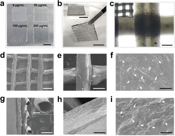
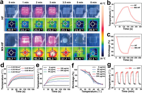
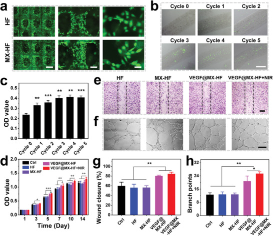
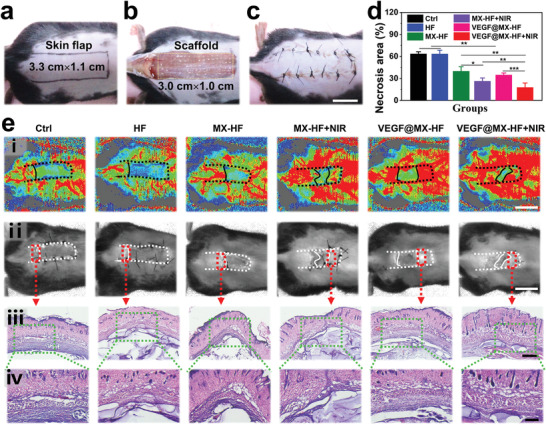
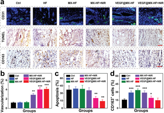
Biological scaffolds hold promising perspectives for random skin flap regeneration, while the practical application is greatly limited by their insufficient vascularization ability and the lack of responsiveness during the dynamical healing process. Herein, a novel MXene-incorporated hollow fibrous (MX-HF) scaffold with dynamically responsive channels is presented for promoting vascularization and skin flap regeneration by using a microfluidic-assisted 3D printing strategy. Benefiting from the photothermal conversion capacity of the MXene nanosheets and temperature-responsive ability of poly(NIPAM) hydrogels in the MX-HF scaffolds, they display a near-infrared (NIR)-responsive shrinkage/swelling behavior, which facilitates the cell penetration into the scaffold channels from the surrounding environment. Moreover, by incorporating vascular endothelial growth factor (VEGF) into the hydrogel matrix for controllable delivery, the MX-HF scaffolds can achieve promoted proliferation, migration, and proangiogenic effects of endothelial cells under NIR irradiation. It is further demonstrated in vivo that the NIR-responsive VEGF@MX-HF scaffolds can effectively improve skin flap survival by promoting angiogenesis, decreasing inflammation, and attenuating apoptosis in skin flaps. Thus, it is believed that such responsive MX-HF scaffolds are promising candidates for clinical random skin flap regeneration as well as other diverse tissue engineering applications.

Keywords: 3D printing; microfluidics; photothermal; regeneration; scaffold; vascularization.


Conflict of interest statement

The authors declare no conflict of interest.

Figures

Figure 1
Figure 1
Schematic illustration of the dynamically responsive scaffolds from microfluidic 3D printing for skin flap regeneration. The MX‐HF scaffolds can be fabricated via a coaxial capillary microfluidic strategy. The MX‐HF scaffolds displays a photothermal‐responsive shrinkage/swelling behavior under the control of NIR irradiation, which can facilitate the infiltration of cells or tissues into the scaffold channels. In addition, the controllable VEGF release from the dynamic channeled scaffolds can exert promotive effects on the formation of new blood vessels and large‐scaled skin flap regeneration.
Figure 2
Figure 2
Characterization of MX‐HF scaffolds. Photographs of a) MX‐HF scaffolds with different MXene contents (MXene concentration in the hydrogel precursor: 0, 50, 100, and 200 µg mL−1). Scale bar indicates 1 cm. b) Photographs of the different views of the hollow fibrous scaffolds (MXene content: 200 µg mL−1). Scale bars indicate 1 cm. c) Optical micrographs of the hollow scaffolds with straight channels. Scale bars indicate 200 and 500 µm (inset). d–f) Top and g) section views of the scanning electron microscope (SEM) images of the freeze‐dried MX‐HF scaffolds at different magnifications. h,i) The high‐resolution SEM images of the scaffold channels indicated the incorporated MXene nanosheets within the MX‐HF scaffolds. The white arrows in (f) and (i) indicate the nanosheets incorporated in the scaffold matrix. Scale bars indicate 500 µm in (d) and (g), 150 µm in (e), 100 µm in (g) (inset), and 5 µm in (h), 2 µm in (f), and 500 nm in (i).
Figure 3
Figure 3
Photothermal responsive performance of the MX‐HF scaffolds. a) Real‐time photographs and thermal images, corresponding b) photothermal heating curves and c) volume shrinkage ratio of HF and MX‐HF scaffolds when exposed to NIR laser irradiation at 0.40 W cm−2 for 3 min (ON), and followed by naturally cooling to room temperature without irradiation for 3 min (OFF). Scale bar indicates 1 cm. Photothermal heating curves of d) MX‐HF scaffolds with MXene contents of 100 µg mL−1 at different laser power densities of 0.30, 0.35, 0.40, 0.45, and 0.50 W cm−2 for 3 min and e) MX‐HF scaffolds with different MXene contents (MXene concentration: 0, 25, 50, 100, and 200 µg mL−1) under continuous 808 nm irradiation at a power intensity of 0.40 W cm−2 for 3 min. f) Volume shrinkage ratio of MX‐HF scaffolds with different MXene contents at different temperatures (n = 4 per group). g) Temperature variation over five ON (red line)/OFF (black line) cycles of NIR irradiation (0.40 W cm−2, 2 min).
Figure 4
Figure 4
In vitro regenerative properties of the NIR‐responsive dynamic scaffolds. a) Live/dead staining of human umbilical vein endothelial cells (HUVECs) adhered to the surface of HF and MX‐HF scaffolds for 24 h. Alive or dead cells were in green or red, respectively. Scale bars indicate 1 mm, 300 µm, and 50 µm from left to right. b) Fluorescent images and c) CCK8 assay of HUVECs enriched in the MX‐HF channels after NIR irradiation (0.40 W cm−2, 2 min for each cycle). Scale bar indicates 200 µm. d) Cell proliferation of HUVECs cultured with different scaffolds for 14 days. e,f) Representative images of e) in vitro scratch assay and f) Matrigel tube formation of HUVECs cultured with different scaffolds. Dotted lines in (e) indicate initial scratch edges. Scale bars indicate 300 µm in (e), and 200 µm in (f). g,h) Quantification of g) closure rates for scratch assay after 15 h and h) Matrigel tube formation after 6 h of HUVECs. n = 6 per group, two‐tailed unpaired Student's t‐tests were performed to calculate the statistical significance between two groups, and *p < 0.05, **p < 0.01, and ***p < 0.001.
Figure 5
Figure 5
The skin flap survival rates after treatment. a) A random skin flap animal model, in which the flap was 1.1 cm (width) × 3.3 cm (length) on the mouse dorsal side with the pedicle at the tail end. b) After flap elevation, an MX‐HF scaffold with 1.0 cm (width) × 3.0 cm (length) × 0.5 mm (height) was implanted into the flap skin. c) The skin flaps were then sutured back in situ using silk sutures. Scale bar indicates 1 cm. d) Flap necrosis area percentages of different groups on day 9. n = 6 per group, two‐tailed unpaired Student's t‐tests were performed to calculate the statistical significance between two groups, and *p < 0.05, **p < 0.01, and ***p < 0.001. e) Histological analysis of the necrotic junction of the skin flaps in different groups: i) The real‐time blood flow images captured with laser speckle contrast imaging; ii) corresponding photos of skin flaps; iii,iv) H&E staining of the necrosis and survival junction area of the skin flaps in different groups. Scale bars indicate 1 cm in (i) and (ii), 500 µm in (iii), and 200 µm in (iv).
Figure 6
Figure 6
Angiogenesis, apoptosis and inflammation in skin flaps. a) Immunohistochemical staining images of skin flaps with anti‐CD31 antibodies (green) and DAPI (blue), TUNEL staining and CD163 staining in different groups. Scale bars indicate 100 µm (top) and 50 µm (middle, bottom). Quantification of the b) CD31 positive vessels, c) cell apoptosis assessed by TUNEL staining and d) CD163 positive macrophages. n = 6 per group, two‐tailed unpaired Student's t‐tests were performed to calculate the statistical significance between two groups, and *p < 0.05, **p < 0.01, and ***p < 0.001 compared with the control group.

References

    1. a) Marks H., Bucknor A., Roussakis E., Nowell N., Kamali P., Cascales J. P., Kazei D., Lin S. J., Evans C. L., Sci. Adv. 2020, 6, eabd1061; - PMC - PubMed
    2. b) Khan Aadil A., Paget James T., McLaughlin M., Kyula Joan N., Wilkinson Michelle J., Pencavel T., Mansfield D., Roulstone V., Seth R., Halle M., Somaiah N., Boult Jessica K. R., Robinson Simon P., Pandha Hardev S., Vile Richard G., Melcher Alan A., Harris Paul A., Harrington Kevin J., Sci. Transl. Med. 2018, 10, eaar2041. - PMC - PubMed
    1. a) Chen H., Cheng Y., Tian J., Yang P., Zhang X., Chen Y., Hu Y., Wu J., Sci. Adv. 2020, 6, eaba4311; - PMC - PubMed
    2. b) Xu P., Xing M., Huang H., Xue K., Chang J., Liu K., Chem. Eng. J. 2021, 423, 130285.
    1. Sun X., Lang Q., Zhang H., Cheng L., Zhang Y., Pan G., Zhao X., Yang H., Zhang Y., Santos H. A., Cui W., Adv. Funct. Mater. 2017, 27, 1604617.
    1. a) Cai Z., Saiding Q., Cheng L., Zhang L., Wang Z., Wang F., Chen X., Chen G., Deng L., Cui W., Bioactive Mater. 2021, 6, 4506; - PMC - PubMed
    2. b) Zhang L., Chen L., Xiang Y., Liu Z., Mao X., Zhang L., Deng L., Zhang Y., Cheng L., Sun X., Cui W., Chem. Eng. J. 2021, 406, 126839;
    3. c) Zhang L., Xiang Y., Zhang H., Cheng L., Mao X., An N., Zhang L., Zhou J., Deng L., Zhang Y., Sun X., Santos H. A., Cui W., Adv. Sci. 2020, 7, 1903553; - PMC - PubMed
    4. d) Mao X., Liu L., Cheng L., Cheng R., Zhang L., Deng L., Sun X., Zhang Y., Sarmento B., Cui W., J. Controlled Release 2019, 297, 91. - PubMed
    1. a) Cui C., Sun S., Wu S., Chen S., Ma J., Zhou F., Eng. Regener. 2021, 2, 82;
    2. b) Li X., Cho B., Martin R., Seu M., Zhang C., Zhou Z., Choi Ji S., Jiang X., Chen L., Walia G., Yan J., Callanan M., Liu H., Colbert K., Morrissette‐McAlmon J., Grayson W., Reddy S., Sacks Justin M., Mao H.‐Q., Sci. Transl. Med. 2019, 11, eaau6210. - PubMed

Publication types

Substances

LinkOut - more resources