. 2023 Jan;98(1):43-53.
doi: 10.1007/s12565-022-00671-w. Epub 2022 Jun 2.

The Corona mortis is similar in size to the regular obturator artery, but is highly variable at the level of origin: an anatomical study

Affiliations

The Corona mortis is similar in size to the regular obturator artery, but is highly variable at the level of origin: an anatomical study

René Heichinger et al. Anat Sci Int. 2023 Jan.

Abstract

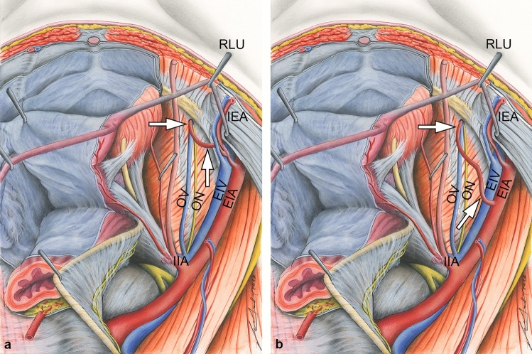
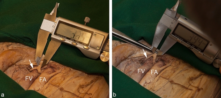
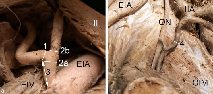
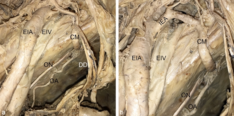
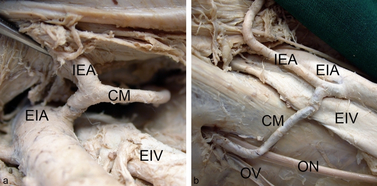
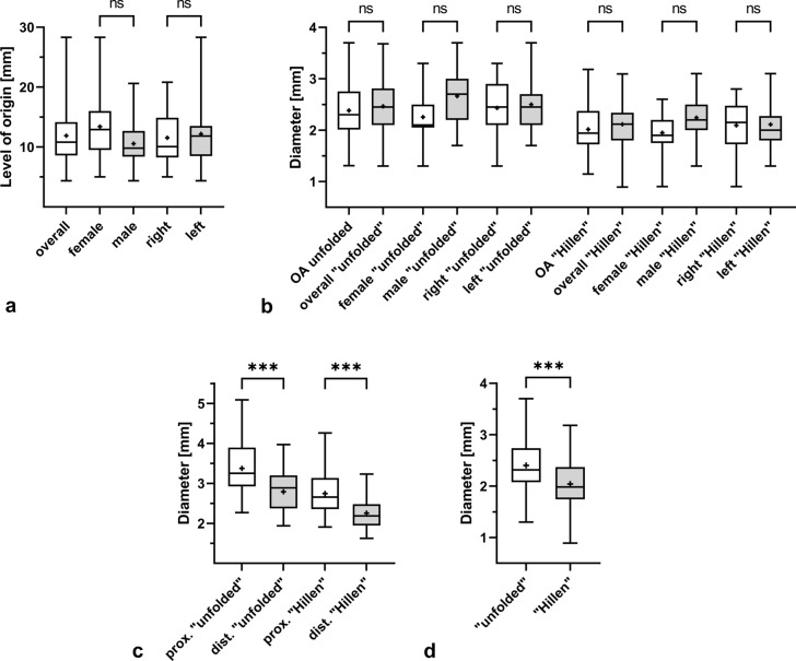
An enlarged anastomosis connecting the vascular territory of the external iliac and the obturator artery may replace most or all of the latter. This relatively common vascular variation, known as Corona mortis, can lead to death in the worst-case scenario if injured. Despite being well-known, exact anthropometric data are lacking. The purpose of this study was to determine diameters of the regular obturator artery, the Corona mortis and the inferior epigastric artery. In addition, the level of origin of the Corona mortis was quantified. The obturator artery and its norm variants were dissected bilaterally in 75 specimens (37 females, 38 males) and measured using two different methods. The Corona mortis was present in 36 of the 150 hemipelves (24%), presenting in one third of all cases bilaterally. Its level of origin measured from the commencement of the inferior epigastric artery was subject to high variability (4.4-28.3 mm). The mean diameters of the Corona mortis (mean 2.5 and 2.1 mm, respectively) and the regular obturator artery (mean 2.4 and 2.0 mm, respectively) were similar for both methods. There were no significant sex nor side differences. The diameter of the inferior epigastric artery was significantly smaller distal to the origin of the Corona mortis. The high incidence, non-predictable level of origin of the Corona mortis and its size similar to the regular obturator artery support its clinical relevance even to date. Clinicians should always be aware of an additional arterial vessel close to the pelvic brim.

Keywords: Arterial variation; Corona mortis; External iliac artery; Inferior epigastric artery; Obturator artery.


Conflict of interest statement

The authors declare that they have no conflict of interest.

Figures

Fig. 1
Fig. 1
Two most common variants of a replaced obturator artery forming a Corona mortis. Most frequently, the Corona mortis (arrows) originates from the inferior epigastric artery (IEA) (a). In rare cases the Corona mortis (arrows) can arise directly from the external iliac artery (EIA) at a variable site within the greater pelvis (b). Dorso-cranial view into the lesser pelvis. In both examples, the Corona mortis joins the obturator nerve (ON) and vein (OV) before entering the obturator canal. EIV external iliac vein, IIA internal iliac artery, RLU round ligament of uterus
Fig. 2
Fig. 2
Methods of vascular diameter measurement. In this example, the inferior epigastric artery was measured within the vascular space distal to the exit of the Corona mortis (white arrow). With the method "unfolded", the outer diameter was measured without any compression of the vessel (a). Using the method "Hillen", the vessel was completely compressed with a forceps and thus half the circumference was determined. The diameter was then calculated from this value (b). FA femoral artery, FV femoral vein
Fig. 3
Fig. 3
Measuring points for vessel diameter and level of origin. The diameter of the Corona mortis (1), the diameter of the inferior epigastric artery proximal (2a) and distal (2b) to the origin of the Corona mortis, and the distance between the origin of the inferior epigastric artery and the Corona mortis (3) were measured within the vascular space. View from distal (a). The diameter of the regular obturator artery (4) was measured half way between its origin and its entrance into the obturator canal. Wall of lesser pelvic, view from ventromedial, for a better overview the internal iliac vein and its branches have been removed (b). EIA external iliac artery, EIV external iliac vein, IIA internal iliac artery, IL inguinal ligament, OIM obturator internus muscle, ON obturator nerve
Fig. 4
Fig. 4
Co-incidence of a Corona mortis and a regular obturator artery. A Corona mortis (CM) and a regular obturator artery (OA) were present together in this left male pelvic hemisphere. Wall of lesser pelvic and vascular space, view from craniomedial, for a better overview the internal iliac vein and its branches have been removed, overview (a), detail (b). DD ductus deferens; EIA external iliac artery, EIV external iliac vein, ON obturator nerve
Fig. 5
Fig. 5
Reversed Corona mortis. In one left male hemipelvis, the Corona mortis (CM) partially replaced the inferior epigastric artery (IEA) instead of the obturator artery. Note the angle of junction and the greater diameter of the inferior epigastric artery (IEA) after the union with the Corona mortis (CM). Vascular space view from distal. EIA external iliac artery, EIV external iliac vein, LL lacunar ligament
Fig. 6
Fig. 6
Corona mortis originating from inferior epigastric artery. An example of a Corona mortis (CM) originating from the inferior epigastric artery (IEA) within the vascular space. View from distal (a). An example of a Corona mortis (CM) originating from the inferior epigastric artery (IEA) proximal to the vascular space. Wall of lesser pelvic, view from medial (b). EIA external iliac artery, EIV external iliac vein, ON obturator nerve, OV obturator vein
Fig. 7
Fig. 7
Corona mortis originating from external iliac artery. An example of a Corona mortis (CM) originating from the external iliac artery (EIA) proximal to the vascular space. Wall of lesser pelvic, view from craniomedial. EIV external iliac vein, ON obturator nerve, OV obturator vein
Fig. 8
Fig. 8
Boxplots summarizing levels of origin and diameters of the regular obturator artery, the Corona mortis, and the inferior epigastric artery. The level of origin of the Corona mortis from the inferior epigastric artery is depicted for all cases (n = 32), for females (n = 15) and males (n = 17), as well as for right (n = 14) and left (n = 18) sides (a). The diameters are given for the regular obturator artery (OA, n = 116) and for the Corona mortis overall (n = 36), in females (n = 17) and males (n = 19), as well as in the right (n = 16) and left (n = 20) side of the body. Thereby, the data obtained by the method “unfolded” are depicted within the left group, the data obtained by the method “Hillen” are depicted within the right group (b). The diameters of the inferior epigastric artery proximal and distal to the origin of the Corona mortis are presented for all cases (n = 32) and both methods (c). The diameters determined by the two methods “unfolded” and “Hillen” are given for all cases, regardless of whether a regular obturator artery or a corona mortis was present (d). The outlines of the boxes indicate the 25% and 75% percentile, the solid black line within the boxes represents the median. Whiskers indicate the minimum and maximum. ns not significant, ***p < 0.001

References

    1. Adachi B. Das Arteriensystem der Japaner. Kyoto: Maruzen; 1928.
    1. Al-Talalwah W. A new concept and classification of corona mortis and its clinical significance. Chin J Traumatol. 2016;19:251–254. doi: 10.1016/j.cjtee.2016.06.004. - DOI - PMC - PubMed
    1. Ates M, Kinaci E, Kose E, et al. Corona mortis: in vivo anatomical knowledge and the risk of injury in totally extraperitoneal inguinal hernia repair. Hernia. 2016;20:659–665. doi: 10.1007/s10029-015-1444-8. - DOI - PubMed
    1. Berberoĝlu M, Uz A, Özmen MM, et al. Corona mortis: an anatomic study in seven cadavers and an endoscopic study in 28 patients. Surg Endosc. 2001;15:72–75. doi: 10.1007/s004640000194. - DOI - PubMed
    1. Braithwaite JL. Variations in origin of the parietal branches of the internal iliac artery. J Anat. 1952;86:423–430. - PMC - PubMed

LinkOut - more resources