. 2022 Jul 8;7(13):e157896.
doi: 10.1172/jci.insight.157896.

ANGPTL4 influences the therapeutic response of patients with neovascular age-related macular degeneration by promoting choroidal neovascularization

Affiliations

ANGPTL4 influences the therapeutic response of patients with neovascular age-related macular degeneration by promoting choroidal neovascularization

Yu Qin et al. JCI Insight. .

Abstract

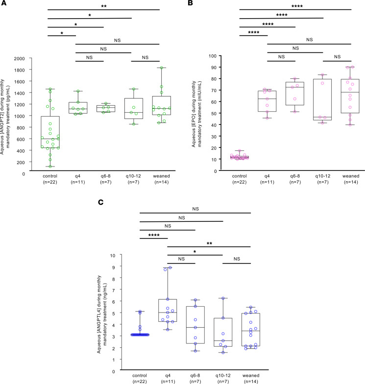
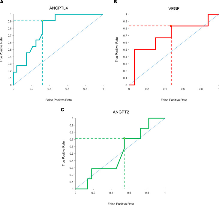
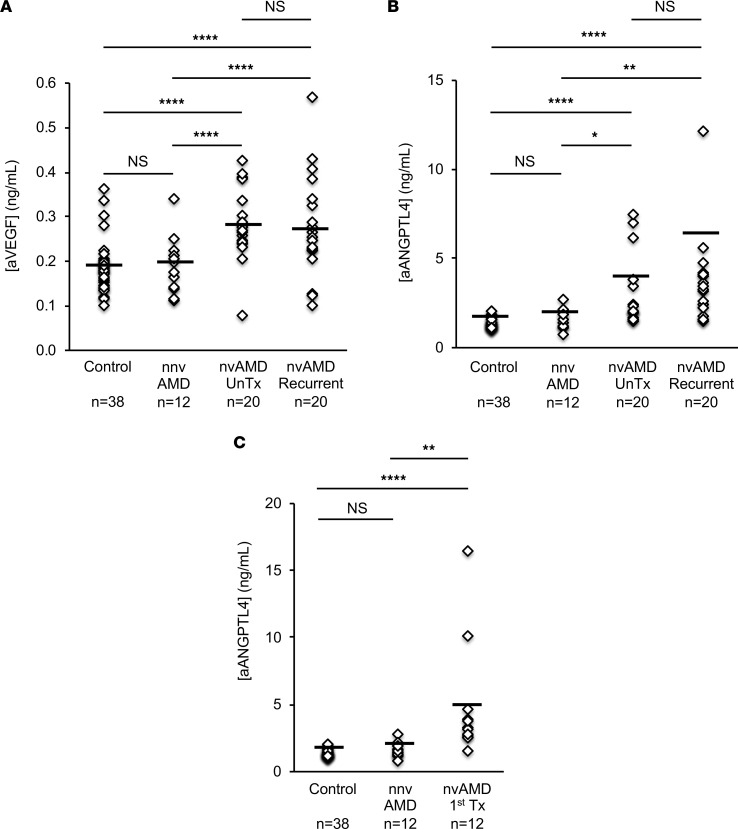
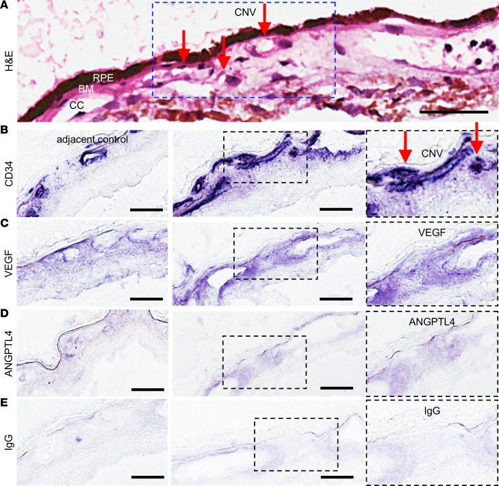
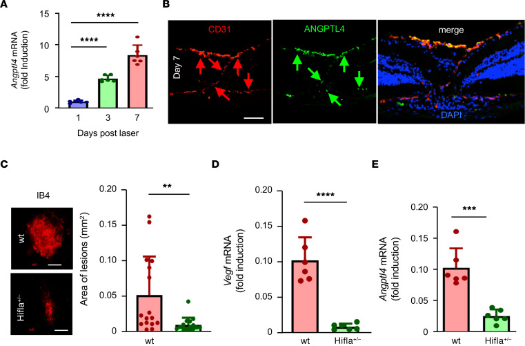
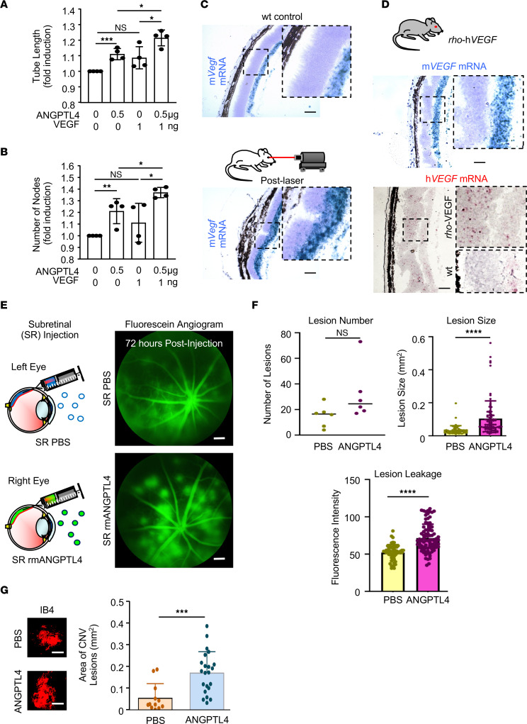
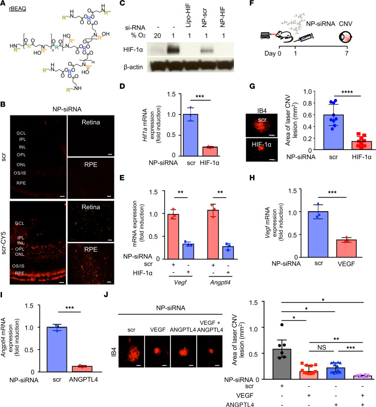
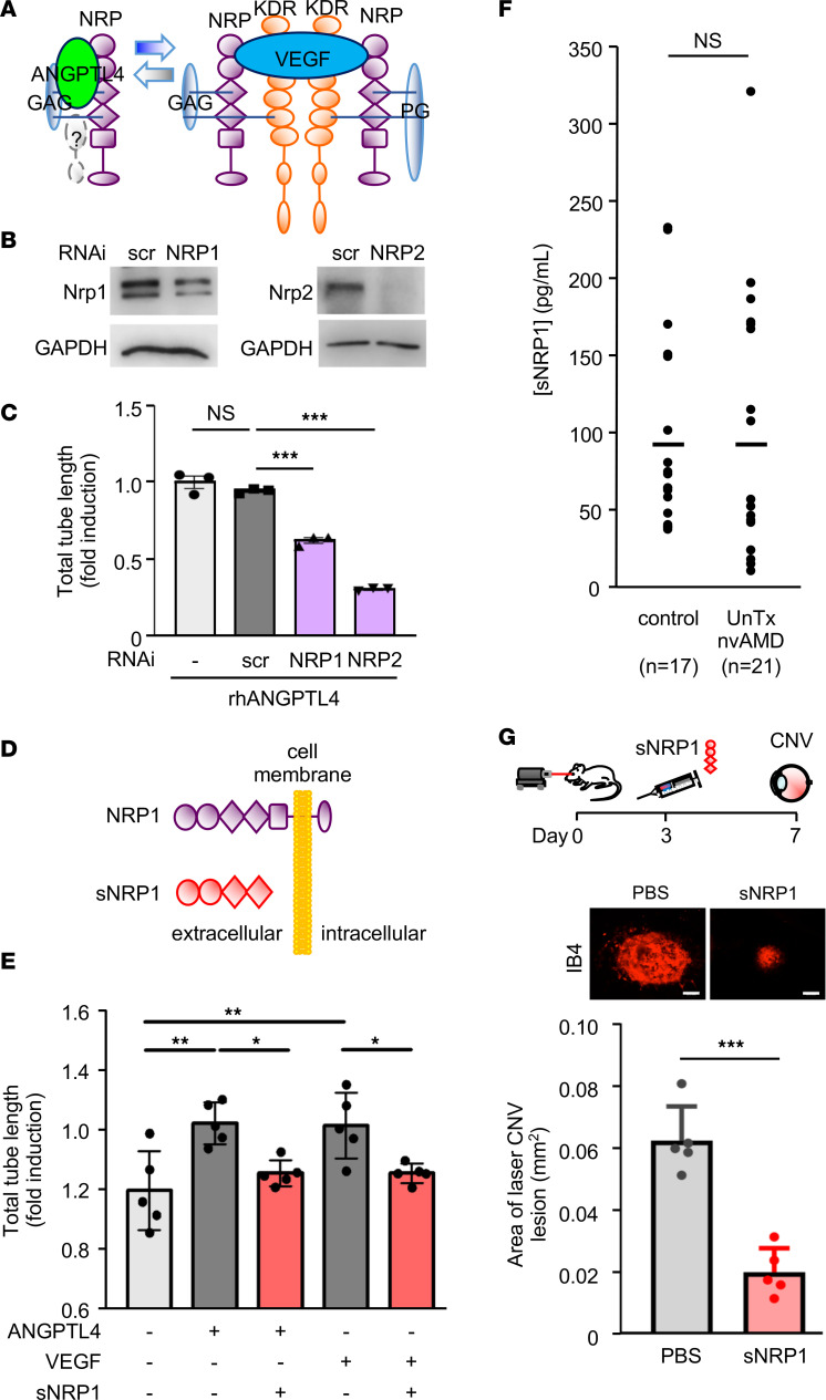
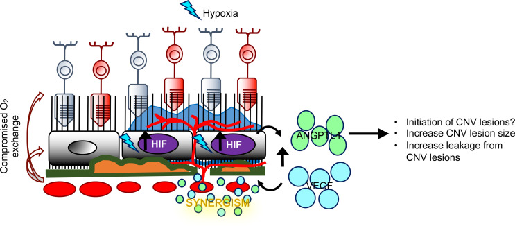
Most patients with neovascular age-related macular degeneration (nvAMD), the leading cause of severe vision loss in elderly US citizens, respond inadequately to current therapies targeting a single angiogenic mediator, vascular endothelial growth factor (VEGF). Here, we report that aqueous fluid levels of a second vasoactive mediator, angiopoietin-like 4 (ANGPTL4), can help predict the response of patients with nvAMD to anti-VEGF therapies. ANGPTL4 expression was higher in patients who required monthly treatment with anti-VEGF therapies compared with patients who could be effectively treated with less-frequent injections. We further demonstrate that ANGPTL4 acts synergistically with VEGF to promote the growth and leakage of choroidal neovascular (CNV) lesions in mice. Targeting ANGPTL4 expression was as effective as targeting VEGF expression for treating CNV in mice, while simultaneously targeting both was more effective than targeting either factor alone. To help translate these findings to patients, we used a soluble receptor that binds to both VEGF and ANGPTL4 and effectively inhibited the development of CNV lesions in mice. Our findings provide an assay that can help predict the response of patients with nvAMD to anti-VEGF monotherapy and suggest that therapies targeting both ANGPTL4 and VEGF will be a more effective approach for the treatment of this blinding disease.

Keywords: Clinical practice; Molecular diagnosis; Molecular pathology; Ophthalmology.


Conflict of interest statement

Conflict of interest: AS and GLS are the cofounders of and hold equity in HIF Therapeutics Inc. AS is a member of the Board of Directors and serves as the CEO of HIF Therapeutics Inc. GLS is also a member of the Board of Directors and serves as the President HIF Therapeutics Inc. This arrangement has been reviewed and approved by the Johns Hopkins University in accordance with its conflict-of-interest policies. JG is an inventor on patent applications related to this work filed by Johns Hopkins University (PCT/US2019/055741 Filed October 10, 2019, 17/282,939 Filed April 5, 2021).

Figures

Figure 1
Figure 1. Aqueous fluid levels of HIF-regulated vasoactive mediators, ANGPT2, EPO, and ANGPTL4 in patients with nvAMD treated with anti-VEGF therapy.
(AC) Aqueous ANGPT2 (A), EPO (B), and ANGPTL4 (C) levels of patients during initial 3 monthly mandatory treatment phase of a treat-and-extend protocol for patients with increasing intervals between treatments at 12 months (from subset of TEP/M patients divided into groups based on the required treatment interval at the end of year 1 using the TEP/M protocol to wean patients from treatment; ref. 8). Patients extended to 12 weeks had treatment paused, and patients were considered “weaned” off treatment if treatment pause reached 30 weeks. Statistical analysis was performed using Wilcoxon rank sum test. control, non-AMD eyes; q4, patients requiring treatment every 4 weeks; q6–8, patients requiring treatment every 6–8 weeks; q10-12, patients requiring treatment every 10-12 weeks. *P < 0.05; **P < 0.01; ****P < 0.0001.
Figure 2
Figure 2. ROC curves for aqueous fluid levels of HIF-regulated vasoactive mediators, ANGPTL4, VEGF, and ANGPT2, in patients who require monthly treatment with anti-VEGF therapy.
(AC) ROC curves for HIF-regulated vasoactive mediators in TEP/M patients when predicting patients who required monthly treatment with anti-VEGF for ANGPTL4 (A), VEGF (B), and ANGPT2 (C). Optimal predictive values are denoted by dashed lines based on selected cutoff concentrations for each mediator: VEGF, 260 pg/mL; ANGPT2, 1100 pg/mL; and ANGPTL4, 4.22 ng/mL.
Figure 3
Figure 3. Expression of VEGF and ANGPTL4 in aqueous fluid from patients with nvAMD treated in the clinic for active CNV.
(A and B) Aqueous fluid levels of VEGF (A) and ANGPTL4 (B) in treatment-naive patients with active nvAMD (i.e., patients with nvAMD who have never received anti-VEGF therapy; nvAMD UnTx) and patients with nvAMD previously treated with anti-VEGF therapy 12 or more weeks prior to sample collection (nvAMD Recurrent) compared with patients with nnvAMD and non-AMD (Control) patients. Aqueous fluid samples with ANGPTL4 > 15 ng/mL are not displayed to adequately demonstrate the variability within the nvAMD samples; see Supplemental Figure 1, A and B, for all samples tested. (C) Aqueous fluid levels of ANGPTL4 in patients with nvAMD treated with their first anti-VEGF therapy within 4–6 weeks of sample collection “nvAMD 1st Tx” compared with patients with nnvAMD and non-AMD (Control) patients. Kruskal-Wallis with Dunn’s multiple-comparison test. *P < 0.05; **P < 0.01; ****P < 0.0001.
Figure 4
Figure 4. Expression of VEGF and ANGPTL4 in autopsy eyes from patients with known nvAMD.
(A) Representative images from H&E staining of a CNV lesion in an autopsy eye from a patient with known nvAMD. Red arrows point to CNV vessels that have broken through (and are anterior to) Bruch’s membrane (BM). (B) Representative images from immunohistochemical analysis for CD31 within the area of the CNV membrane (CNV; right) or adjacent tissue without active CNV (adjacent control; left). (C and D) Representative images from immunohistochemical analysis for VEGF and ANGPTL4 within the area of the CNV membrane or in adjacent control. Inset demonstrates magnified view of staining within CNV lesion. (E) IgG is used as a negative control. RPE, retinal pigment epithelium; BM, Bruch’s membrane; CC, choriocapillaris. Scale bars: 25 μm.
Figure 5
Figure 5. HIF-dependent expression of ANGPTL4 in laser-induced CNV lesions in mice.
(A) Expression of Angptl4 mRNA (qPCR) in RPE/choroidal lysates from laser CNV eyes over time. (B) Expression of ANGPTL4 protein (green arrows) in laser CNV lesions (labeled with CD31; red arrows) 7 days following laser treatment. (C) Size of CNV lesions in Hif1a+/– mice compared with WT littermate controls. (D and E) Expression of Vegf (D) and Angptl4 (E) mRNA (qPCR) in RPE/choroidal lysates from laser CNV eyes in Hif1a+/– mice compared with WT littermate controls. n = 3–6 animals. Blue nuclear staining with DAPI. Scale bars: 60 μm (B) or 200 μm (C). Student’s t test. **P < 0.01; ***P < 0.001; ****P < 0.0001.
Figure 6
Figure 6. ANGPTL4 enhances the ability of VEGF to promote CNV lesions in mice.
(A and B) Endothelial cell tubule length (A) and number of nodes (B) in response to treatment with rhANGPTL4, rhVEGF, or both. (C) Expression of murine Vegf (mVegf) mRNA (in situ hybridization) in control (WT) animals (above) and 7 days following treatment with laser (below). (D) Expression of mVegf (blue) and human VEGF (hVEGF; red) mRNA in rho-hVEGF transgenic mice. (E) Schematic (left) demonstrating subretinal injection with PBS (above) or rmANGPTL4 (below) in rho-hVEGF mice. Fluorescein angiogram (FA) demonstrating fluorescein leakage from CNV lesions 72 hours following subretinal injection of PBS (above) or rmANGPTL4 (below). (F) The number and size of, and leakage from CNV lesions on FA. (G) Size of CNV lesions on choroidal flat mounts stained with isolectin 72 hours after following subretinal injection of PBS or rmANGPTL4. n = 6–9 animals. Scale bars: 100 μm (C and D [above]) and 200 μm (D [below], E, and G). Wilcoxon rank sum test and Student’s t test. *P < 0.05; **P < 0.01; ***P < 0.001; ****P < 0.0001.
Figure 7
Figure 7. In vivo nanoparticle-mediated siRNA targeting ANGPTL4 and VEGF is more effective than targeting either angiogenic factor alone for the treatment of laser CNV mice.
(A) rBEAQ polymer for in vivo delivery of siRNA. (B) Expression of fluorophore in cross section (left) and neurosensory retina (Retina) or RPE/choroidal (RPE) flat mounts (right) of mice 1 day following intravitreal injection with NP-scr or NP-scr conjugated to Cy5. (C) Expression of HIF-1α protein (WB) in lysates from primary mouse RPE cells treated with 1% O2 in the presence of nanoparticle-encapsulated siRNA targeting Hif1a (NP-HIF) versus scrambled control (NP-scr) for 72 hours. Standard transfection with Lipofectamine RNAiMAX-encapsulated siRNA targeting Hif1a (Lipo-HIF) was used as a positive control. (D and E) Hif1a (D), Vegf, and Angptl4 (E) mRNA expression (qPCR) in RPE/choroidal lysates 5 days following a single intravitreal injection with siRNA targeting Hif1a (NP-HIF) versus NP-scr in mice. (F) Schematic of laser CNV model in which a single dose of NP-HIF or NP-scr control is administered by intravitreal injection 1 day after laser treatment; eyes were enucleated for analysis on day 7. (G) Size of CNV lesion in mice treated with NP-scr or NP-HIF. (H and I) Vegf (H) and Angptl4 (I) mRNA expression (qPCR) in RPE/choroidal lysates 5 days following a single intravitreal injection with siRNA targeting Vegf (NP-VEGF) or Angptl4 (NP-ANGPTL4), respectively. (J) Size of CNV lesion in mice treated with NP-scr, NP-VEGF, NP-ANGPTL4, or both NP-VEGF and NP-ANGPTL4 administered by intravitreal injection 1 day after laser treatment; eyes were enucleated for analysis on day 7. n = 3–6 animals. GCL, ganglion cell layer; IPL, inner plexiform layer; INL, inner nuclear layer; OPL, outer plexiform layer; ONL, outer nuclear layer; IS/OS, inner/outer segments; RPE, retinal pigment epithelium. Scale bars:25 μm (B) and 100 μm (G and J). Student’s t test (D, E, and GI) and Kruskal-Wallis with Dunnett’s T3 correction (J). *P < 0.05; **P < 0.01; ***P < 0.001; ****P < 0.0001.
Figure 8
Figure 8. sNRP1 reduces the promotion of angiogenesis by ANGPTL4 and inhibits CNV in mice.
(A) Schematic demonstrating binding of ANGPTL4 and VEGF to the endothelial cell receptors, neuropilin 1 (NRP1) and NRP2. (B and C) Knockdown of NRP1 or NRP2 (B) inhibits the promotion of human umbilical vein endothelial cell (HUVEC) tubule formation by rhANGPTL4 (C). (D) Schematic comparing NRP1 and sNRP1; the latter lacks the transmembrane domain and is, therefore, soluble. (E) sNRP1 inhibits the promotion of iREC tubule formation by rhANGPTL4 and rhVEGF. (F) Expression of sNRP1 in the aqueous fluid of patients with treatment-naive (UnTx) nvAMD compared with non-AMD controls. (G) Schematic of laser CNV model in which a single dose of rhsNRP1 control is administered by intravitreal injection 3 days after laser treatment; eyes were enucleated for analysis on day 7. Size of CNV lesion in mice treated with PBS or rhsNRP1. n = 3–6 animals. Scale bars: 100 μm. One-way ANOVA with Bonferroni correction (C and E) and Student’s t test (F and G). *P < 0.05; **P < 0.01; ***P < 0.001.
Figure 9
Figure 9. Schematic demonstrating the synergistic contribution of VEGF and ANGPTL4 in the development of CNV in patients with nvAMD.
In the aging eyes of patients with AMD, collapse of the choriocapillaris combined with thickening of Bruch’s membrane and drusen deposits impedes delivery of oxygen to the overlying RPE. This, in turn, results in relative ischemia, the accumulation of HIF-1α, and increased expression of VEGF and ANGPTL4. VEGF and ANGPTL4 cooperate to promote the development of CNV.

Similar articles

Cited by

References

    1. Harvey PT. Common eye diseases of elderly people: identifying and treating causes of vision loss. Gerontology. 2003;49(1):1–11. doi: 10.1159/000066507. - DOI - PubMed
    1. Jager RD, et al. Age-related macular degeneration. N Engl J Med. 2008;358(24):2606–2017. doi: 10.1056/NEJMra0801537. - DOI - PubMed
    1. Nguyen DH, et al. Current therapeutic approaches in neovascular age-related macular degeneration. Discov Med. 2013;15(85):343–348. - PubMed
    1. Spooner KL, et al. The burden of neovascular age-related macular degeneration: a patient’s perspective. Clin Ophthalmol. 2018;12:2483–2491. doi: 10.2147/OPTH.S185052. - DOI - PMC - PubMed
    1. Malik D, et al. Factors associated with a patient’s decision to select a cost-effective vs the most effective therapy for their own eye disease. JAMA Netw Open. 2021;4(2):e2037880. doi: 10.1001/jamanetworkopen.2020.37880. - DOI - PMC - PubMed

Publication types

MeSH terms

Substances