. 2022 Jun 2;17(6):e0268378.
doi: 10.1371/journal.pone.0268378. eCollection 2022.

Surgical and medical management in the treatment of proximal tibial metaphyseal fracture in immature dogs

Affiliations

Surgical and medical management in the treatment of proximal tibial metaphyseal fracture in immature dogs

Carly Sullivan et al. PLoS One. .

Abstract

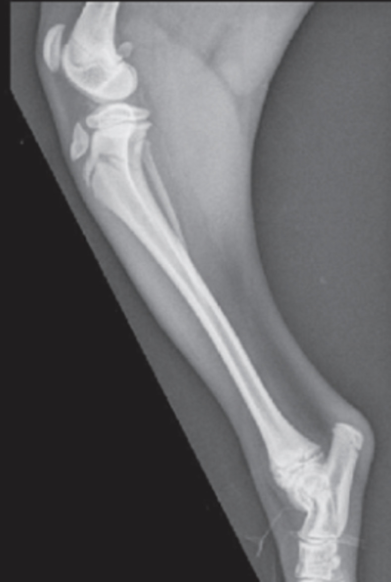
The purpose of this study was to report approaches to surgical and medical management of proximal tibial metaphyseal fractures (PTMF) and short-term case outcome. Medical records of immature dogs with PTMF were reviewed and data were collected including history, signalment and side affected. Data pertaining to surgical and medical management including radiographic evaluation and short-term complications were recorded. Forty-five dogs with a total of 47 PTMF identified and treated between 2007-2019 were included in this study. Six cases were managed with external coaptation alone. Forty-one cases were treated surgically with constructs including K-wires in different configurations, bone plate and screws, and external skeletal fixation. Of the cases managed conservatively, 4 developed complications, including bandage sores, diffuse osteopenia of the tarsus/metatarsus, and angular limb deformities. Surgical complications including pin migration necessitating removal, osteopenia, and screw placement in the proximal tibial growth plate or into the stifle joint were found in 16 cases. PTMF treated with surgery had a subjectively more predictable outcome compared to those treated with external coaptation alone. Conservative management may result in complications including development of excessive tibial plateau angle (TPA) as well as distal tibial valgus.


Conflict of interest statement

The authors have declared that no competing interests exists.

Figures

Fig 1
Fig 1. Characteristic curvilinear configuration of the proximal tibia seen with PTMF.
Fig 2
Fig 2. Mediolateral radiograph of a PTMF demonstrating cranial displacement of the distal fragment and caudal tipping of the proximal tibia resulting in an increased tibial plateau angle.
Fig 3
Fig 3. Case 34 managed with a splint bandage alone.
A and B: Mediolateral and craniocaudal views at time of injury. C and D: Mediolateral and craniocaudal views 3 weeks post injury demonstrating valgus deviation of the distal tibia. Ultimately development of genu varum, medially luxating patella, tibial tuberosity avulsion fracture, patella alta, tarsal osteopenia, and fibular malunion led to an amputation.
Fig 4
Fig 4. Case 8 managed with locking T-plate for stabilization of PTMF.
A and B: Preoperative mediolateral and craniocaudal views. C and D: Immediate postoperative mediolateral and craniocaudal views. E and F: 6 weeks postoperative mediolateral and craniocaudal views.
Fig 5
Fig 5. Case 47 managed using a locking TPLO plate.
A and B: Mediolateral and craniocaudal views. C and D: Immediate postoperative mediolateral and craniocaudal views. E and F: 4 weeks postoperative mediolateral and craniocaudal views.
Fig 6
Fig 6. Intramedullary pin and modified type 1a external fixator (Case 2).
A and B: Preoperative mediolateral and craniocaudal views. C and D: 8 weeks postoperative radiographs: mediolateral and craniocaudal views.
Fig 7
Fig 7. Case 40a managed with a splint bandage alone.
A and B: Mediolateral and craniocaudal views at time of injury. C: Mediolateral view 2 weeks post injury. D and E: Mediolateral and craniocaudal views 6 weeks post injury demonstrating excessive TPA that can result from treatment with external coaptation alone.
Fig 8
Fig 8. Development of diffuse osteopenia of the tarsus and metatarsus after a bandage was placed for 4 weeks postoperatively (case 37).
A, B and C: Bilateral mediolateral and craniocaudal views immediately postoperative. D and E: Mediolateral and craniocaudal views 4 weeks postoperative. There is progressive healing of the proximal tibial fractures. Severe osteopenia is present affecting the tarsal cuboidal bones and proximal metatarsal bones (white arrows). F, G and H: Mediolateral and craniocaudal views 8 weeks postoperative. The proximal tibial fractures have healed appropriately. Mild to moderate osteopenia of distal limbs, but improved compared to radiographs at 4 weeks after surgery (white arrows).
Fig 9
Fig 9. Inadvertent placement of the most proximal screw through the proximal tibial physis resulting in valgus deviation of the proximal tibia (Case 6).
A and B: Mediolateral and craniocaudal views at time of fracture diagnosis. C and D: Immediate postoperative mediolateral and craniocaudal views. E and F: 6 weeks postoperative mediolateral and craniocaudal views.
Fig 10
Fig 10. Case 41 in which the proximal screw violated the proximal tibial physis immediately postoperatively on radiographs, but was not within the proximal tibial physis at radiographic evaluation 4 weeks after surgery.
A and B: Mediolateral and craniocaudal views at time of injury. C and D: Immediate postoperative mediolateral and craniocaudal views. E and F: 4 weeks postoperative radiographs mediolateral and craniocaudal views.
Fig 11
Fig 11. Inadvertent place of the most proximal screw through the proximal tibial physis and into stifle joint after the case was first medically managed with a splint bandage (Case 17).
A and B: Mediolateral and craniocaudal views performed 1 day after the initial injury: C and D: Mediolateral and craniocaudal views performed 3 weeks post splint placement. There was concern for collapsing of the lateral tibial cortex resulting in increased tibial angulation. The patient was still grade 4/4 lame on exam, so surgical stabilization was elected. E and F: Mediolateral and craniocaudal views immediately postoperatively showing screw placement into the joint. G and H: Mediolateral and craniocaudal views performed 6 weeks postoperatively with screw placement still within the joint, but patient was not lame or painful on exam.

References

    1. Seamon J.A. and Simpson A.M. Tibial fractures. Clin Tech Small Anim Pract. 2005; 19: 151–167 - PubMed
    1. Gorse M.J. Using external skeletal fixation for fractures of the radius and ulna and tibia. Vet Med 1998; 93: 463–467
    1. Marretta S.M. and Schrader S.C. Physeal injuries in the dog: a review of 135 cases. JAVMA 1983; 182: 708–710 - PubMed
    1. Schmokel H. Weber U. and Hartmeier G. Salter-II fracture of the proximal tibia with avulsion of the tuberositas tibiae in the dog. Schwizer Archiv fur Tierheilkunde 1995; 137: 124–128 - PubMed
    1. Pratt J.N. Avulsion of the tibial tuberosity with separation of the proximal tibial physis in seven dogs. Vet Rec 2001; 149; 352–356 doi: 10.1136/vr.149.12.352 - DOI - PubMed