. 2022 Aug 8;61(32):e202201083.
doi: 10.1002/anie.202201083. Epub 2022 Jun 24.

Fast Magic-Angle-Spinning NMR Reveals the Evasive Hepatitis B Virus Capsid C-Terminal Domain

Affiliations

Fast Magic-Angle-Spinning NMR Reveals the Evasive Hepatitis B Virus Capsid C-Terminal Domain

Morgane Callon et al. Angew Chem Int Ed Engl. .

Abstract

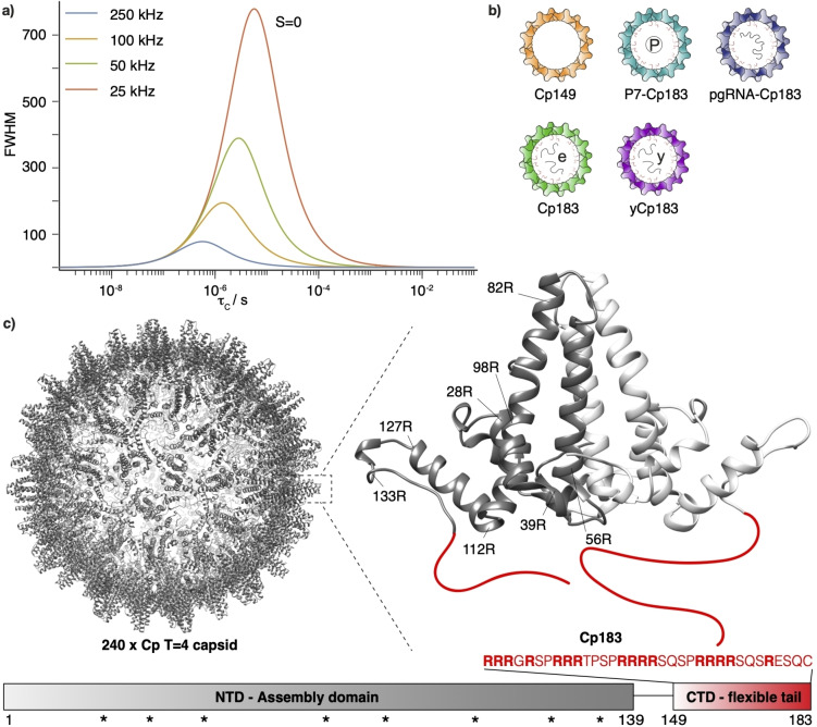
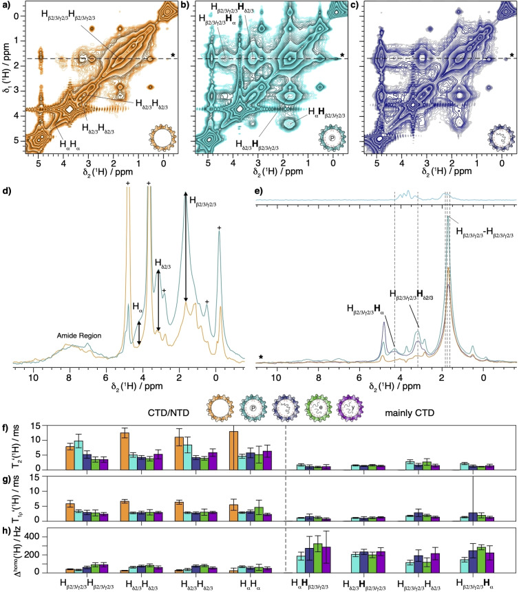
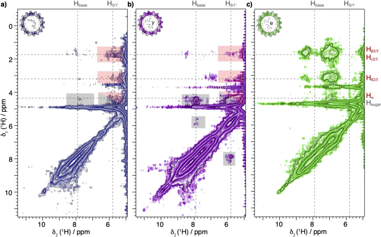
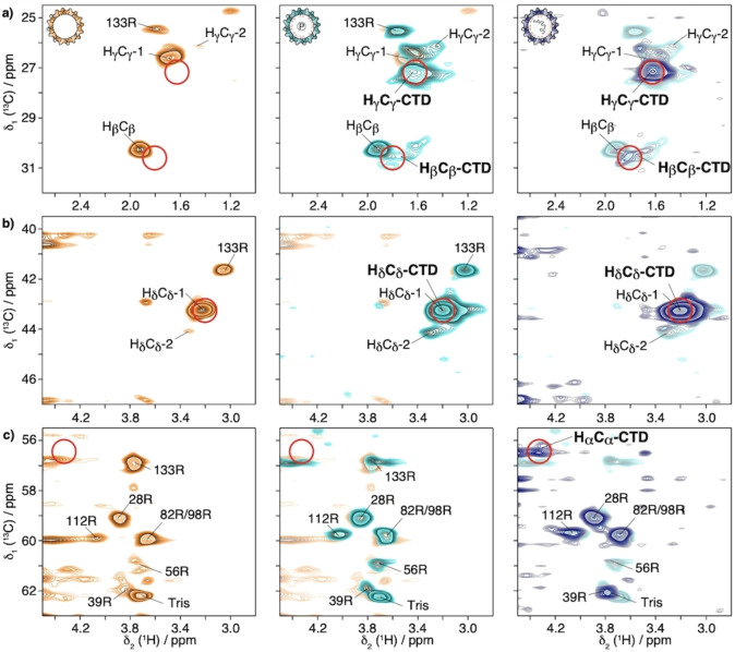
Experimentally determined protein structures often feature missing domains. One example is the C-terminal domain (CTD) of the hepatitis B virus capsid protein, a functionally central part of this assembly, crucial in regulating nucleic-acid interactions, cellular trafficking, nuclear import, particle assembly and maturation. However, its structure remained elusive to all current techniques, including NMR. Here we show that the recently developed proton-detected fast magic-angle-spinning solid-state NMR at >100 kHz MAS allows one to detect this domain and unveil its structural and dynamic behavior. We describe the experimental framework used and compare the domain's behavior in different capsid states. The developed approaches extend solid-state NMR observations to residues characterized by large-amplitude motion on the microsecond timescale, and shall allow one to shed light on other flexible protein domains still lacking their structural and dynamic characterization.

Keywords: Dynamics; Hepatitis B Virus; Relaxation; Solid-State NMR; Viral Capsid.


Conflict of interest statement

The authors declare no conflict of interest.

Figures

Figure 1
Figure 1
a) Incoherent proton FWHM as function of the motional correlation time of the NH vector (τ C) simulated for an isolated 1H‐15N two‐spin system with the parameters of an amide group in a protein and an order parameter of S=0 (strongest broadening) at MAS frequencies between 25 and 250 kHz. Microsecond time scales of the underlying dynamics can lead to incoherent line broadening, but can be reduced at faster MAS frequencies. The equation describing the curve is provided in the Supporting Information, Equation 3. b) Schematic representation of the different capsid forms investigated: truncated Cp149 for reference of capsids devoid of the CTD; empty hyperphosphorylated P7‐Cp183; pg‐RNA containing reassembled capsids; E. coli RNA containing capsids; and yeast‐RNA containing reassembled capsids. The molecular mass of Cp149 is 4.0 MDa, of Cp183 is 5.1 MDa and of P7‐Cp183 is 5.2 MDa. c) Representation of the hepatitis B virus capsid 3D structure (PDB: 1qgt [35] ), with a zoom on the dimeric subunit structure, and its domain organization with the amino‐acid sequence of the CTD in red. The X‐ray structure was determined on the isolated NTD. The stars show positions of the arginine residues in the NTD.
Figure 2
Figure 2
1H‐1H spectroscopy on selectively Arginine‐labelled samples based on 1H‐1H homonuclear mixing by spin diffusion or NOE effects. EXSY spectra using 50 ms mixing time of a) Cp149, b) P7‐Cp183 and c) Cp183‐pgRNA recorded at 100 kHz MAS and 850 MHz proton frequency. d) Overlay of the first FIDs of EXSY spectra of Cp149 and P7‐Cp183 recorded with a mixing time of 10 ms. The peaks labelled with a cross correspond to buffer signals. e) Overlay of extracted traces from EXSY spectra at δ1(1H)=1.67 ppm with the projection of a standard hCH CP based experiment recorded on P7‐Cp183 (Figure S9b) on top. The dashed lines indicate random‐coil chemical shift positions of Arg. f) T2' (1H) and g) T1ρ (1H) values recorded from the EXSY spectra for all five capsid samples shown above. h) T2' (1H) corresponding homogeneous line width Δhomo (1H). In the figure the proton of the cross peak where the relaxation takes place is labelled in bold.
Figure 3
Figure 3
RNA‐CTD interaction is detected in EXSY spectra. EXSY spectra using 50 ms 1H‐1H homonuclear mixing time of a) Cp183‐pgRNA, b) yCp183 and c) Cp183 samples recorded at 100 kHz MAS and 850 MHz proton frequency. The RNA proton chemical shifts are indicated by vertical dashed lines while the horizontal ones indicate the positions of the cross peaks observed in the 2D EXSY spectra (Figure 2a–c). The red‐shaded boxes highlight protein‐RNA interactions, the grey‐shaded boxes RNA‐RNA interactions. We attribute the presence of stronger amide proton‐sidechain proton cross peaks in Cp183 to a lower level of deuteration at exchangeable positions.
Figure 4
Figure 4
2D hCH INEPT‐based spectra with INEPT delays τ 1=τ 2=1 ms of Cp149 (orange), P7‐Cp183 (cyan) and Cp183‐pgRNA (steel blue) in D2O buffer recorded at 100 kHz MAS and 850 MHz proton frequency. Zooms of the a) Hβ2/3Cβ/Hγ2/3Cγ , b) Hδ2/3Cδ and c) HαCα regions of 2D‐hCH INEPT‐based spectra. The NTD assignment is transferred from. For sake of simplicity Hγ2/3Cγ , Hβ2/3Cβ and Hδ2/3Cδ correlations from the CTD are labelled HγCγ , HβCβ and HδCδ . Except for R133, the CTD Arg side chains HγCγ , HβCβ and HδCδ are not resolved. The random‐coil chemical‐shift positions are shown with red circles. Spectra are overlaid with the spectrum from the previous panel for better comparison.
Figure 5
Figure 5
INEPT‐EXSY NMR experiments. 2D hCH INEPT‐based spectra with τ 1=τ 2=1 ms followed by 50 ms 1H‐1H mixing time of a) Cp149 b) P7‐Cp183 and c) Cp183‐pgRNA in D2O buffer recorded at 100 kHz MAS and 850 MHz proton frequency. The Arg 1H random coil chemical shift positions observed in the EXSY spectra in Figure 2 are shown in red, and correspond to the observed cross peaks here. Spectra are overlaid with the spectrum from the previous panel for better comparison.

References

    1. Mittermaier A. K., Kay L. E., Trends Biochem. Sci. 2009, 34, 601–611. - PubMed
    1. Matlahov I., van der Wel P. C. A., Methods 2018, 148, 123–135. - PMC - PubMed
    1. Siemer A., Arnold A., Ritter C., Westfeld T., Ernst M., Riek R., Meier B. H., J. Am. Chem. Soc. 2006, 128, 13224–13228. - PubMed
    1. Siemer A. B., Solid State Nucl. Magn. Reson. 2020, 106, 101643. - PMC - PubMed
    1. Cousin S. F., Kadeřávek P., Haddou B., Charlier C., Marquardsen T., Tyburn J.-M., Bovier P.-A., Engelke F., Maas W., Bodenhausen G., Pelupessy P., Ferrage F., Angew. Chem. Int. Ed. 2016, 55, 9886–9889; - PubMed
    2. Angew. Chem. 2016, 128, 10040–10043.

Publication types

MeSH terms

LinkOut - more resources