. 2022 Jun 7;119(23):e2204557119.
doi: 10.1073/pnas.2204557119. Epub 2022 Jun 2.

The aryl hydrocarbon receptor instructs the immunomodulatory profile of a subset of Clec4a4+ eosinophils unique to the small intestine

Affiliations

The aryl hydrocarbon receptor instructs the immunomodulatory profile of a subset of Clec4a4+ eosinophils unique to the small intestine

Wei-Le Wang et al. Proc Natl Acad Sci U S A. .

Abstract

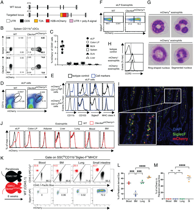
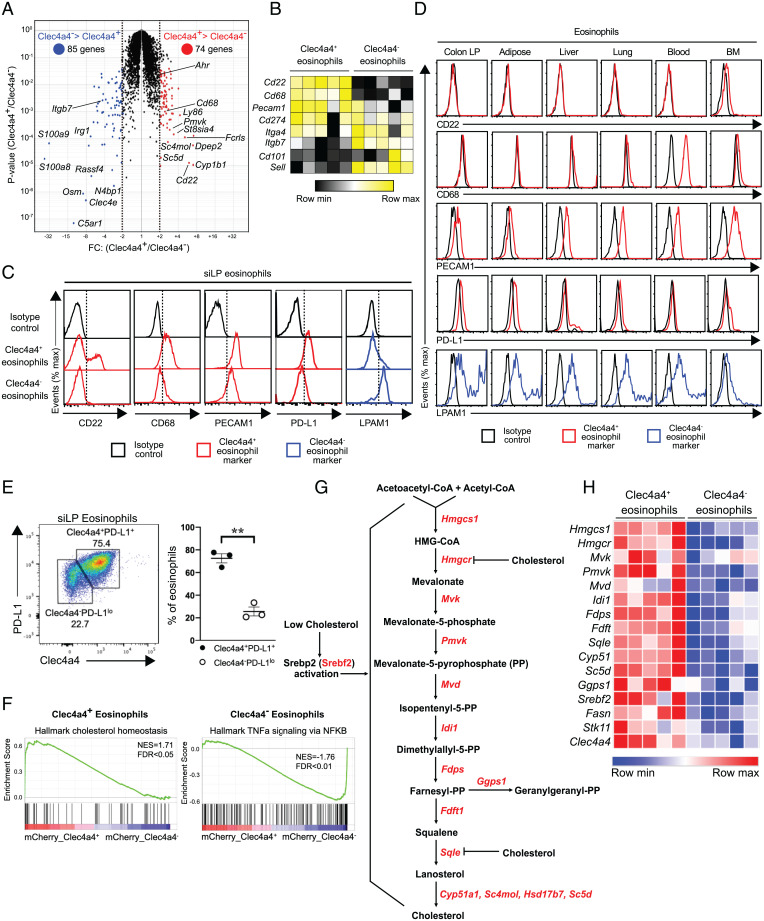
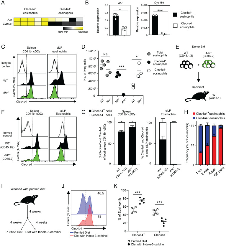
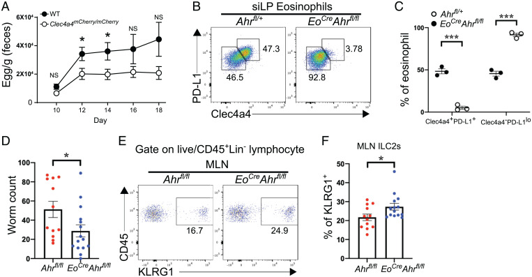
C-type lectin domain family 4, member a4 (Clec4a4) is a C-type lectin inhibitory receptor specific for glycans thought to be exclusively expressed on murine CD8α− conventional dendritic cells. Using newly generated Clec4a4-mCherry knock-in mice, we identify a subset of Clec4a4-expressing eosinophils uniquely localized in the small intestine lamina propria. Clec4a4+ eosinophils evinced an immunomodulatory signature, whereas Clec4a4− eosinophils manifested a proinflammatory profile. Clec4a4+ eosinophils expressed high levels of aryl hydrocarbon receptor (Ahr), which drove the expression of Clec4a4 as well as other immunomodulatory features, such as PD-L1. The abundance of Clec4a4+ eosinophils was dependent on dietary AHR ligands, increased with aging, and declined in inflammatory conditions. Mice lacking AHR in eosinophils expanded innate lymphoid cells of type 2 and cleared Nippostrongylus brasiliensis infection more effectively than did wild-type mice. These results highlight the heterogeneity of eosinophils in response to tissue cues and identify a unique AHR-dependent subset of eosinophils in the small intestine with an immunomodulatory profile.

Keywords: allergy; aryl hydrocarbon receptor; eosinophil; helminth; intestine.


Conflict of interest statement

Competing interest statement: M. Colonna receives research support from Pfizer, Aclaris, Ono, NGM Biopharmaceutical, Oncorus, and VigilNeuro; serves on the scientific advisory boards of Vigil Neuro and NGM Biopharmaceutical; and is consultant for Cell Signaling Technologies. All other authors have nothing to disclose.

Figures

Fig. 1.
Fig. 1.
A heterogeneous population of Clec4a4+ and Clec4a4 eosinophils resides in the small intestine. (A) Targeting strategy to generate Clec4a4mCherry/+ mice. CDS, coding sequence; H2B, human histone H2B; T2A, T2A self-cleaving peptide; UTR, untranslated region. (B) Expression of Clec4a4 and mCherry in splenic CD11b+ cDCs. (C) Frequency of mCherry+ cells among CD45+ cells (gated on live cells) in the indicated tissues (n = 3 to 7). DLN, skin-draining lymph node; LP, lamina propria; MLN, mesenteric lymph node. (D) mCherry expression on CD11chigh and CD11clow CD45+ cells derived from siLP (gated on live cells). (E) Expression of CD11b, CD103, Siglec-F, and MHC class II on mCherry+ CD11chigh CD45+ cells and mCherry+ CD11clow CD45+ cells derived from siLP (gated on live cells). (F) mCherry expression in Siglec-F+ eosinophils derived from siLP (gated on SSChigh MHC class II CD11b+ cells). (G) Representative pictures of FACS-sorted mCherry+ and mCherry eosinophils from siLP. (H) CCR3 expression in mCherry+ and mCherry eosinophils in siLP. (I) Representative image showing localization of Siglec-F+mCherry+ eosinophils in siLP villi of Clec4a4mCherry/+ mice. Inset is an enlargement of I. DAPI, 4′,6-diamidino-2-phenylindole. (J) mCherry expression in eosinophils in the indicated tissues. (K) Parabiosis of CD45.1 WT and CD45.2 Clec4a4mCherry/+ mice indicates that eosinophils recirculate in siLP, blood, and lung but much less so in BM; Clec4A4 mCherry is only expressed by eosinophils in the siLP. (L) Frequency of parabiont-derived eosinophils in the indicated tissues. (M) Frequency of parabiont-derived mCherry+ eosinophils in the indicated tissues. Results are shown as mean ± SEM. P values were calculated using one-way ANOVA and the Tukey's multiple comparisons test or the two-tailed Student’s t test. ns, not significant. ***P < 0.001; ****P < 0.0001.
Fig. 2.
Fig. 2.
Transcriptomic profiling and characterization of Clec4a4+ and Clec4a4 eosinophil subsets. (A) Volcano plot of differentially expressed genes in mCherry+ and mCherry eosinophils (n = 5 replicates in each group). Transcripts with FC (fold change) > 2 and P < 0.05 in mCherry+ and mCherry eosinophils are depicted in red and blue, respectively. (B) Heat map of selected genes specific to Clec4a4+ and Clec4a4 eosinophils. (C) Differential expression of cell surface (CD22, PECAM1, PD-L1, and LPAM1) and intracellular markers (CD68) by Clec4a4+ and Clec4a4 siLP eosinophils. (D) Expression of the markers indicated in C by eosinophils from the indicated tissues. LP, lamina propria. (E, Left) Expression of Clec4a4 and PD-L1 in eosinophils derived from siLP. (E, Right) Frequency of Clec4a4+PD-L1+ and Clec4a4PD-L1lo eosinophils in the siLP. (F) Enrichment plots demonstrating up-regulated cholesterol homeostasis and TNFα signaling via NF-κB in mCherry+ and mCherry eosinophils, respectively. NES, normalized enrichment score; FDR, false discovery rate. (G) Biochemical pathways for de novo cholesterol synthesis. CoA, coenzyme A; HMG, hydroxymethylglutaryl. (H) Heat map of genes associated with cholesterol synthesis pathways in mCherry+ and mCherry eosinophils. Results are shown as mean ± SEM. P values were calculated using the two-tailed Student’s t test. **P < 0.01.
Fig. 3.
Fig. 3.
Development of the Clec4a4+ eosinophil subset is dependent on aging and on the AHR pathway. (A) Heat map of Ahr and Cyp1b1 in mCherry+ and mCherry eosinophils. (B) qRT-PCR of Ahr and Cyp1b1 transcripts in Clec4a4+ and Clec4a4 eosinophils (n = 4 replicates). (C) Clec4a4 expression in splenic CD11b+ cDCs and siLP eosinophils from WT and Ahr−/− mice. (D) Absolute numbers of Clec4a4+ and Clec4a4 eosinophils in WT and Ahr−/− mice (n = 3 in each group). (E) Experimental strategy for mixed BM chimeras. (F) Clec4a4 expression in splenic CD11b+ cDCs and siLP eosinophils derived from WT and Ahr−/− BM cells after reconstitution. (G) Frequency of Clec4a4+ and Clec4a4 splenic CD11b+ cDCs and siLP eosinophils in BM chimeras. (H) Frequency of Clec4a4+ and Clec4a4 siLP eosinophils in WT mice at 1, 2, and 10 to 12 wk of age (n = 3, 6, and 7 replicates, respectively) and in GF mice (n = 5). (I) Experimental time line for feeding mice with a purified diet free of AHR ligands or a purified diet containing the AHR ligand I3C. (J) Representative Clec4a4 expression in siLP eosinophils derived from mice fed with a purified diet free of AHR ligands or a purified diet containing AHR ligands. (K) Frequency of Clec4a4+ and Clec4a4 siLP eosinophils in mice fed with a purified diet free of AHR ligands (n = 4) or a purified diet containing AHR ligands (n = 5). Results are shown as mean ± SEM. P values were calculated using the two-tailed Student’s t test. NS, not significant. *P < 0.05; ***P < 0.001.
Fig. 4.
Fig. 4.
Clec4a4+ eosinophils modulate type 2 immunity during helminth infection. (A) Egg counts in feces of WT and Clec4a4-deficient (Clec4a4mCherry/mCherry) mice (n = 13 in each group) at the indicated time points during H. polygyrus infection. (B) Expression of Clec4a4 and PD-L1 in siLP eosinophils derived from Ahrfl/+ and EoCreAhrfl/fl mice. (C) Frequency of Clec4a4+PD-L1+ and Clec4a4PD-L1lo siLP eosinophils in Ahrfl/+ and EoCreAhrfl/fl mice (n = 3 each group). (D) Worm counts in Ahrfl/fl (n = 12) and EoCreAhrfl/fl (n = 15) mice at day 6 postinfection with N. brasiliensis. (E) Representative percentages of KLRG1+ ILC2s among CD45+ cells in mesenteric lymph nodes (MLNs) of Ahrfl/fl and EoCreAhrfl/fl mice. A gate was applied on lineage-negative cells. Lineage markers included CD3, CD4, CD8, CD19, and NK1.1. (F) Frequency of ILC2s in MLNs from Ahrfl/fl (n = 12) and EoCreAhrfl/fl (n = 14) mice at day 6 postinfection with N. brasiliensis. Results are shown as mean ± SEM. P values were calculated using the two-tailed Student’s t test or one-way ANOVA and the Tukey's multiple comparisons test. NS, not significant. *P < 0.05; ***P < 0.001.

References

    1. Weller P. F., Spencer L. A., Functions of tissue-resident eosinophils. Nat. Rev. Immunol. 17, 746–760 (2017). - PMC - PubMed
    1. Rothenberg M. E., Hogan S. P., The eosinophil. Annu. Rev. Immunol. 24, 147–174 (2006). - PubMed
    1. Gleich G. J., Adolphson C. R., Leiferman K. M., The biology of the eosinophilic leukocyte. Annu. Rev. Med. 44, 85–101 (1993). - PubMed
    1. Acharya K. R., Ackerman S. J., Eosinophil granule proteins: Form and function. J. Biol. Chem. 289, 17406–17415 (2014). - PMC - PubMed
    1. Hamann K. J., et al. , In vitro killing of microfilariae of Brugia pahangi and Brugia malayi by eosinophil granule proteins. J. Immunol. 144, 3166–3173 (1990). - PubMed

Publication types