. 2022 Jun 7;119(23):e2201301119.
doi: 10.1073/pnas.2201301119. Epub 2022 Jun 2.

Structural and mechanistic basis of σ-dependent transcriptional pausing

Affiliations

Structural and mechanistic basis of σ-dependent transcriptional pausing

Chirangini Pukhrambam et al. Proc Natl Acad Sci U S A. .

Abstract

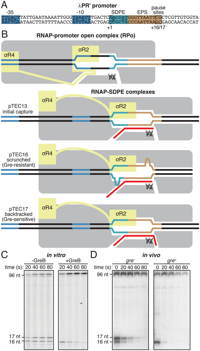
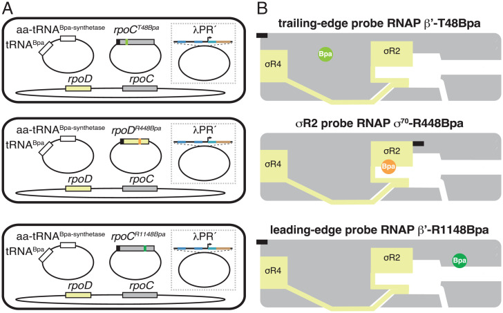
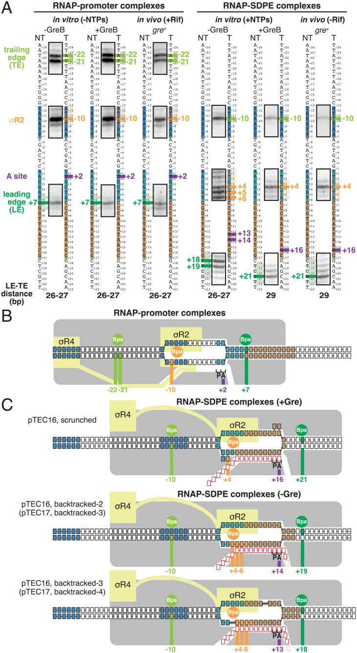
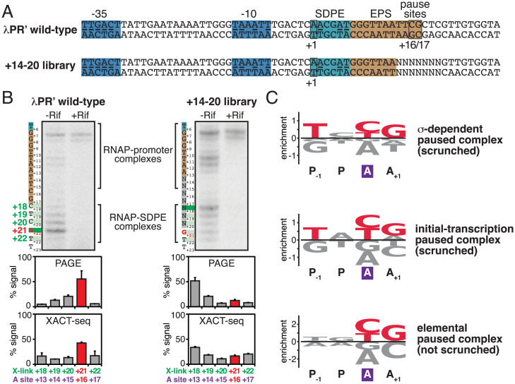
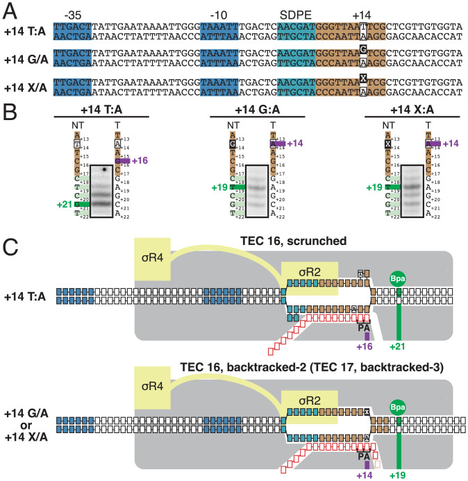
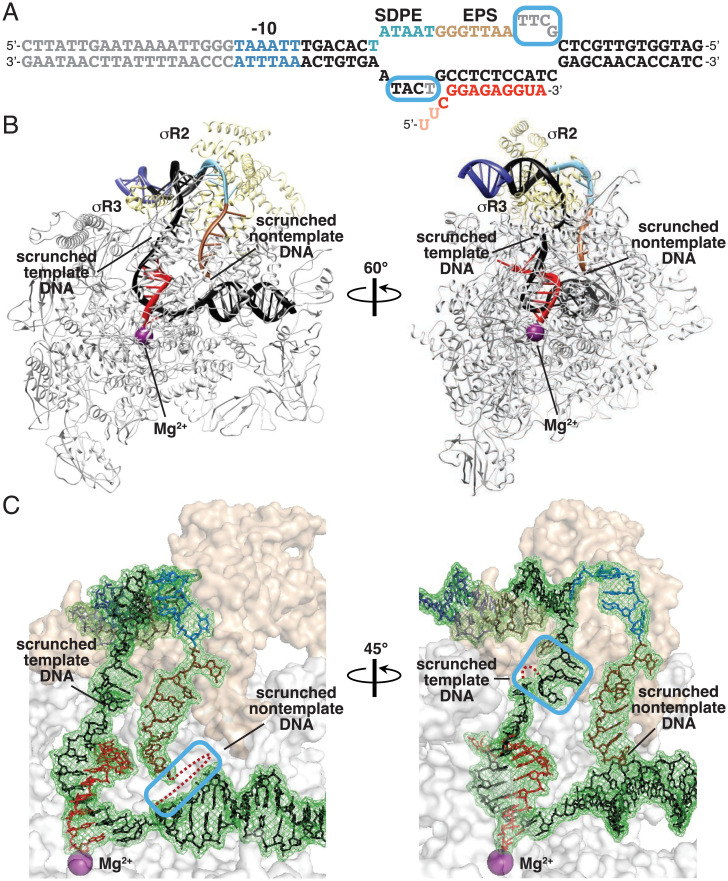
In σ-dependent transcriptional pausing, the transcription initiation factor σ, translocating with RNA polymerase (RNAP), makes sequence-specific protein–DNA interactions with a promoter-like sequence element in the transcribed region, inducing pausing. It has been proposed that, in σ-dependent pausing, the RNAP active center can access off-pathway “backtracked” states that are substrates for the transcript-cleavage factors of the Gre family and on-pathway “scrunched” states that mediate pause escape. Here, using site-specific protein–DNA photocrosslinking to define positions of the RNAP trailing and leading edges and of σ relative to DNA at the λPR′ promoter, we show directly that σ-dependent pausing in the absence of GreB in vitro predominantly involves a state backtracked by 2–4 bp, and σ-dependent pausing in the presence of GreB in vitro and in vivo predominantly involves a state scrunched by 2–3 bp. Analogous experiments with a library of 47 (∼16,000) transcribed-region sequences show that the state scrunched by 2–3 bp—and only that state—is associated with the consensus sequence, T−3N−2Y−1G+1, (where −1 corresponds to the position of the RNA 3′ end), which is identical to the consensus for pausing in initial transcription and which is related to the consensus for pausing in transcription elongation. Experiments with heteroduplex templates show that sequence information at position T−3 resides in the DNA nontemplate strand. A cryoelectron microscopy structure of a complex engaged in σ-dependent pausing reveals positions of DNA scrunching on the DNA nontemplate and template strands and suggests that position T−3 of the consensus sequence exerts its effects by facilitating scrunching.

Keywords: DNA scrunching; RNA polymerase; pausing; sigma; transcription elongation.


Conflict of interest statement

The authors declare no competing interest.

Figures

Fig. 1.
Fig. 1.
σ-Dependent pausing at λPR’: scrunched and backtracked states. (A) λPR′ promoter. Blue, –35 element and –10 element; light blue, SDPE; brown; elemental pause site (EPS); black rectangles, transcription start site (+1) and pause sites (+16/+17); underlining, consensus nucleotides of sequence elements. (B) Initiation complexes and paused complexes at λPR′. Four complexes are shown: 1) initiation complex, RPo; 2) initial-capture σ-dependent paused complex, pTEC13 (where “pTEC” denotes paused TEC and “13” denotes 13 nt RNA product); 3) scrunched σ-dependent paused complex, pTEC16; and 4) backtracked σ-dependent paused complex, pTEC17. Gray, RNAP core; yellow, σ; red, RNA product; P and A, RNAP active-center product and addition sites; blue, –35 element and –10 element; light blue, SDPE; brown, EPS; black, other DNA (nontemplate-strand above template-strand). Scrunching of DNA strands is indicated by bulges in DNA strands. (C and D) RNA product length and Gre-factor sensitivity in pTECs. (C) RNA product distributions in vitro for transcription reactions in absence or presence of GreB at indicated times after addition of NTPs. (D) RNA product distributions in vivo for gre or gre+ cells at indicated times after addition of rifampin (Rif). Positions of pTEC-associated RNA products (16 nt and 17 nt) and full-length RNA product (96 nt) are indicated.
Fig. 2.
Fig. 2.
Use of site-specific protein–DNA photocrosslinking to define positions of RNAP TEs and LEs and of σ relative to DNA at λPR′: approach. (A) Two-plasmid (for in vitro studies) or three-plasmid (for in vivo studies) merodiploid system for coproduction, in E. coli cells, of decahistidine-tagged RNAP-β′ T48Bpa, RNAP-β′ R1148Bpa, or RNAP-σ70 R448Bpa in the presence of untagged wild-type RNAP holoenzyme. First plasmid carries gene for decahistidine-tagged RNAP β′ subunit (gray rectangle) with nonsense codon at position 48 (olive green; Top row); decahistidine-tagged σ70 (light yellow rectangle) with nonsense codon at position 448 (orange; Center row); or gene for decahistidine-tagged RNAP β′ subunit with nonsense codon at position 1148 (forest green; Bottom row). Second plasmid carries genes for engineered Bpa-specific nonsense-suppressor tRNA and aminoacyl-tRNA synthetase (white rectangles). Third plasmid (shown inside dashed box), when present, carries λPR′ promoter or λPR′ promoter derivative. Chromosome (shown below plasmids) carries genes for wild-type RNAP β′ subunit and σ70. Black rectangles, decahistidine-tag coding sequence. (B) Bpa-modified RNAPs. Olive green circle, TE Bpa; orange circle, σR2 Bpa; forest green circle, LE Bpa. Black rectangles, decahistidine-tag. Other colors and symbols as in Fig. 1 A and B.
Fig. 3.
Fig. 3.
Use of site-specific protein–DNA photocrosslinking to define positions of RNAP TEs and LEs and of σ relative to DNA at λPR′: results. (A) Positions of RNAP TEs and LEs and of σR2 in RNAP-promoter complexes (Left) or RNAP-SDPE complexes (pTECs) (Right) at λPR′. For each experimental condition in vitro and in vivo, identified at top, figure shows segments of gel images for primer-extension mapping of crosslinking sites (full gel images in SI Appendix, Figs. S2–S4), nontemplate- and template-strand sequences of λPR′ (to Left and Right, respectively, of gel images; –35 element, –10 element, SDPE, and EPS colored as in Fig. 1A), observed crosslinking sites (olive green for RNAP TE, forest green for RNAP LE, and orange for σR2), inferred positions of RNAP-active-center A site (violet), and inferred modal TE/LE distances. (B) Mechanistic interpretation of data for RNAP-promoter complexes at λPR′ (A, Left). Olive green circle and olive green vertical lines denote Bpa site at RNAP TE and observed crosslinking sites in DNA for Bpa at RNAP TE, forest green circle and forest green vertical line denote Bpa site at RNAP LE and observed crosslinking site in DNA for Bpa at RNAP LE, and orange circle and orange vertical line denote Bpa site in σR2 and observed crosslinking site in DNA for Bpa in σR2. Violet vertical line denotes inferred position of RNAP-active-center A site. Gray, RNAP core; yellow, σ; P and A, RNAP active-center product and addition sites; black boxes with blue fill, –35 element and –10 element nucleotides; black boxes with light blue fill, SDPE nucleotides; black boxes with brown fill, EPS nucleotides; other black boxes, other DNA nucleotides (nontemplate-strand nucleotides above template-strand nucleotides). (C) Mechanistic interpretation of data for RNAP-SDPE complexes at λPR′ (pTEC; A, Right). Three complexes are shown: (1) a scrunched σ-dependent paused complex with 16 nt RNA product and RNAP-active-center A-site at position +16 (pTEC16 scrunched; Top row); (2) a backtracked σ-dependent paused complex with 16- or 17-nt RNA product and RNAP-active-center at position +14 (pTEC16, backtracked-2 or pTEC17, backtracked-3; Center row); and (3) a backtracked σ-dependent paused complex with 16 or 17 nt RNA product and RNAP-active-center at position +13 (pTEC16, backtracked-3 or pTEC17, backtracked-4; Bottom row). Red boxes, RNA nucleotides; pink boxes, additional RNA nucleotide present in 17 nt RNA product. Other colors as in B. Scrunched segments of nontemplate and template DNA strands in pTEC16 shown as bulges.
Fig. 4.
Fig. 4.
Sequence determinants for scrunching in σ-dependent pausing. (A) DNA templates containing wild-type λPR′ or +14 to +20 library. NNNNNNN, randomized nucleotides of +14 to +20 library. Other colors as in Fig. 1A. (B) Positions of RNAP LE in wild-type λPR’ and +14 to +20 library in vivo. Top, PAGE analysis of crosslinking. For each experimental condition, identified at top, figure shows gel image for primer-extension mapping of crosslinking sites, nontemplate-strand sequence (to left of gel image; SDPE and EPS colored as in Fig. 1A), and observed crosslinking sites (forest green). Center, quantitation (mean ± SD) of PAGE analysis of crosslinking. Bottom, quantitation (mean ± SD) of XACT-seq analysis of crosslinking. In all subpanels, the observed major crosslinking site for pTEC at λPR′ (position +21) and inferred major RNAP-active-center A-site position for pTEC at λPR′ (position +16) are highlighted in red. (C) Sequence logos quantifying formation and/or stability of scrunched σ-dependent paused complex (Top; this work); formation and/or stability of scrunched initial-transcription paused complex (Center) (65); and elemental pausing in transcription elongation (Bottom) (–72). Positions are labeled relative to RNAP-active-center A site (violet rectangle) and P-site. Red, most highly preferred DNA nucleotides. Logos were generated using Logomaker (93) as described in SI Appendix, Materials and Methods.
Fig. 5.
Fig. 5.
Strand-dependence of sequence determinants for scrunching in σ-dependent pausing. (A) λPR′ promoter derivatives containing consensus nucleotide T at nontemplate strand position +14 (Top row; +14 T/A), nonconsensus nucleotide G at nontemplate strand position +14 (Center row; +14 G/A), or abasic site at nontemplate strand position +14 (Bottom row; +14 X/A). Raised black-filled box, nonconsensus nucleotide, or abasic site. Other colors as in Fig. 1A. (B) Positions of RNAP LE on λPR′ promoter derivatives of A. For each promoter derivative, figure shows gel image for primer-extension mapping of crosslinking sites, nontemplate- and template-strand sequences (to left and right of gel image; EPS colored as in Fig. 1A), observed crosslinking sites (forest green), and inferred RNAP-active-center A-site position. (C) Mechanistic interpretation of data in B. Colors as in Fig. 3 B and C.
Fig. 6.
Fig. 6.
Structural basis for scrunching in σ-dependent pausing. (A) Nucleic-acid scaffold. DNA, black (–10 element, SDPE, EPS, and disordered nucleotides in blue, light blue, brown, and gray, respectively; noncomplementary region corresponding to unwound transcription bubble indicated by raised and lowered letters); RNA, red (disordered nucleotides in pink); cyan boxes, nontemplate- and template-strand DNA nucleotides disordered or repositioned due to DNA scrunching. (B) Cryo-EM structure of scrunched σ-dependent paused TEC (pTEC; two orthogonal view orientations). Violet sphere, RNAP-active-center catalytic Mg2+. Other colors as in A. (C) Cryo-EM density and atomic model, showing interactions of RNAP and σ with DNA and RNA. Cyan boxes, nontemplate-strand (Left) and template-strand (Right) DNA nucleotides disordered or repositioned due to DNA scrunching; red dots, DNA nucleotides disordered due to DNA scrunching. Other colors as in A.

References

    1. Winkelman J. T., Nickels B. E., Ebright R. H., “The transition from transcription initiation to transcription elongation: Start-site selection, initial transcription, and promoter escape” in RNA Polymerase as a Molecular Motor, Landick R., Wang J., Strick T. R., Eds. (RSC Publishing, Cambridge, UK, ed. 2, 2021).
    1. Chen J., Boyaci H., Campbell E. A., Diverse and unified mechanisms of transcription initiation in bacteria. Nat. Rev. Microbiol. 19, 95–109 (2021). - PMC - PubMed
    1. Mazumder A., Kapanidis A. N., Recent advances in understanding σ70-dependent transcription initiation mechanisms. J. Mol. Biol. 431, 3947–3959 (2019). - PMC - PubMed
    1. Ruff E. F., M. T. Record, Jr, Artsimovitch I., Initial events in bacterial transcription initiation. Biomolecules 5, 1035–1062 (2015). - PMC - PubMed
    1. Feklístov A., Sharon B. D., Darst S. A., Gross C. A., Bacterial sigma factors: A historical, structural, and genomic perspective. Annu. Rev. Microbiol. 68, 357–376 (2014). - PubMed

Publication types

LinkOut - more resources