. 2022 Aug 3;110(15):2422-2437.e9.
doi: 10.1016/j.neuron.2022.05.009. Epub 2022 Jun 1.

Neuronal NLRP3 is a parkin substrate that drives neurodegeneration in Parkinson's disease

Affiliations

Neuronal NLRP3 is a parkin substrate that drives neurodegeneration in Parkinson's disease

Nikhil Panicker et al. Neuron. .

Abstract

Parkinson's disease (PD) is mediated, in part, by intraneuronal accumulation of α-synuclein aggregates andsubsequent death of dopamine (DA) neurons in the substantia nigra pars compacta (SNpc). Microglial hyperactivation of the NOD-like receptor protein 3 (NLRP3) inflammasome has been well-documented in various neurodegenerative diseases, including PD. We show here that loss of parkin activity in mouse and human DA neurons results in spontaneous neuronal NLRP3 inflammasome assembly, leading to DA neuron death. Parkin normally inhibits inflammasome priming by ubiquitinating and targeting NLRP3 for proteasomal degradation. Loss of parkin activity also contributes to the assembly of an active NLRP3 inflammasome complex via mitochondrial-derived reactive oxygen species (mitoROS) generation through the accumulation of another parkin ubiquitination substrate, ZNF746/PARIS. Inhibition of neuronal NLRP3 inflammasome assembly prevents degeneration of DA neurons in familial and sporadic PD models. Strategies aimed at limiting neuronal NLRP3 inflammasome activation hold promise as a disease-modifying therapy for PD.

Keywords: Caspase-1; NLRP3; PARIS; Parkinson’s disease; ZNF746; inflammasome; neurodegeneration; parkin; ubiquitination.


Conflict of interest statement

Declaration of interests The value of patents owned by Valted could be affected by the study described in this article. Drs. T.D. and V.D. are founders of Valted and hold an ownership equity interest in the company. This arrangement has been reviewed and approved by the Johns Hopkins University in accordance with its conflict-of-interest policies.

Figures
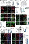
Figure 1:
Figure 1:. DA neuron specific parkin deletion spontaneously activates the neuronal NLRP3 inflammasome.
A, Schematic of the AAV GFP- or AAV GFP-Cre-injection model in parkinflx/flx mice. B, Adult-onset parkin ablation in SNpc DA neurons was sufficient to induce NLRP3 induction as well as Casp1 cleavage, as verified by immunoblot analysis of AAV GFP-and AAV GFP-Cre-injected parkinflx/flx ventral midbrain lysates. Bars represent mean ± s.e.m. n=7). C, Representative IHC showing upregulated Casp1 activity, assayed using FLICA staining, in TH positive DA neurons of the SNpc, not Iba-1 positive microglia in AAV GFP-Cre-injected parkinflx/flx mice. Bars represent mean ± s.e.m (n=5). D, Representative IHC showing ASC speck formation within TH positive SNpc DA neurons, but not within Iba-1 positive microglia in AAV GFP-Cre-injected parkinflx/flx mice. Bars represent mean ± s.e.m (n=5). E, q-RT PCR shows unchanged NLRP3 and Casp1 mRNA levels in AAV GFP- or AAV GFP-Cre-injected parkinflx/flx mouse ventral midbrain tissues. Bars represent mean ± s.e.m. (n=5) F, Representative immunoblots showing that microglia depletion in vivo does not affect loss of parkin-mediated NLRP3 induction and Casp1 cleavage. Bars represent mean ± s.e.m. (n=6). G, Representative IHC showing no change in DA neuron FLICA activity following microglia depletion in adult-onset DA neuron parkin ablated mouse brains. Bars represent mean ± s.e.m. (n=6). H, Representative immunoblot showing increased NLRP3 and cleaved Casp1 in PARKIN deficient hDA neurons when compared to iCont DA neurons. Bars represent mean ± s.e.m. (n=4). I, Increased ASC oligomerization in PARKIN deficient hDA neurons when compared to iCont DA neurons, as verified by DSS-crosslinking assay. Bars represent mean ± s.e.m. (n=3). J, Representative Immunofluorescence images showing upregulated Casp1 activity, assayed using FLICA staining in PARKIN deficient hDA neurons. Bars represent mean ± s.e.m (n=7).
Figure 2:
Figure 2:. SNpc DA neuron specific NLRP3 inflammasome activation is sufficient to drive DA neuron death.
A, Schematic of the bilateral AAV GFP- and AAV GFP-Cre-injection model in NLRP3A350V mice. B, Verification of SNpc DA neuron targeting of AAV GFP- and AAV GFP-Cre-injected NLRP3A350V mice via triple staining with TH, GFP, and Iba-1. Bars represent mean ± s.e.m. (n=4). C, Higher magnification image showing GFP expression in TH positive DA neurons, not Iba-1 positive microglia. D, Representative immunoblot showing increased inflammasome activation in AAV GFP-Cre-injected ventral midbrain lysates of NLRP3A350V mice. Bars represent mean ± s.e.m. (n=6). E, Representative ventral midbrain section of NLRP3A350V mouse bilaterally injected in the SNpc with AAV GFP and AAV GFP-Cre stained 3 months post injection with TH and Nissl. F, Stereological counting of TH and Nissl positive cells in the SNpc of NLRP3A350V mice subjected to aforementioned-bilateral injection model, showing loss of SNpc DA neurons mediated by SNpc DA neuron specific inflammasome activation. Bars represent mean ± s.e.m. (n=5).
Figure 3:
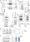
Figure 3:. NLRP3 is a parkin polyubiquitination substrate.
A, GST pull-down assay demonstrating direct interaction of recombinant NLRP3 and parkin proteins. Bars represent mean ± s.e.m. (n=3). B, Co-immunoprecipitation of parkin and NLRP3 in WT (C57BL6), but not parkin or NLRP3 deficient mouse ventral midbrain lysates. Bars represent mean ± s.e.m. (n=3). C, Myc-tagged WT parkin, but not C431S inactive-mutant parkin ubiquitinates FLAG-NLRP3, as evidenced by an in-cell ubiquitination assay. HA-tagged WT Ub, but not (lysine-mutated) KO Ub can be incorporated into polyubiquitinated NLRP3 chains. Bars represent mean ± s.e.m. (n=3). D, In vitro ubiquitination assay shows that Tc PINK1-activated WT, but not C431S recombinant parkin could ubiquitinate NLRP3. Kinase-deficient Tc PINK1 fails to elicit parkin autoubiquitination/activation and subsequent NLRP3 polyubiquitination. Bars represent mean ± s.e.m. (n=3). E, TUBE assay in iCont and PARKIN-deficient hDA neurons shows polyubiquitination of NLRP3 is abolished upon PARKIN-depletion. Bars represent mean ± s.e.m. (n=3) F, TUBE assay in AAV GFP- and AAV GFP-Cre-injected parkinflx/flx ventral midbrain lysates shows polyubiquitination of NLRP3 is abolished upon in vivo DA neuron parkin depletion. Bars represent mean ± s.e.m. (n=4). G, Overexpression of WT myc-tagged parkin hastens FLAG-NLRP3 clearance, whereas overexpression of the C431S myc-tagged parkin prevents FLAG-NLRP3 clearance, evidenced by a cycloheximide chase assay. Bars represent mean ± s.e.m. (n=3). H, Cycloheximide chase assay in iCont and PARKIN-deficient hDA neurons shows that the absence of PARKIN prevents NLRP3 clearance. Bars represent mean ± s.e.m. (n=3).
Figure 4:
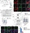
Figure 4:. PARIS facilitates inflammasome activation in parkin-depleted DA neurons.
A, Schematic for stereotaxic injection paradigm B, In vivo FLICA assay demonstrates near complete abrogation of DA neuron Casp1 activity in AAV GFP-Cre-injected parkinflx/flx mice that were co-injected with AAV shRNA-PARIS to prevent PARIS induction. Bars represent mean ± s.e.m. (n=5). C, Immunoblot analysis reveals that preventing PARIS induction in the adult-onset DA neuron specific parkin deletion model did not affect inflammasome priming (no significant change in NLRP3 induction), but completely prevented Casp1 activation, evidenced by significantly reduced Casp1 p20 levels as well as Gasdermin-D cleavage. Bars represent mean ± s.e.m. (n=4 to 6). D, CRISPR/Cas9-mediated PARIS knockdown prevents ASC oligomerization in PARKIN deficient hDA neurons, as assayed using DSS-crosslinking assay. Input samples reveal reduced Casp1 and Gasdermin-D cleavage and unchanged NLRP3 induction following PARIS knockdown. Bars represent mean ± s.e.m. (n=3) E, PARIS knockdown prevents Casp1 activation in PARKIN deficient hDA neurons, as assayed using the FLICA assay. Live iCont, PARKIN deficient, and PARKIN deficient + PARIS KD DA neurons were stained with the FLICA reagent, then fixed and counterstained with TH. Bars represent mean ± s.e.m. (n=4).
Figure 5:
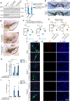
Figure 5:. Preventing inflammasome activation protects against loss of parkin-associated neuron death.
A, Immunoblots showing that knocking down of NLRP3 expression via shRNA AAV abrogates Casp1 activation in adult onset, DA neuron parkin depleted mice. Bars represent mean ± s.e.m. (n=4). B, C, Representative images, and stereological counting of TH and Nissl positive cells shows significant neuroprotection in AAV NLRP3-shRNA-injected adult onset, DA-neuron parkin depleted mice, when compared to AAV con-shRNA injected control mice. Bars represent mean ± s.e.m. (n=6–8). D, E, Representative images and stereological counting of TH and Nissl positive cells shows significant neuroprotection in adult onset, parkin depleted parkinflx/flx/ Casp1−/− mice, when compared to littermate control parkinflx/flx/ Casp1+/+ mice. Bars represent mean ± s.e.m. (n=7). F, H, Immunofluorescence for TH in iCont, PARKIN−/− and PARKIN−/−+Casp1 KD hDA neurons shows that knockdown of Casp1 prevents the loss of PARKIN-associated reduction in TH levels. Bars represent mean ± s.e.m. (n=4). G, I, PI staining in iCont, PARKIN−/− and PARKIN−/−+Casp1 KD hDA neurons shows that knockdown of Casp1 prevents the onset of loss of PARKIN-associated DA neuron death. Bars represent mean ± s.e.m. (n=10).
Figure 6:
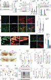
Figure 6:. Loss of parkin activity licenses DA neuron NLRP3 inflammasome-dependent neurodegeneration in the α-Syn PFF model.
A, Immunoprecipitation of parkin from PBS and α-Syn PFF-injected mouse ventral midbrain lysates reveals reduced parkin autoubiquitination following α-Syn PFF-subjection, indicating reduction in parkin activity. Bars represent mean ± s.e.m. (n=4). B, Immunoblot analysis reveals NLRP3 inflammasome priming and activation in the ventral midbrain lysates of α-Syn PFF-injected mice. Bars represent mean ± s.e.m. (n=5). C, D, FLICA staining followed by double immunostaining reveals increased Casp1 activity within TH positive SNpc DA neurons in α-Syn PFF-injected mice. Counterstaining for p-S129-α-Syn shows that many of the neurons harboring α-Syn aggregates are FLICA positive. Bars represent mean ± s.e.m. (n=5). E, Immunoblot analysis showing that α-Syn PFF-induced Casp1 activity is significantly reduced in the ventral midbrain lysates of PARIS−/− mice. Bars represent mean ± s.e.m. (n=5). F, FLICA staining followed by TH immunostaining reveals abrogation of Casp1 activity in the SNpc DA neurons of α-Syn PFF-injected PARIS−/− mice. Bars represent mean ± s.e.m. (n=5). G, H, Stereological counting of TH and Nissl positive cells shows significant neuroprotection in α-Syn PFF-injected Casp1−/− mice, when compared to Casp1+/+ littermate control mice. Bars represent mean ± s.e.m. (n=8). I, J, α-Syn PFF-injected Casp1−/− mice exhibit significantly diminished motor deficits, as assayed by performance on the pole test and the grip-strength test. Bars represent mean ± s.e.m. (n=6–7). K, Casp1−/− mice are protected from α-Syn PFF-induced TH reduction in the striatum, as well as pathologic p-S129-α-Syn and oligomeric α-Syn accumulation in Triton-X insoluble ventral midbrain lysates, as evidenced by immunoblots. Bars represent mean ± s.e.m. (n=4–5).
Figure 7:
Figure 7:. NLRP3 inflammasome activation in Human PD postmortem DA neurons.
A, Immunoblot analysis reveals Parkin inactivation, NLRP3 inflammasome priming and activation in the SNpc lysates of postmortem PD brains. Bars represent mean ± s.e.m. (n=6). B, IHC analysis reveals NLRP3 induction within surviving DA neurons in SNpc brain sections from PD patients, compared to age-matched controls. Bars represent mean ± s.e.m. (n=6). C, In vivo FLICA assay followed by double IHC for TH and ASC showing increased Casp1 activation and ASC speck formation within TH-positive SNpc DA neurons in PD patient SNpc brain sections. Bars represent mean ± s.e.m. (n=6). D, Double IHC for TH and ASC shows that SNpc DA neurons (indicated with white arrowhead) as well as other, non-DA cells (indicated with yellow arrowheads) contain ASC specks.

Comment in

References

    1. Brahmachari S, Lee S, Kim S, Yuan C, Karuppagounder SS, Ge P, Shi R, Kim EJ, Liu A, Kim D, et al. (2019). Parkin interacting substrate zinc finger protein 746 is a pathological mediator in Parkinson’s disease. Brain 142, 2380–2401. 10.1093/brain/awz172. - DOI - PMC - PubMed
    1. Brydges SD, Mueller JL, McGeough MD, Pena CA, Misaghi A, Gandhi C, Putnam CD, Boyle DL, Firestein GS, Horner AA, et al. (2009). Inflammasome-mediated disease animal models reveal roles for innate but not adaptive immunity. Immunity 30, 875–887. 10.1016/j.immuni.2009.05.005. - DOI - PMC - PubMed
    1. Dauer W, and Przedborski S (2003). Parkinson’s disease: mechanisms and models. Neuron 39, 889–909. 10.1016/s0896-6273(03)00568-3. - DOI - PubMed
    1. Di Maio R, Barrett PJ, Hoffman EK, Barrett CW, Zharikov A, Borah A, Hu X, McCoy J, Chu CT, Burton EA, et al. (2016). alpha-Synuclein binds to TOM20 and inhibits mitochondrial protein import in Parkinson’s disease. Sci Transl Med 8, 342ra378. 10.1126/scitranslmed.aaf3634. - DOI - PMC - PubMed
    1. Exner N, Lutz AK, Haass C, and Winklhofer KF (2012). Mitochondrial dysfunction in Parkinson’s disease: molecular mechanisms and pathophysiological consequences. EMBO J 31, 3038–3062. 10.1038/emboj.2012.170. - DOI - PMC - PubMed

Publication types

MeSH terms