. 2022 Jul 21;29(7):1209-1217.e4.
doi: 10.1016/j.chembiol.2022.05.004. Epub 2022 Jun 1.

Visualizing Staphylococcus aureus pathogenic membrane modification within the host infection environment by multimodal imaging mass spectrometry

Affiliations

Visualizing Staphylococcus aureus pathogenic membrane modification within the host infection environment by multimodal imaging mass spectrometry

William J Perry et al. Cell Chem Biol. .

Abstract

Bacterial pathogens have evolved virulence factors to colonize, replicate, and disseminate within the vertebrate host. Although there is an expanding body of literature describing how bacterial pathogens regulate their virulence repertoire in response to environmental signals, it is challenging to directly visualize virulence response within the host tissue microenvironment. Multimodal imaging approaches enable visualization of host-pathogen molecular interactions. Here we demonstrate multimodal integration of high spatial resolution imaging mass spectrometry and microscopy to visualize Staphylococcus aureus envelope modifications within infected murine and human tissues. Data-driven image fusion of fluorescent bacterial reporters and matrix-assisted laser desorption/ionization Fourier transform ion cyclotron resonance imaging mass spectrometry uncovered S. aureus lysyl-phosphatidylglycerol lipids, localizing to select bacterial communities within infected tissue. Absence of lysyl-phosphatidylglycerols is associated with decreased pathogenicity during vertebrate colonization as these lipids provide protection against the innate immune system. The presence of distinct staphylococcal lysyl-phosphatidylglycerol distributions within murine and human infections suggests a heterogeneous, spatially oriented microbial response to host defenses.

Keywords: MALDI imaging; Staphylococcus aureus; host-pathogen interactions; image fusion; imaging mass spectrometry; infectious disease; infective endocarditis; multimodal imaging; staph infection.


Conflict of interest statement

Declaration of interests The authors declare no interests.

Figures

Fig. 1:
Fig. 1:. MALDI IMS spatially informed by fluorescence microscopy of S. aureus, PmntArfp maps chemical information from staphylococcal colonies.
A: H&E staining of a 7 DPI murine renal tissue section shows an abscess at the top right. B: A fluorescence micrograph of the same tissue section as A and C prior to MALDI IMS highlights the location of SACs (red fluorescent protein (RFP)). C: An ion overlay image highlights the various molecular constituents of the abscess and chemical complexity of the tissue section. These distributions also highlight kidney tissue substructures, such as the medulla and cortex. D: Lysyl-phosphatidylglycerol lipid species were isolated using image fusion data mining strategies (relative slopes shown) and identified by subsequent MALDI MS/MS experiments. The red crosshair is present to use as a reference point across the fluorescence micrograph zoom and ion images. E: A zoom of single extracted mass spectrum shows signals corresponding to lysyl-PG species. See also Figs. S1 – S4.
Fig. 2:
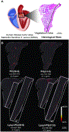
Fig. 2:. MALDI IMS of lipids from infective endocarditis tissue reveals S. aureus PGs and lysyl-PGs.
A) A graphic depicts infected human heart tissue lesions. A tissue stain of an infected heart lesion is annotated for normal and diseased tissue. B) Ion images of S. aureus PGs show distributions across the diseased tissue. Increased presence of lysyl-PGs can be observed near valve tissue (zoom).
Fig. 3:
Fig. 3:. MALDI IMS of intact proteins from an infective endocarditis vegetation reveals CAMPs colocalizing with diseased tissue
A) Ion images of host CAMPs, α-defensin 2 and α-defensin 1 show high abundances within vegetations. B) LC-MS/MS spectra from bottom-up proteomics workflows show peptides from α-defensin 2 and α-defensin 1.

References

    1. Beceiro A; Tomás M; Bou G (2013). Antimicrobial Resistance and Virulence: a Successful or Deleterious Association in the Bacterial World? Clin. Microbiol. Rev 26 (2), 185–230. - PMC - PubMed
    1. Casadevall A; Pirofski L.-a. (2000). Host-Pathogen Interactions: Basic Concepts of Microbial Commensalism, Colonization, Infection, and Disease. Infect. Immun 68 (12), 6511–6518. - PMC - PubMed
    1. Kohanski MA; Dwyer DJ; Collins JJ (2010). How antibiotics kill bacteria: from targets to networks. Nat. Rev. Microbiol 8 (6), 423–435. - PMC - PubMed
    1. Muthaiyan A; Silverman JA; Jayaswal RK; Wilkinson BJ (2008). Transcriptional Profiling Reveals that Daptomycin Induces the Staphylococcus aureus Cell Wall Stress Stimulon and Genes Responsive to Membrane Depolarization. Antimicrob. Agents Chemother 52 (3), 980–990. - PMC - PubMed
    1. Slavetinsky C; Kuhn S; Peschel A (2017). Bacterial aminoacyl phospholipids – Biosynthesis and role in basic cellular processes and pathogenicity. Biochim. Biophys. Acta – Mol. Cell Biol. Lipids 1862 (11), 1310–1318. - PubMed

Publication types

Substances