. 2022 Jun 2;13(1):3064.
doi: 10.1038/s41467-022-30721-w.

A supramolecular photosensitizer derived from an Arene-Ru(II) complex self-assembly for NIR activated photodynamic and photothermal therapy

Affiliations

A supramolecular photosensitizer derived from an Arene-Ru(II) complex self-assembly for NIR activated photodynamic and photothermal therapy

Gang Xu et al. Nat Commun. .

Abstract

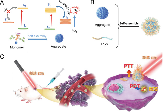
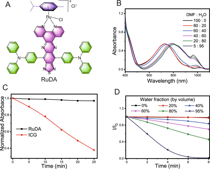
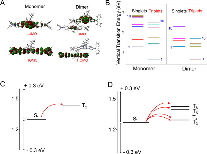
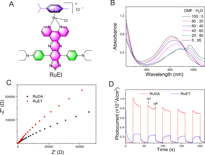
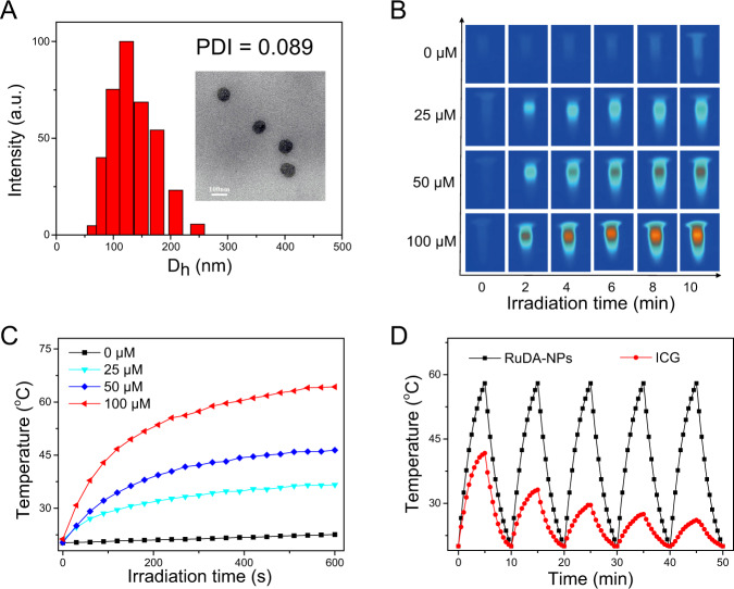
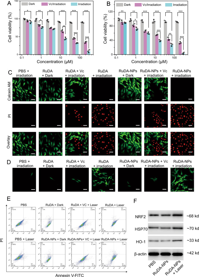
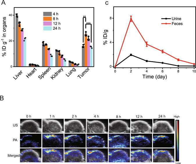
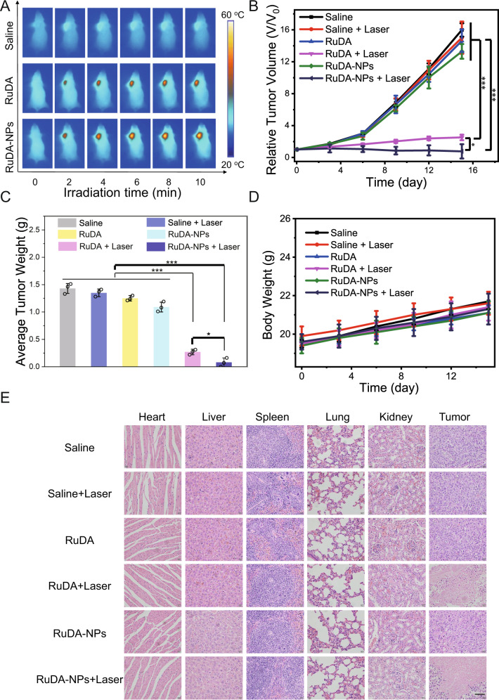
Effective photosensitizers are of particular importance for the widespread clinical utilization of phototherapy. However, conventional photosensitizers are usually plagued by short-wavelength absorption, inadequate photostability, low reactive oxygen species (ROS) quantum yields, and aggregation-caused ROS quenching. Here, we report a near-infrared (NIR)-supramolecular photosensitizer (RuDA) via self-assembly of an organometallic Ru(II)-arene complex in aqueous solution. RuDA can generate singlet oxygen (1O2) only in aggregate state, showing distinct aggregation-induced 1O2 generation behavior due to the greatly increased singlet-triplet intersystem crossing process. Upon 808 nm laser irradiation, RuDA with excellent photostability displays efficient 1O2 and heat generation in a 1O2 quantum yield of 16.4% (FDA-approved indocyanine green: ΦΔ = 0.2%) together with high photothermal conversion efficiency of 24.2% (commercial gold nanorods: 21.0%, gold nanoshells: 13.0%). In addition, RuDA-NPs with good biocompatibility can be preferably accumulated at tumor sites, inducing significant tumor regression with a 95.2% tumor volume reduction in vivo during photodynamic therapy. This aggregation enhanced photodynamic therapy provides a strategy for the design of photosensitizers with promising photophysical and photochemical characteristics.


Conflict of interest statement

Southeast University has applied for a Chinese patent (Patent Number: CN 111808144 A) of RuDA reported here with J.Z. listed as one of the inventors. The remaining authors declare no competing interests.

Figures

Fig. 1
Fig. 1. Schematic illustration of RuDA-NPs for phototherapy.
A Schematic illustration of the photophysical mechanism of RuDA in monomer and aggregate forms for cancer phototherapy, B synthesis of RuDA-NPs, and C RuDA-NPs for NIR-activated PDT and PTT.
Fig. 2
Fig. 2. Photo-physicochemical properties of RuDA.
A Chemical structure of RuDA. B Absorption spectra of RuDA in mixtures of DMF and water at various ratios. C Time-dependent variation in the normalized absorbance values of RuDA (800 nm) and ICG (779 nm) under 808 nm laser irradiation at 0.5 W cm−2. D Photodegradation of ABDA indicative of 1O2 generation induced by RuDA in DMF/H2O mixtures with different water fractions under 808 nm laser irradiation at 0.5 W cm−2.
Fig. 3
Fig. 3. Quantum-chemical calculations of RuDA.
A Calculated HOMOs and LUMOs of RuDA in monomeric and dimeric forms. B Singlet and triplet energy levels of of RuDA in monomer and dimer, respectively. C Calculated energy levels and possible ISC channels of RuDA in monomeric C and dimeric D forms. The arrows refer to the possible ISC channels.
Fig. 4
Fig. 4. Photoelectrochemical properties of RuDA and RuET.
A Chemical structure of RuET. B Absorption spectra of RuET in mixtures of DMF and water at various ratios. C EIS Nyquist plots of RuDA and RuET. D Photocurrent responses of RuDA and RuET under 808 nm laser irradiation.
Fig. 5
Fig. 5. The characterization and photothermal properties of RuDA-NPs.
A DLS analysis and TEM image (inset) of RuDA-NPs. B Thermal images of RuDA-NPs at different concentrations under 808 nm (0.5 W cm-2) laser irradiation. C Photothermal conversion profile of RuDA-NPs at different concentrations, which is the quantitative data of B. D Temperature elevation of RuDA-NPs and ICG during five cycles of heating-cooling processes.
Fig. 6
Fig. 6. In vitro phototherapy efficiencies of RuDA and RuDA-NPs.
A RuDA- and B RuDA-NPs-dose dependent cell viabilities in MDA-MB-231 cells in the presence or absence of Vc (0.5 mM), respectively. Error bars, mean ± SD (n = 3). Unpaired, two-sided t tests *p < 0.05, **p < 0.01, and ***p < 0.001. C Live/dead cell staining assays using calcein AM and propidium iodide as fluorescence probes. Scale bars: 30μm. A representative image of three biological replicates from each group is shown. D Confocal fluorescence images of ROS generation in MDA-MB-231 cells under different treatment conditions. Green fluorescence from DCF indicates the presence of ROS. Irradiation was performed using 808 nm laser at 0.5 W cm−2 for 10 min (300 J cm−2). Scale bars: 30 μm. A representative image of three biological replicates from each group is shown. E Flow cytometry analysis for apoptosis of MDA-MB-231 cells treated with RuDA-NPs (50 μM) or RuDA (50 μM) in the presence and absence of Vc (0.5 mM), and irradiated with or without 808 nm laser (0.5 W cm−2) for 10 min. A representative image of three biological replicates from each group is shown. F Cellular Nrf-2, HSP70, and HO-1 expressions of MDA-MB-231 cells treated with RuDA-NPs (50 μM) with or without 808 nm laser irradiation (0.5 W cm−2, 10 min, 300 J cm−2). A representative image of two biological replicates from each group is shown.
Fig. 7
Fig. 7. Ex vivo biodistribution and in vivo PA imaging.
A Ex vivo tissue distribution of RuDA-NPs in mice determined by the content of Ru (% injected dose (ID) of Ru per gram of tissues) at different post injection time. The data represent the mean ± SD (n = 3). Unpaired, two-sided t tests *p < 0.05, **p < 0.01, and ***p < 0.001. B In vivo PA images of tumor sites under excitation at 808 nm after intravenous injection of RuDA-NPs (10 μmol kg−1) at different time points. C Ru excreted out of the mice body via urine and feces after intravenous administration of RuDA-NPs (10 μmol kg−1) at different time intervals. The data represent the mean ± SD (n = 3).
Fig. 8
Fig. 8. In vivo phototherapy efficacies of RuDA and RuDA-NPs on tumors.
A IR thermal images of MDA-MB-231 tumor-bearing mice irradiated with 808 nm laser for different time at 8 h post injection. A representative image of four biological replicates from each group is shown. B Relative tumor volume and C average tumor weights for different groups of mice during the therapeutic process. D Body weight curves of different groups of mice. Irradiation was performed with 808 nm laser at 0.5 W cm−2 for 10 min (300 J cm−2). Error bars, mean ± SD (n = 3). Unpaired, two-sided t tests *p < 0.05, **p < 0.01, and ***p < 0.001. E H&E staining images of major organs and tumors from different treatment groups, including Saline, Saline + Laser, RuDA, RuDA + Laser, RuDA-NPs, and RuDA-NPs + Laser groups. Scale bars: 60 μm.

References

    1. Li J, Pu K. Development of organic semiconducting materials for deep-tissue optical imaging, phototherapy and photoactivation. Chem. Soc. Rev. 2019;48:38–71. doi: 10.1039/C8CS00001H. - DOI - PubMed
    1. Zhao X, Liu J, Fan J, Chao H, Peng X. Recent progress in photosensitizers for overcoming the challenges of photodynamic therapy: from molecular design to application. Chem. Soc. Rev. 2021;50:4185–4219. doi: 10.1039/D0CS00173B. - DOI - PubMed
    1. Dai J, et al. Efficient near-infrared photosensitizer with aggregation-induced emission for imaging-guided photodynamic therapy in multiple xenograft tumor models. ACS Nano. 2020;14:854–866. doi: 10.1021/acsnano.9b07972. - DOI - PubMed
    1. Yang B, Chen Y, Shi J. Reactive oxygen species (ROS)-based nanomedicine. Chem. Rev. 2019;119:4881–4985. doi: 10.1021/acs.chemrev.8b00626. - DOI - PubMed
    1. Guo Z, et al. Bifunctional platinated nanoparticles for photoinduced tumor ablation. Adv. Mater. 2016;28:10155–10164. doi: 10.1002/adma.201602738. - DOI - PubMed

Publication types