. 2022 Jun 2;12(1):9213.
doi: 10.1038/s41598-022-13189-y.

HER2 G776S mutation promotes oncogenic potential in colorectal cancer cells when accompanied by loss of APC function

Affiliations

HER2 G776S mutation promotes oncogenic potential in colorectal cancer cells when accompanied by loss of APC function

Yosuke Mitani et al. Sci Rep. .

Abstract

Clinical cancer genome sequencing detects oncogenic variants that are potential targets for cancer treatment, but it also detects variants of unknown significance. These variants may interact with each other to influence tumor pathophysiology, however, such interactions have not been fully elucidated. Additionally, the effect of target therapy for those variants also unclarified. In this study, we investigated the biological functions of a HER2 mutation (G776S mutation) of unknown pathological significance, which was detected together with APC mutation by cancer genome sequencing of samples from a colorectal cancer (CRC) patient. Transfection of the HER2 G776S mutation alone slightly increased the kinase activity and phosphorylation of HER2 protein, but did not activate HER2 downstream signaling or alter the cell phenotype. On the other hand, the HER2 G776S mutation was shown to have strong oncogenic potential when loss of APC function was accompanied. We revealed that loss of APC function increased Wnt pathway activity but also increased RAS-GTP, which increased ERK phosphorylation triggered by HER2 G776S transfection. In addition, afatinib, a pan-HER tyrosine kinase inhibitor, suppressed tumor growth in xenografts derived from HER2 G776S-transfected CRC cells. These findings suggest that this HER2 mutation in CRC may be a potential therapeutic target.


Conflict of interest statement

The authors declare no competing interests.

Figures

Figure 1
Figure 1
Genetic characteristics of a case of colorectal cancer harboring HER2 G776S mutation detected by clinical NGS. (A) Summary of the patient's clinical profile. (B) Comprehensive genomic analysis report of the cancer of the patient. Positive biomarkers are shown. (C) Sanger sequencing of a part of HER2 gene of the cancer genome DNA. The amino acid (a.a.) sequence from 774 to 778 is shown. The sequence of the corresponding wild-type (WT) site is also presented. The red arrow indicates the mutation position of G776S. (D) The mutation position of G776 in HER2 protein domains based on the COSMIC database. The nucleotide sequences of the WT, G776S and G776 > VC are described. The underlined part indicates a substitution mutation and the yellow background site indicates a short insertion. Recep_L, receptor L domain; Furin-like, furin-like cysteine-rich region; GF_recep_IV, growth factor receptor domain IV. In the lower part of the figure, the number of reports in the COSMIC database and annotations from OnkoKB (https://www.oncokb.org/) are shown. The data are current as of April 2021.
Figure 2
Figure 2
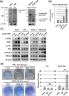
Effects of HER2 mutations on kinase activity and phosphorylation, and their functional evaluation in classical cell-based transfection assays. (A) Western blot results showing recombinant proteins of HER2 WT and mutants (G776S and G776 > VC). HeLa cells were transiently transfected with the HER2 expression vectors, and 48 h after the transfection, the cells were lysed and purified by immunoprecipitation using HER2 antibody. (B) Measurement of the kinase activity of 100 ng of recombinant HER2 protein using the kinase assay. Kinase activity was measured in triplicate, and the data were standardized to the mean HER2 WT activity. One-way ANOVA: P < 0.01, *P < 0.05, **P < 0.01. (C) Western blot results for the expression and phosphorylation of HER2 and EGFR in HeLa cells transfected with the HER2 expression vectors. β-Actin served as an internal control. The bar graph indicates the normalized ratios of densitometric values of phosphorylated to total HER2 protein. (D) Focus formation assays using NIH/3T3 cells stably transfected with the indicated plasmids. (E) Ba/F3 transformation assays using Ba/F3 (interleukin 3-dependent cells) stably transfected with the indicated plasmids. The bar graph shows the fold changes in number of viable cells. ns, not significant. One-way ANOVA: P < 0.01, **P < 0.01 (n = 3).
Figure 3
Figure 3
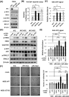
Effects of HER2 G776S mutation on the HER2 signaling pathway and anchorage-independent growth in several cell lines. (A) Detection of APC proteins in each cell by Western blotting. Full-length APC was detected with APC antibody (#2504, Cell Signaling Technology), and truncated APC was detected with APC antibody (sc-9998, Santa Cruz Biotechnology) raised against the N-terminus of APC. (B) The activity of the Wnt/β-catenin signaling pathway measured using the TCF/LEF luciferase reporter assay. The assay was performed in triplicate and the results were standardized to the mean of the activity of HeLa cells. (C) Phosphorylation and expression of HER2 and the downstream signaling in WT HER2- or HER2 G776S-transfected HeLa and colon cells (FHC, CACO-2 and COLO-320). (D) Soft agar colony-forming assays showing the effects of stable transfection with HER2 WT or HER2 G776S in FHC (WT APC) and COLO-320 (mutant APC) cells. The cells were seeded into six-well plates in triplicate, and the number of colonies per well was counted. ns, not significant. **P < 0.01 HER2 WT vs HER2 G776S.
Figure 4
Figure 4
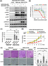
Effects of APC KO on HER signaling in HeLa cells (with WT APC). (A) Confirmation of APC KO and its effect on the HER2–ERK pathway in APC-KO HeLa cells using Western blotting. β-Actin served as a loading control. (B) Activity of the Wnt/β-catenin signaling pathway measured using the TCF/LEF luciferase reporter assay. Nontargeting control cells (NTC) incubated with Wnt3a (100 ng/ml) for 24 h were used as a positive control (rightmost bar). The data were standardized to the mean of the activity of NTC cells. One-way ANOVA: P < 0.01, *P < 0.05, **P < 0.01 vs NTC cells (n = 3). (C) Measurement of activated RAS (RAS–GTP) using the G-LISA assay. NTC plus Wnt3a (100 ng/ml) was used as a control (rightmost bar). The data were standardized to the mean of the activity of NTC cells. One-way ANOVA: P < 0.05, *P < 0.05 vs NTC cells (n = 3). (D) Western blot results showing the effects of HER2 WT or HER2 G776S transfection on the HER2 signaling pathway in APC-KO cells. APC-KO cells or NTC cells were transfected with mock or HER2 expression vectors, and the experiments were performed 48 h after transfection. β-Actin served as a loading control. (E) Measurement of RAS-GTP using the G-LISA assay performed 48 h after HER2 expression vector transfection. The data were standardized to the mean of the activity of NTC cells transfected with mock vectors. Two-way ANOVA: interaction P < 0.01, *P < 0.05, **P < 0.01 (n = 3). (F) Colony-forming assay results showing the effects of stable transfection with HER2 WT or HER2 G776S in APC-KO cells. The number of colonies was counted in six random low-power fields. Scale bar, 200 μm. Two-way ANOVA: interaction P < 0.01, **P < 0.01.
Figure 5
Figure 5
Effects of WT APC overexpression on the HER2 signaling pathway in COLO-320 cells (with mutant APC). (A) Confirmation of APC overexpression in cells transfected with pCMV_APC vector using Western blotting. (B) Activity of the Wnt/β-catenin pathway in COLO-320 cells transfected with pCMV_APC measured using the TCF/LEF luciferase reporter assay. The reporter vectors were cotransfected into COLO-320 cells along with the pCMV empty vector (mock) or pCMV-APC. The data were standardized to the mean of the activity of mock cells. **P < 0.01 vs mock (n = 3). (C) Measurement of activated RAS using the G-LISA assay performed 48 h after the transfection with pCMV. The data were standardized to the mean of the activity of cells transfected with mock vectors. *P < 0 .05 vs mock (n = 3) (D) Western blot results showing the effects of APC overexpression on the HER2 signaling pathway in COLO-320 cells. The cells were cotransfected with pcDNA_HER2-WT/G776S vectors and/or pCMV_APC vectors. Western blotting was performed 48 h after transfection. β-Actin served as a loading control. (E) Measurement of activated RAS using the G-LISA assay performed 48 h after transfection. The data were standardized to the mean of the activity of cells transfected with mock vectors. Two-way ANOVA: interaction P < 0.01, **P < 0.01 (n = 3). (F) Activity of the Wnt/β-catenin pathway in COLO-320 cells measured 24 h after addition of ICG-001 at different concentrations. The assay was performed in triplicate. (G) Measurement of activated RAS using the G-LISA assay in COLO-320 cells 24 h after addition of ICG-001 at different concentrations. The assay was performed in triplicate.
Figure 6
Figure 6
Efficacy of afatinib treatment in COLO-320 cells stably transfected with HER2 G776S. (A) Western blot results show the effects of gefitinib (1 μM) or afatinib (0.1 μM) treatment in COLO-320 cells transfected with WT HER2 or HER2 G776S. One day after seeding, the cells were treated for 24 h. (B) Cell proliferation assay (WST-1 assay) results show the effects of afatinib treatment in COLO-320 cells transfected with HER2 expression vectors. The cells were exposed to afatinib at the indicated concentrations for 24 h. (C) Number of colonies per well in the colony-forming assay treated with DMSO, gefitinib (1 μM) or afatinib (0.1 μM). Cells were seeded into six-well plates in triplicate and treated for 10 days. The number of colonies was counted in six random low-power fields. ns, not significant. **P < 0.01 vs DMSO treatment. (D) The efficacy of afatinib treatment in HER2 WT or HER2 G776S stably expressing COLO-320 xenograft tumors. Xenograft tumors were treated with DMSO or afatinib (25 mg/kg/day p.o.). Two-way ANOVA: interaction P < 0.01, ns, not significant. *P < 0.05 (n = 6). (E) Images of hematoxylin and eosin staining of xenograft tumor tissues in each treatment group and images of immunohistochemical staining for Ki-67 on day 18. Scale bar, 100 μm. (F) Rates of Ki-67-positively stained cells observed in six random fields. ns, not significant. Two-way ANOVA: interaction P < 0.01, *P < 0.05, **P < 0.01.
Figure 7
Figure 7
Schema of the relationship between HER2 mutations and APC mutations in colorectal cancer cells. HER2 G776S is a mutation that increases HER2 kinase activity, although the activity is weak. APC loss-of-function increases Wnt/β-catenin pathway activation but also increases the amount of RAS-GTP, which helps the HER2 G776S mutation to activate the HER2-ERK pathway.

Similar articles

Cited by

References

    1. Biesecker LG, Green RC. Diagnostic clinical genome and exome sequencing. N. Engl. J. Med. 2014;371(12):1170. doi: 10.1056/NEJMc1409040. - DOI - PubMed
    1. Tsimberidou AM, Wen S, Hong DS, Wheler JJ, Falchook GS, Fu S, et al. Personalized medicine for patients with advanced cancer in the phase I program at MD Anderson: Validation and landmark analyses. Clin. Cancer Res. 2014;20(18):4827–4836. doi: 10.1158/1078-0432.CCR-14-0603. - DOI - PMC - PubMed
    1. Kou T, Kanai M, Yamamoto Y, Kamada M, Nakatsui M, Sakuma T, et al. Clinical sequencing using a next-generation sequencing-based multiplex gene assay in patients with advanced solid tumors. Cancer Sci. 2017;108(7):1440–1446. doi: 10.1111/cas.13265. - DOI - PMC - PubMed
    1. Jiang J, Greulich H, Janne PA, Sellers WR, Meyerson M, Griffin JD. Epidermal growth factor-independent transformation of Ba/F3 cells with cancer-derived epidermal growth factor receptor mutants induces gefitinib-sensitive cell cycle progression. Cancer Res. 2005;65(19):8968–8974. doi: 10.1158/0008-5472.CAN-05-1829. - DOI - PubMed
    1. Ross JS, Fakih M, Ali SM, Elvin JA, Schrock AB, Suh J, et al. Targeting HER2 in colorectal cancer: The landscape of amplification and short variant mutations in ERBB2 and ERBB3. Cancer. 2018;124(7):1358–1373. doi: 10.1002/cncr.31125. - DOI - PMC - PubMed

Publication types