. 2022 Jun 2;13(1):3075.
doi: 10.1038/s41467-022-30831-5.

YAP inhibits ERα and ER+ breast cancer growth by disrupting a TEAD-ERα signaling axis

Affiliations

YAP inhibits ERα and ER+ breast cancer growth by disrupting a TEAD-ERα signaling axis

Xu Li et al. Nat Commun. .

Abstract

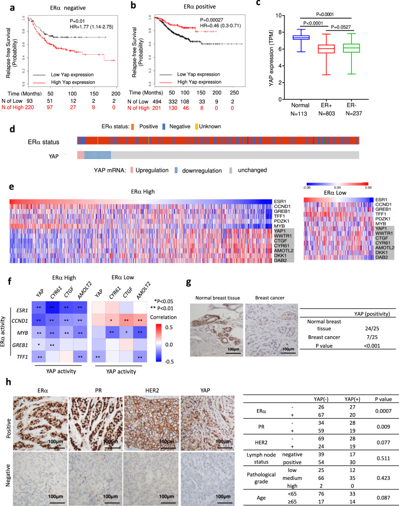
Hippo signaling restricts tissue growth by inhibiting the transcriptional effector YAP. Here we uncover a role of Hippo signaling and a tumor suppressor function of YAP in estrogen receptor positive (ER+) breast cancer. We find that inhibition of Hippo/MST1/2 or activation of YAP blocks the ERα transcriptional program and ER+ breast cancer growth. Mechanistically, the Hippo pathway transcription factor TEAD physically interacts with ERα to increase its promoter/enhancer occupancy whereas YAP inhibits ERα/TEAD interaction, decreases ERα occupancy on its target promoters/enhancers, and promotes ERα degradation by the proteasome. Furthermore, YAP inhibits hormone-independent transcription of ERα gene (ESR1). Consistently, high levels of YAP correlate with good prognosis of ER+ breast cancer patients. Finally, we find that pharmacological inhibition of Hippo/MST1/2 impeded tumor growth driven by hormone therapy resistant ERα mutants, suggesting that targeting the Hippo-YAP-TEAD signaling axis could be a potential therapeutical strategy to overcome endocrine therapy resistance conferred by ERα mutants.


Conflict of interest statement

The authors declare no competing interests.

Figures

Fig. 1
Fig. 1. The expression level and prognostic effect of YAP in ERα-positive breast tumors.
a Kaplan–Meier graph of relapse-free survival shows that high YAP relates to poor prognosis in ERα negative breast tumor. b Kaplan–Meier graph of relapse-free survival shows that high YAP favors the survival in ERα positive breast cancer patients. c Analysis of TCGA database shows that YAP mRNA level is decreased in both ER+ and ER− breast cancer samples compared with normal breast tissues. Normal group, minima = 5.669927, maxima = 8.34457571, mean = 7.280, n = 113, ER+ group, minima = 2.9289187, maxima = 7.78269181, mean = 5.931, n = 803, ER− group, minima = 3.02059779, maxima = 7.84498136, mean = 6.055, n = 237. p-value calculated by one way ANOVA. d YAP expression pattern in ERα positive and negative breast cancer cells. Data were derived from TCGA database (https://www.cbioportal.org). e Heatmap for the expression of ERα signaling and YAP signaling pathway target genes (shaded) in TCGA ERα high (left) and ERα low (right) breast cancer datasets (in the diagram, red represents high gene expression levels, blue represents low gene expression levels). f Heatmap of the correlation between ERα target genes and YAP target genes in TCGA ER high (left) and ERα low (right) breast cancer datasets. The horizontal and vertical coordinates represent genes, and different colors represent correlation coefficients (in the diagram, red represents positive correlation, blue represents negative correlation), the darker the color represents the stronger the correlation. Asterisks represent levels of significance (*p < 0.05, **p < 0.01). g YAP expression is decreased in human breast cancers compared with normal breast tissues in immunohistochemistry analysis (P < 0.001). Examples of YAP staining in normal and ER+ breast tumor samples were shown at ×40 magnification. Results are representative of one set of experiments. h YAP expression is reversely correlated with ERα/PR positivity (P = 0.0007; P = 0.009 respectively). Examples of positive/negative YAP, ERα, PR, and HER2 staining in breast tumor samples were shown at ×40 magnification. Results are representative of one set of experiments. Two-sided log-rank test were used for a and b. Chi-squared test were used for g and h.
Fig. 2
Fig. 2. YAP inhibits ERα-positive breast cancer cell growth.
a XMU-MP-1 inhibits MCF-7 and T47D cancer cell proliferation but facilitates MDA-MB-231 cancer cell growth. b Comparison of inhibitory efficacy between tamoxifen and XMU-MP-1 on MCF-7 and T47D cancer cell growth. c YAP siRNA blocked the inhibitory effect of XMU-MP-1 on MCF-7 and T47D cancer cell growth as well as the stimulatory effect of XMU-MP-1 on MDA-MB-231 cancer cell growth. d YAP depletion promoted MCF-7 and T47D but inhibited MDA-MB-231 cancer cell growth. e Overexpression of either wild-type YAP or YAP-5SA inhibited MCF-7 and T47D but facilitated MDA-MB-231 cancer cell growth. f XMU-MP-1 inhibited ER-positive breast cancer growth in xenografts. Female NOD scid gamma (NSG) mice bearing MCF-7 tumors were treated daily with vehicle or XMU-MP-1 at the indicated concentrations. Tumor growth curve (left), photograph of tumor samples (middle), and quantification of tumor weight (right) at the end of treatment were shown, each group n = 7. g YAP-5SA but not YAP-5SAS94A inhibited ER-positive breast cancer growth in xenografts bearing MCF-7 cells that stably expressed the indicated constructs. The mice were injected i.p. with PBS containing Dox (20 mg/kg) or PBS daily for the indicated period of time. Tumor growth curve (left), photograph of tumor samples (middle), and quantification of tumor weight (right) at the end of treatment were shown, each group n = 7. h XMU-MP-1 inhibited cell proliferation of ER-positive breast cancer samples but did not inhibit ER-negative breast cancer patient samples in patient-derived explant (PDEx) assay. The patient-derived tumor samples were cultured ex vivo on gelatin sponges for 48 h with 10% FBS in the presence of 3 μM XMU-MP-1 or vehicle. The tumor samples were fixed and stained with ERα, PR, HER2, and Ki67 via IHC analysis (left). The dynamic change of Ki67 positive cells were counted and shown (right). Scale bars are 100 μm. Results shown in ae are representative of three independent experiments while results shown in fh are based on one set of experiments. Data are means ± s.d. Two-side, unpaired t-test for ah. Source data are provided in the Source Data file.
Fig. 3
Fig. 3. YAP suppresses ERα signaling activity in breast cancer cells.
a, b XMU-MP-1 inhibited ERα target gene expression in MCF-7 (a) and T47D (b) cells. Cells grown in hormone deletion medium were treated for 3 μM XMU-MP-1 for 8 h and then treated with 10 nM 17β-Estradiol (E2) or vehicle for 6 h, followed by RT-qPCR analysis of GREB1, PS2, and PDZK1 expression. c YAP depletion rescued ERα target gene expression inhibited by XMU-MP-1. MCF-7 cells were transfected with control or YAP siRNA for 48 h in hormone deletion medium. The cells were treated for 3 μM XMU-MP-1 for 8 h and then treated with 10 nM E2 or vehicle for 6 h, followed by RT-qPCR. d YAP depletion increased ERα target gene expression. MCF-7 cells were transfected with control or YAP siRNA for 48 h under hormone depletion condition and then treated with 10 nM E2 or vehicle for 6 h, followed by RT-qPCR. e, f Ectopic expression of YAP inhibited ERα target gene expression. MCF-7 (e) or T47D (f) cells were stably infected with YAP or control virus. After 48 h, cells were treated with 10 nM E2 or vehicle for 6 h, followed by RT-qPCR. g Ectopic expression of YAP or YAP-5SA inhibited ERα target gene expression. MCF-7 or T47D cells were stably infected with wild-type YAP, YAP-5SA, or control virus. After 48 h, the total RNA was extracted for RT-qPCR analysis of GREB1, PS2 and PDZK1 expression. h Top 10 signaling pathways significantly decreased (top) or increased (bottom) in MCF-7 cells treated with XMU-MP-1. The cells were treated with vehicle or 3 μM XMU-MP-1 for 8 h. Threshold P < 0.001 and fold change > 2. n = 3. i Gene set enrichment analysis (GSEA) shows a depletion of estrogen-responsive signature genes in MCF-7 cells treated with XMU-MP-1. j Volcano plot shows the opposite effects of XMU-MP-1 treatment on estrogen-responsive signature genes (blue) and the Hippo pathway signature genes (red) in MCF-7 cells. Threshold P < 0.05 and fold change > 1.5. k, l GSEA shows a depletion of estrogen-responsive signature genes (k) and enrichment of YAP signature genes (l) in MCF-7 cells expressing an inducible YAP-5SA. Results shown in ag are representative of three independent experiments. Data are means ± s.d. Two-side, unpaired t-test for ag. Source data are provided in the Source Data file.
Fig. 4
Fig. 4. XMU-MP-1 and YAP inhibits ERα binding to its target gene promoters/enhancers.
a XMU-MP-1 decreased the half-life of ERα. MCF-7 cells were treated with vehicle or 3 μM XMU-MP-1 in the presence of cycloheximide for the indicated times, followed by western blot analysis. b XMU-MP-1 downregulated ERα through proteosome. MCF-7 cells were treated with vehicle or 3 μM XMU-MP-1 in the absence or presence of 10 μM MG132, followed by western blot analysis with the indicated antibodies. c XMU-MP-1-promoted ERα degradation was inhibited by Leptomycin B (LMB). MCF-7 cells were treated with vehicle or 3 μM XMU-MP-1 in the absence or presence of 50 nM LMB, followed by western blot analysis. d, e XMU-MP-1 inhibited ERα target gene expression (d), inhibited ERα binding to its target gene promoters/enhancers (e) even when ERα degradation was inhibited by LMB. MCF-7 cells were treated with vehicle or 3 μM XMU-MP-1 in the absence or presence of 50 nM LMB, followed by RT-qPCR analysis of GREB1, PS2, PDZK1, and ESR1 expression (d) and ChIP-qPCR analysis for ERα binding to the promoter/enhancer regions of PS2, GREB1, and PDZK1. fh Time course experiments showed that XMU-MP-1 inhibited ERα transcriptional activity and binding to its target promoters/enhancers prior to ERα protein downregulation. MCF-7 cells were treated with 3 μM XMU-MP-1 for the indicated time, followed by western blot analysis for ERα (f), RT-qPCR analysis for ERα target gene expression (g), and ChIP-qPCR analysis for ERα binding to the promoter/enhancer regions of its target genes (h). in Induced expression of YAP-5SA but not YAP-5SAS94A blocked ERα transcriptional activity and binding to its target promoters/enhancers. MCF-7 cells expressing Tet-O-YAP5SA (ik) or Tet-O-YAP5SAS94A (In) were treated with 0.2 μg/ml doxycycline (Dox) for the indicated time, followed by western blot analysis for YAP1 and ERα (i, l), ChIP-qPCR analysis for ERα binding to its target gene promoter/enhancer regions (j, m), and RT-qPCR analysis for ERα target gene expression (k, n). Results shown in an are representative of three independent experiments. Data are means ± s.d. Two-sided, unpaired t-test for d, e, g, h, j, k, m, n. Source data are provided in the Source Data file.
Fig. 5
Fig. 5. TEAD interacts with ERα and promotes ERα signaling activity.
a High levels of TEAD4 correlate with poor survival in endocrine therapy breast cancer patients based on https://kmplot.com/analysis/. b The effect of TEAD4 RNAi on TEAD4 and ERα expression in MCF-7. c, d TEAD4 RNAi inhibited ER+ breast cancer cell growth. MCF-7 (c) and T47D (d) cells transfected with control or TEAD4 siRNA were analyzed using the WST-1 assay at indicated time points. e TEAD4 depletion reduced ERα target gene expression. MCF-7 cells transfected with TEAD4 siRNA were grown for 48 h, followed by RT-qPCR analysis of the indicated ERα target genes. f ChIP-qPCR assay showed that TEAD4 RNAi decreased ERα recruitment to its target gene promoters/enhancers. g Western blot analysis of ERα and TEAD4 expression in control or MCF-7 cells stably expressing RNAi-insensitive TEAD4 (TEAD4INS) and transfected with control or TEAD4 siRNA. h TEAD4INS restored the expression of ERα target genes in TEAD4 depleted MCF-7 cells. i ChIP-qPCR assay showed that TEAD4 and ERα co-occupied on ERα target promoters/enhancers in MCF-7 cells. j Co-IP experiments revealed that TEAD4 formed a complex with ERα in MCF-7 cells. k, l TEAD4 (k) or ERα (l) domain structure and deletion mutants used for Co-IP experiments. mo TEAD4 interacted with the AF2 domain of ERα through its YAP-binding domain. HEK293T cells were transfected with the indicated TEAD4 and ERα constructs, followed by Co-IP and western blot analyses. p ERα recruitment to its target gene promoters/enhancers is enhanced by TEAD41-434 but not by TEAD4 deletion mutants TEAD41-220 or TEAD4131-434. HEK293T cells were transfected with HA-ERα together and the indicated TEAD4 constructs, followed by ChIP-qPCR analysis of HA-ERα binding to ERα target promoters/enhancers. q, r AF2 is required for TEAD4 to facilitate ERα binding to its target promoters/enhancers. HEK293T cells were transfected with GFP-TEAD4 and the indicated ERα constructs, followed by ChIP-qPCR analysis of occupancy of ERα or its deletion mutants on ERα target promoters/enhancers. Two-sided log-rank test were used for a. Results shown in bj, mr are representative of three independent experiments. Data are means ± s.d. Two-sided, unpaired t-test for cf, h, i, pr. Source data are provided in the Source Data file.
Fig. 6
Fig. 6. YAP inhibits ERα-TEAD interaction and hormone-independent ESR1 expression.
a Coexpression of YAP with ERα and TEAD4 disrupted their association. HEK293T cells were transfected with fixed amounts of GFP-TEAD4 and HA- ERα and increasing amounts of Flag-YAP, followed by Co-IP and western blot analyses. b, c YAP inhibited TEAD4-mediated enhancement of ERα binding to its target promoters/enhancers. HEK293T cells were transfected with the indicated constructs, followed by western blot analysis (b) or ChIP-qPCR analysis for HA-ERα binding to its target promoters/enhancers (c). d XUM-MP-1 dissociated ERα from TEAD4 while promoted YAP/TEAD4 association. MCF-7 cells were treated with 2 μM XMU-MP-1 for the indicated time, followed by Co-IP and western blot analyses. e YAP inhibited ERα-TEAD4 association by binding to TEAD4. Control or MCF-7 cells expressing Tet-O-YAP5SA or Tet-O-YAP5SAS94A were treated with 0.2 μg/ml Dox for 10 h, followed by Co-IP and western blot analyses. fh XUM-MP-1 increased the promoter/enhancer occupancy of YAP to ERα target genes including ESR1. MCF-7 cells were treated with 3 μM XMU-MP-1 for 6 h, followed by ChIP-qPCR analysis for ERα (f), TEAD4 (g), YAP (h) binding to ERα target promoters/enhancers. i Ectopic expression of active form of YAP increased YAP binding to ERα target promoters/enhancers. MCF-7 cells expressing Tet-O-YAP5SA were treated with 0.2 μg/ml Dox for the indicated time, followed by ChIP-qPCR analysis for YAP binding to ERα target promoters/enhancers. j, k Aggregate plots showing the normalized tag density of ERα (j) and YAP1 (k) ChIP-seq data for the ERα/TEAD sites in MCF-7 cells treated with vehicle or XMU-MP-1. l XUM-MP-1 inhibited hormone-independent transcription of ESR1 and ERα target genes. MCF-7 cells grown in hormone-depleted medium were treated 3 μM XMU-MP-1 for 6 h, followed by RT-qPCR analysis of the indicated genes. m YAP5SA but not YAP5SAS94A inhibited hormone-independent transcription of ESR1 and other ERα target genes. Control or MCF-7 cells expressing Tet-O-YAP5SA or Tet-O-YAP5SAS94A were treated with 0.2 μg/ml Dox for 16 h, followed by RT-qPCR analysis of the indicated genes. Results shown in ai and l, m are representative of three independent experiments. Data are means ± s.d. Two-sided, unpaired t-test for c, fi, l, m. Source data are provided in the Source Data file.
Fig. 7
Fig. 7. MST1/2 inhibition overcomes hormone therapy resistance.
a Western blot analysis to show the expression ERα in control and MCF-7 cells transfected with lentivirus carrying wild-type (WT) or mutant (Y537S) ERα. b XMU-MP-1 downregulated both ERα-WT and ERα-Y537S. MCF-7 cells expressing ERα-WT or ERα-Y537S were treated with 3 μM XMU-MP-1 or 1 μM tamoxifen for 24 h, followed by western blot analysis. c XMU-MP-1 but not tamoxifen (TAM) suppressed ERα-Y537S activity. Control and MCF-7 cells expressing ERα-WT or ERα-Y537S were treated with 3 μM XMU-MP-1 or 1 μM tamoxifen for 24 h, followed by RT-qPCR analysis of ERα target gene expression. d, e XMU-MP-1 but not tamoxifen inhibited the growth of ERα-Y537S-expressing cells. MCF-7 cells expressing ERα-WT or ERα-Y537S were seeded into 96-well plates and treated with tamoxifen (d) or XMU-MP-1 (e) at the indicated concentrations, followed by the WST-1 assay. f XMU-MP-1 inhibited TEAD4/ERα-Y537S interaction. MCF-7 cells expressing ERα-Y537S were treated with 3 μM XMU-MP-1 for 2 h, followed by Co-IP and western blot analysis with the indicated antibodies. gj XMU-MP-1 inhibited ERα-Y537S-expressing breast cancer tumor growth in xenograft. Female NSG mice carrying ERα-Y537S-expressing MCF-7 tumors were treated daily with vehicle or 3 mg/kg XMU-MP-1 for the indicated time. Tumor growth curve (g), photograph of tumor samples (h), quantification of tumor weight (i) and analysis of ERα target gene expression (j) at the end of treatment were shown. n = 7 for g, h. km, XMU-MP-1 inhibited hormone therapy-resistant breast cancer tumor in patient-derived xenograft model. Female NSG mice carrying WHIM20 tumors were treated daily with vehicle or 3 mg/kg XMU-MP-1 for the indicated time. Tumor growth curve (k), photograph of tumor samples (l), and quantification of tumor weight (m) at the end of treatment were shown. Each group n = 7. n YAP and TEAD have opposite effects on ERα transcriptional activity. See text for details. Results shown in bf are representative of three independent experiments, results shown in gm are from one set of experiments. Data are means ± s.d. Two-sided, unpaired t-test for ce, g, i, j, k, m. Source data are provided in the Source Data file.

References

    1. Zhang L, Yue T, Jiang J. Hippo signaling pathway and organ size control. Fly. 2009;3:68–73. doi: 10.4161/fly.3.1.7788. - DOI - PMC - PubMed
    1. Halder G, Johnson RL. Hippo signaling: growth control and beyond. Development. 2011;138:9–22. doi: 10.1242/dev.045500. - DOI - PMC - PubMed
    1. Zhao B, Li L, Lei Q, Guan KL. The Hippo-YAP pathway in organ size control and tumorigenesis: an updated version. Genes Dev. 2010;24:862–874. doi: 10.1101/gad.1909210. - DOI - PMC - PubMed
    1. Wu S, Huang J, Dong J, Pan D. hippo encodes a Ste-20 family protein kinase that restricts cell proliferation and promotes apoptosis in conjunction with salvador and warts. Cell. 2003;114:445–456. doi: 10.1016/S0092-8674(03)00549-X. - DOI - PubMed
    1. Harvey KF, Pfleger CM, Hariharan IK. The Drosophila Mst ortholog, Hippo, restricts growth and cell proliferation and promotes apoptosis. Cell. 2003;114:457–467. doi: 10.1016/S0092-8674(03)00557-9. - DOI - PubMed

Publication types