. 2022 Jun;23(6):960-970.
doi: 10.1038/s41590-022-01222-1. Epub 2022 Jun 2.

Targeted isolation of diverse human protective broadly neutralizing antibodies against SARS-like viruses

Affiliations

Targeted isolation of diverse human protective broadly neutralizing antibodies against SARS-like viruses

Wan-Ting He et al. Nat Immunol. 2022 Jun.

Abstract

The emergence of current severe acute respiratory syndrome coronavirus 2 (SARS-CoV-2) variants of concern (VOCs) and potential future spillovers of SARS-like coronaviruses into humans pose a major threat to human health and the global economy. Development of broadly effective coronavirus vaccines that can mitigate these threats is needed. Here, we utilized a targeted donor selection strategy to isolate a large panel of human broadly neutralizing antibodies (bnAbs) to sarbecoviruses. Many of these bnAbs are remarkably effective in neutralizing a diversity of sarbecoviruses and against most SARS-CoV-2 VOCs, including the Omicron variant. Neutralization breadth is achieved by bnAb binding to epitopes on a relatively conserved face of the receptor-binding domain (RBD). Consistent with targeting of conserved sites, select RBD bnAbs exhibited protective efficacy against diverse SARS-like coronaviruses in a prophylaxis challenge model in vivo. These bnAbs provide new opportunities and choices for next-generation antibody prophylactic and therapeutic applications and provide a molecular basis for effective design of pan-sarbecovirus vaccines.


Conflict of interest statement

Competing interests: W.H., R.M., G.S., K.D., T.F.R., D.R.B. and R.A. are listed as inventors on pending patent applications describing the sarbecovirus broadly neutralizing antibodies isolated in this study. A.B.W, I.A.W and D.R.B. receive research funding from Adagio. RSB and LEG have ongoing collaborations with Adagio. All other authors have no competing interests to declare.

Figures

Extended Data Fig. 1.
Extended Data Fig. 1.. Serum neutralization and flow cytometry B cell profiling and sorting strategies.
a. Neutralization by sera from COVID-19, 2 x mRNA-spike-vaccinated and SARS-CoV-2 recovered/mRNA vaccinated donors with pseudotyped SARS-CoV-2, SARS-CoV-2 variants of concern [B.1.1.7 (Alpha), B.1.351 (Beta), P.1 (Gamma), B.1.617.2 (Delta) and B.1.1.529 (Omicron)], as well as other sarbecoviruses (Pang17, SARS-CoV-1, and WIV1). ID50 neutralization titers are shown. Prior to vaccination, the sera from infected-vaccinated donors were tested for neutralization and the ID50 neutralization titers are shown for comparison. b. Gating strategy for analysis of IgG+ B cell populations that bind SARS-CoV-1 S-protein only (CD19+CD20+CD3CD4CD8CD14IgMIgG+CoV2BV421CoV2AF488CoV1+), SARS-CoV-2 S-protein only (CD19+CD20+CD3CD4CD8CD14IgMIgG+ CoV2BV421+CoV2AF488+CoV1), or both SARS-CoV-1 and SARS-CoV-2 S-proteins (CD19+CD20+CD3CD4CD8CD14IgMIgG+CoV2BV421+CoV2AF488+CoV1+). c. Gating strategy used to isolate single cross-reactive IgG+ B cells (indicated in red).
Extended Data Fig. 2.
Extended Data Fig. 2.. Binding, neutralization and immunogenetics information of isolated mAbs.
A total of 107 mAbs from two SARS-CoV-2 recovered-vaccinated donors CC25 (n = 56 mAbs) and CC84 (n = 51 mAbs) were isolated by single B cell sorting using SARS-CoV-1 and SARS-CoV-2 S-proteins as baits. MAbs were expressed and tested for antigen binding, pseudovirus neutralization, and analyzed for immunogenetic properties. Germline, lineage, somatic hypermutation (SHM), ELISA binding to S-proteins and RBDs, neutralization of ACE2-utilizing sarbecoviruses and breadth are colored according to the key. Paired gene information, including heavy chain CDRH3 and light chain CDRL3 sequences are represented for each mAb.
Extended Data Fig. 3.
Extended Data Fig. 3.. Immunoglobulin heavy and light chain germline gene enrichments in isolated mAbs compared to a reference human germline database.
Baseline germline frequencies of heavy chain genes (IGHV (a), IGHD (b) and IGHJ (c) genes) and light chain genes (IGKV and IGLV (d), IGKJ and IGLJ (e) genes) are shown in grey, and mAb, bnAbs and cross-reactive mAbs in a-e panels are colored according to the key in (a and d). Arrows indicate gene enrichments compared to human baseline germline frequencies. The gene usage enrichments in panels a-e are shown for all unique clone mAbs isolated from CC25 and CC84 donors. Data are shown as bar plots.
Extended Data Fig. 4.
Extended Data Fig. 4.. mAb supernatant binding to SARS-CoV-2 RBD and SARS-CoV-2 S and association with SHM, binding and neutralization breadth.
a. Supernatants from Expi293F cell-expressed mAbs were screened for BLI binding with SARS-CoV-2 RBD and SARS-CoV-2 S-protein. Binding kinetics (KD (monomeric SARS-CoV-2 RBD) KDApp (SARS-CoV-2 S-protein), kon and koff constants) of antibodies with human proteins are shown. Binding kinetics were obtained using the 1:1 binding kinetics fitting model on ForteBio Data Analysis software. b. Correlations of mAb binding (KD or KDApp (M) values) to SARS-CoV-2 RBD or S-protein with heavy chain SHMs, neutralization breadth (neutralization against 4 ACE2-using-sarbecovirus panel), and sarbecovirus RBD breadth (binding against all 12 RBDs of clades 1a, 1b, 2 and 3) are determined by nonparametric Spearman correlation two-tailed test with 95% confidence interval. The Spearman correlation coefficient (r) and p-value are indicated
Extended Data Fig. 5.
Extended Data Fig. 5.. Binding of select mAbs to RBDs from sarbecovirus clades.
BLI binding kinetics of select CC25 and CC84 mAbs to monomeric RBDs derived from sarbecovirus clades: clade 1b (SARS-CoV-2, RatG13, Pang17), clade 1a (SARS-CoV-2, WIV1, SHC014), clade 3 (BM-4831, BtKY72) and clade 2 ((RmYN02, Rf1, Rs4081, Yun11). Binding kinetics were obtained using the 1:1 binding kinetics fitting model on ForteBio Data Analysis software and maximum binding responses, dissociations constants (KD) and on-rate (kon) and off-rate constants (koff) for each antibody protein interaction are shown. KD, kon and koff values were calculated only for antibody-antigen interactions where a maximum binding response of 0.05nm was obtained.
Extended Data Fig. 6.
Extended Data Fig. 6.. Neutralization of RBD bnAbs against SARS-CoV-2 and major variants of concern (VOCs).
IC50 neutralization titers of RBD bnAbs against SARS-CoV-2 (WT) and five major SARS-CoV-2 VOCs: (B.1.1.7 (Alpha), B.1.351 (Beta), P.1 (Gamma), B.1.617.2 (Delta) and B.1.1.529 (Omicron)). The IC50 neutralization fold-change of RBD bnAbs with SARS-CoV-2 VOCs compared to the WT virus. CC12.1, DH1047 and DEN mAbs were used as controls.
Extended Data Fig. 7.
Extended Data Fig. 7.. Epitope binning of mAbs using a competition assay.
30 select mAbs (19 mAbs from donor CC25 and 11 mAbs from donor CC84) were assayed in BLI competition binning to evaluate epitope properties shared with previously isolated human (CC12.1, CC12.19, CR3022, DH1047 and S309) and macaque (K398.22) mAbs with known epitope specificities. His-tagged SARS-CoV-2 RBD protein (200nM) was captured on anti-His biosensors and incubated with the indicated mAbs at a saturating concentration of 100μg/mL for 10 mins and followed by nAb incubation for 5 min at a concentration of 25μg/mL. All BLI measurements were performed on an Octet RED384 system. BLI traces are shown for each binding. The binding inhibition % is calculated with the formula: percent (%) of inhibition in the BLI binding response = 1- (response in presence of the competitor antibody / response of the corresponding control antibody without the competitor antibody).
Extended Data Fig. 8.
Extended Data Fig. 8.. Epitope mapping of bnAbs using negative stain Electron Microscopy (ns-EM).
Electron microscopy (EM) images of sarbecovirus cross-neutralizing antibody Fabs with SARS-CoV-2 S-protein. 2D class averages of S-protein bound Fabs for each mAbs are shown. One of the group 2 bnAb Fabs, CC25.56, had some destabilizing effect on the S-protein trimer (indicated in yellow circles). The antibody S-protein complexing experiments were performed twice and similar results were obtained.
Extended Data Fig. 9.
Extended Data Fig. 9.. Neutralization of replication competent sarbecoviruses by select RBD bnAbs.
a. Neutralization of replication competent viruses encoding SARS-CoV-2 (SARS2-nLuc), SARS-CoV-1 (SARS1-nLuc) and SHC014 (SHC014-nLuc) by 3 select RBD bnAbs, CC25.54, CC25.36 and CC25.53. DEN3 antibody was a negative control for the neutralization assay. Data are presented as mean values +/− SD. b-c. Comparison of IC50 neutralization titers of 3 RBD bnAbs with replication-competent (b) and pseudoviruses (c) of SARS-CoV-2, SARS-CoV-1 and SHC014 ACE2-utilizing sarbecoviruses. Data are shown as scatter dot plots with line representing the mean.
Extended Data Fig. 10.
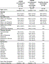
Demographic information of human donors.
Fig. 1.
Fig. 1.. Plasma neutralization and B cell responses in convalescent recovered, vaccinated, and recovered-vaccinated donors.
a. SARS-CoV-2 pseudovirus neutralization of plasma samples from COVID-19 convalescent recovered donors (blue: recovered), vaccinated donors (green: 2X vaccinated) or vaccinated donors with a prior history of SARS-CoV-2 infection (red: recovered-vaccinated). COVID-19 recovered donors (n = 21); spike-mRNA vaccinated (2X) donors (n = 10); COVID-19 recovered-vaccinated donors (n = 15). Data are shown as scatter dot plots with a line at geomatic mean value. Statistical comparisons between the two groups were performed using a Mann-Whitney two-tailed test, (*p < 0.05, ****p < 0.0001). b. Plasma neutralization for all three groups against distantly related sarbecoviruses. Pang17 (cyan), SARS-CoV-1 (yellow), and WIV1 (violet) are shown. RBDs are colored pink for all spikes. In contrast to infected-only and vaccinated-only donors, approximately half of the recovered-vaccinated donors have neutralizing titers against SARS-CoV-1 above background (Extended Data Fig. 1). Data are shown as scatter dot plots and the bar height represents median. c. Plasma neutralizing activity against SARS-CoV-2 (Wuhan) and SARS-CoV-2 variants of concern (B.1.1.7 (Alpha), B.1.351 (Beta), P.1 (Gamma), B.1.617.2 (Delta) and B.1.1.529 (Omicron)). Data are shown as scatter dot plots and the bar height represents median. d. Flow cytometric analysis of IgG+ B cells from PBMCs of human donors CC25 and CC84 isolated at the indicated timepoints (see Extended Data Fig. 2 for gating strategy). Numbers indicate percentage of cells binding to SARS-CoV-1 and SARS-CoV-2 spike proteins, respectively. e. Quantification of SARS-CoV-1-specific, SARS-CoV-2-specific, and cross-reactive IgG+ B cells from individual donor CC25 (top) and individual donor CC84 (bottom).
Fig. 2.
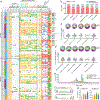
Fig. 2.. Binding, neutralization and immunogenetic properties of sarbecovirus mAbs.
A total of 107 mAbs were isolated, 56 mAbs from donor CC25 and 51 mAbs from donor CC84. MAbs were isolated by single B cell sorting using SARS-CoV-1 and SARS-CoV-2 S-proteins as baits. a. Heatmap showing IGHV germline gene usage (colored: VH1–46 (magenta), VH1–69 (spring) and VH3–30 (plum) and other V-genes (grey)), lineage information (unique (sky) and expanded (tangerine) lineages) and V-gene nucleotide somatic mutations (SHMs). ELISA binding activity of mAbs with SARS-CoV-2, SARS-CoV-1 and other β- and α-HCoV derived S-proteins and domains of SARS-CoV-2 S-protein (NTD, RBD-SD1, RBD-SD1–2) (LOD <0.5 OD405). Binding of mAbs with clade 1a (SARS-CoV-2 related: SARS-CoV-2, RatG13, Pang17), clade 1b (SARS-CoV-1 related: SARS-CoV-1, WIV1, SHC014), clade 2 (RmYN02, Rf1, Rs4081, Yun11) and clade 3 (BM4831, BtKY72) sarbecovirus S-protein derived monomeric RBDs. Percent neutralization of ACE2-utilizing sarbecoviruses (SARS-CoV-2, Pang17, SARS-CoV-1 and WIV1) by mAb supernatants (cut-off <60%). Breadth (%) of binding to 12 sarbecovirus RBDs and breadth (%) of neutralization with 4 ACE2 sarbecoviruses is indicated for each mAb. MAb expression levels in the supernatants were quantified by total IgG ELISA and the concentrations were approximately comparable overall. For reproducibility, the binding and neutralization assays were all performed twice with mAb supernatants expressed independently twice. b. Number of mAbs (unique clones) neutralizing SARS-CoV-2 and other sarbecoviruses. 40 mAbs neutralized all 4 ACE2 sarbecoviruses tested. c. Pie plots showing IGHV gene usage distribution of isolated mAbs (n = 93) with enriched gene families colored, VH1–46 (magenta) VH1–69 (spring) and VH3–30 (plum). Dot plots showing % nucleotide mutations (SHMs) in the heavy chain (VH) of isolated mAbs. SHMs are shown as scatter dot plots with a line at geomatic mean value. The mAbs are grouped by neutralization with sarbecoviruses. d. Pie and dot plots depicting IGHV gene distribution and VH nucleotide SHMs respectively. The mAbs (n = 93) are grouped by binding to sarbecovirus-derived RBDs. SHMs are shown as scatter dot plots with a line at geomatic mean value. e. CDRH3 length distributions of isolated mAbs (n = 93) across broadly neutralizing and broadly cross-reactive mAb groups compared to human baseline germline reference. MAbs with 20- and 21- amino acid-CDRH3s are highly enriched. f. Sequence conservation logos of 20 (n = 17) and 21 (n = 11) amino acid long CDRH3-bearing mAbs show enrichment of D-gene derived residues, including IGHD2–15 germline D-gene encoded two cysteines in 20 amino acid long CDRH3-bearing mAbs that may potentially form a disulfide bond. g. Enrichment of IGHD2–15 and IGHD3–22 germline D-genes in sarbecovirus broadly neutralizing or broadly cross-reactive mAbs compared to corresponding human baseline germlines.
Fig. 3.
Fig. 3.. Binding and neutralization of mAbs in terms of affinity/potency and breadth.
A total of 19 mAbs from donor CC25 and 11 mAbs from donor CC84 were selected to determine specificity, relative affinities, and neutralization of sarbecoviruses and SARS-CoV-2 VOCs. a. Heatmap of binding responses (nm) determined by BLI using SARS-CoV-1 and SARS-CoV-2 S and S-protein domains and subdomains with IGHV gene usage for each mAb indicated. b. Heatmap of dissociation constants (KD (M) values) for mAb binding to spike-derived monomeric RBDs from four sarbecovirus clades: clade 1b (n = 3); clade 1a (n = 3); clade 2 (n = 4); clade 3 (n = 2). Binding kinetics were obtained using the 1:1 binding kinetics fitting model on ForteBio Data Analysis software. c. IC50 neutralization of mAbs against SARS-CoV-2, SARS-CoV-1, Pang17, WIV1, and SHC014 determined using pseudotyped viruses. d, Neutralization of 20 bnAbs against SARS-CoV-2 (Wuhan) and five major SARS-CoV-2 variants of concern (B.1.1.7 (Alpha), B.1.351 (Beta), P.1 (Gamma), B.1.617.2 (Delta) and B.1.1.529 (Omicron)). SARS-CoV-2 Abs, CC12.1, DH1047, and Dengue Ab, DEN3 were used as controls. Data are presented as mean values +/− SD. The experiments were independently performed twice and consistent results were obtained.
Fig. 4.
Fig. 4.. Epitope specificities of sarbecovirus bnAbs.
a. Heatmap summary of epitope binning of sarbecovirus bnAbs based on BLI competition of bnAbs with human (CC12.1, CC12.19, CR3022, DH1047 and S309) and macaque (K398.22) RBD-specific nAbs. IGHV gene usage for each mAb is indicated. Geomean neutralization potency and breadth (calculated from Fig. 3c) and RBD binding breadth with clade 2 or all clade sarbecoviruses (calculated from Fig. 3b) for each mAb are indicated. The BLI competition was performed with monomeric SARS-CoV-2 RBD, and the competition levels are indicated as red (strong), orange (moderate), light blue (weak) and grey (very weak) competition. Based on competition with human and one macaque nAb of known specificities, the sarbecovirus bnAbs were divided into groups-1 and −2. b. Binding of human nAbs to SARS-CoV-2 RBD. The RBD is shown as a black chain trace, whereas antibodies are represented by solid surfaces in different colors: CC12.1 (pink, PDB 6XC2), CR3022 (yellow, PDB 6W41), S309 (orange, PDB 7R6W), DH1047 (cyan, 7LD1), and K398.22 (blue, PDB: 7TP4) . c. Electron microscopy 3D reconstructions of sarbecovirus bnAb Fabs with SARS-CoV-2 S-protein. Fabs of group-1 (n = 9) and group-2 (n = 2) were complexed with SARS-CoV-2 S-protein and 3D reconstructions were generated from 2D class averages. Fabs are shown in different colors and the spike S1 and S2 subunits (grey) and the RBD sites are labelled. d. The epitope of each antibody is outlined in different colors corresponding to panel b. Epitope residues are defined by buried surface area (BSA) > 0 Å2 as calculated by PDBePISA (https://www.ebi.ac.uk/msd-srv/prot_int/pistart.html). Putative epitope regions of group-1 bnAbs based on the competitive binding assay are indicated by red circles. e. Conservation of 12 sarbecovirus RBDs. Gray surface represents conserved regions, whereas blue represents variable regions. The conservation was calculated by ConSurf (https://consurf.tau.ac.il/). The putative epitope region targeted by group-1 bnAbs is circled as in panel d.
Fig. 5.
Fig. 5.. Immunogenetic properties of Group 1 and 2 RBD bnAbs.
a. VH gene family usage: IGHV1 (blue), IGHV3 (orange), IGHV4 (violet), and IGHV5 (red). b. CDRH3 length distribution (amino acids) in Groups 1 (red) and 2 (cyan). n = 30. CDRH3 lengths of group 1 and 2 bnAbs are shown as box plots with a line at geomatic mean value. P-values computed using the Kruskal-Wallis test and denoted as follows: P<0.05:*, P<0.005:**, P<0.00005:****. c. CDRH3 use of YYDxxG motif in group 1 and 2 RBD bnAbs: with (green) and without (gray). n = 30. d. CDRH3 length distribution in RBD bnAbs with (green) and without (gray) YYDxxG motifs. n = 30. Data are shown as box plots with a line at geomatic mean value. P-values computed using the Kruskal-Wallis test and denoted as follows: P<0.05:*, P<0.005:**, P<0.00005:****. e. Phylogenetic tree of heavy chain sequences of 30 RBD bnAbs. Each sequence is colored according to its group (left bar), IGHV gene family (middle bar), and the presence of the YYDxxD motif in the HCDR3 (right bar). Colors of heavy chain characteristics are consistent with panels a-d. Here and further, the phylogenetic tree is computed using Clustal Omega . f. Phylogenetic tree combining IGHD gene fragments of CDRH3s of nine mAbs with YYDxxG motifs and the amino acid translation of the germline sequence of IGHD3–22-containing YYGSSG. Each sequence is colored according to the amino acid following YYD: S (violet), R (orange), or others (gray). The alignment corresponding to the tree is shown below. Dots represent amino acids matching the germline amino acids. Germline amino acids truncated in CDRH3s are shown by dashes. g. Frequencies of IGHD3–22 germline genes with differing characteristics in naive heavy chain repertoires. From left to right: all IGHD3–22 in all CDRH3s; IGHD3–22 with YYDxxG motif in all CDRH3s; IGHD3–22 with YYDxxG motif in CDRH3s derived from IGHD3–22; distribution of lengths (in amino acids) of CDRH3s with (green) and without (gray) YYDxxG motifs. The fraction statistics were computed using ten Rep-seq libraries representing ten donors from the study by : ERR2567178–ERR2567187. The distribution of CDRH3 lengths was computed for library ERR2567178. Data are shown as box plots with a line at geomatic mean value. P-values computed using the Kruskal-Wallis test and denoted as follows: P<0.05:*, P<0.005:**, P<0.00005:****.
Fig. 6.
Fig. 6.. Prophylactic treatment of aged mice with RBD bnAbs protects against challenge with diverse SARS-like viruses.
a. Three RBD bnAbs (CC25.54, CC25.36 and CC25.53) individually, or a dengue DEN3 control antibody were administered intra-peritoneally (i.p.) at 300μg per animal into 12 groups of aged mice (10 animals per group). Each group of animals was challenged intra-nasally (i.n.) 12h after antibody infusion with one of 3 mouse-adapted (MA) sarbecoviruses, (MA10 = SARS-CoV-2; 1 × 10³ plaque forming units (PFU), MA15 = SARS-CoV-1; 1 × 10³ PFU or MA15-SHC = SARS-CoV MA15-SHC014 chimera; 1 × 10⁵ PFU). As a control, groups of mice were exposed only to PBS in the absence of virus. b,f,j. Percent weight change in RBD bnAbs or DEN3 control antibody-treated animals after challenge with mouse-adapted sarbecoviruses. Percent weight change was calculated from day 0 starting weight for all animals. n = 10 individuals for each group were used. Data are presented as mean values +/− SEM. Statistical significance was calculated with Dunnett’s multiple comparisons test between each experimental group and the DEN3 control Ab group. (**p <0.01, ***p <0.001; ****p < 0.0001; ns- p >0.05). One-way ANOVA was used. c,g,k. Day 2 post-infection Hemorrhage (Gross Pathology score) scored at tissue harvest in mice prophylactically treated with RBD bnAbs or DEN3 control mAb. n = 5 individuals for each group. Data are presented as mean values +/− SEM. d,h,l. Day 2 post-infection pulmonary function (shown as Penh score) was measured by whole body plethysmography in mice prophylactically treated with RBD bnAbs or DEN3 control mAb. n = 5 individuals for each group. Data are shown as box and whisker plots showing data points from Min to Max. e,i,m. Lung virus titers (PFU per Lung) were determined by plaque assay of lung tissues collected at day 2 or day 4 after infection. n = 5 individuals per timepoint for each group. Data are shown as scatter dot plots with bar heights representing the mean.

Update of

Comment in

References

    1. Baden LR et al. Efficacy and Safety of the mRNA-1273 SARS-CoV-2 Vaccine. The New England journal of medicine 384, 403–416, doi: 10.1056/NEJMoa2035389 (2020). - DOI - PMC - PubMed
    1. Polack FP et al. Safety and Efficacy of the BNT162b2 mRNA Covid-19 Vaccine. The New England journal of medicine 383, 2603–2615, doi: 10.1056/NEJMoa2034577 (2020). - DOI - PMC - PubMed
    1. Khoury DS et al. Neutralizing antibody levels are highly predictive of immune protection from symptomatic SARS-CoV-2 infection. Nature medicine 27, 1205–1211, doi: 10.1038/s41591-021-01377-8 (2021). - DOI - PubMed
    1. Gilbert PB et al. Title: Immune Correlates Analysis of the mRNA-1273 COVID-19 Vaccine Efficacy Trial. Science 375, 43–50, doi: 10.1101/2021.08.09.21261290 (2021). - DOI - PMC - PubMed
    1. Earle KA et al. Evidence for antibody as a protective correlate for COVID-19 vaccines. Vaccine 39, 4423–4428, doi: 10.1016/j.vaccine.2021.05.063 (2021). - DOI - PMC - PubMed

Publication types

Substances

Supplementary concepts