Multicenter Study
. 2022 Jul;45(7):992-1000.
doi: 10.1007/s00270-022-03169-0. Epub 2022 Jun 2.

Image-Guided Embolotherapy of Arteriovenous Malformations of the Face

Affiliations
Multicenter Study

Image-Guided Embolotherapy of Arteriovenous Malformations of the Face

Vanessa F Schmidt et al. Cardiovasc Intervent Radiol. 2022 Jul.

Abstract

Purpose: To evaluate the safety and outcome of image-guided embolotherapy of extracranial arteriovenous malformations (AVMs) primarily affecting the face.

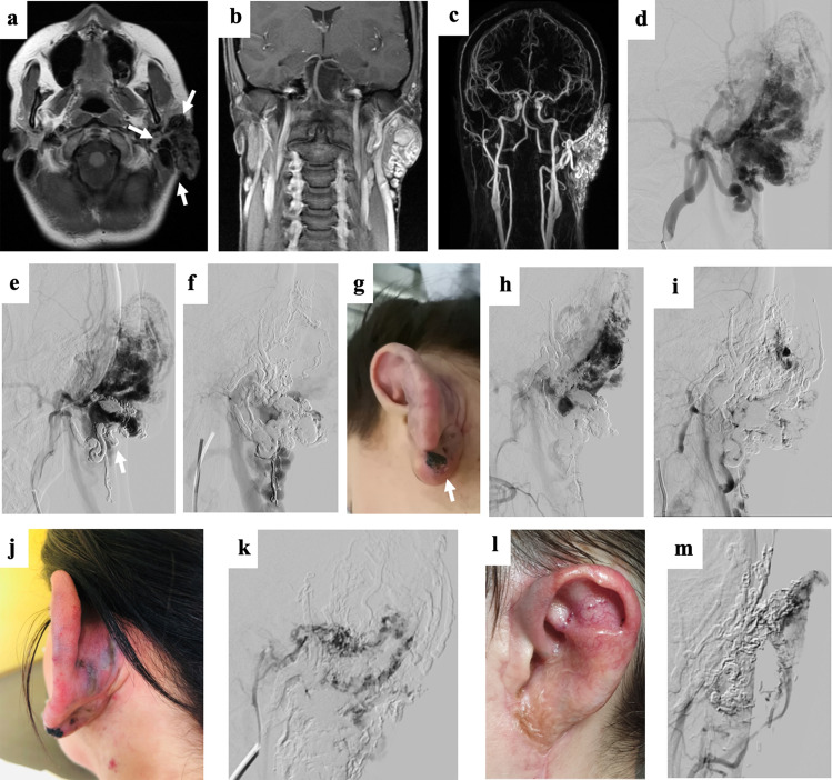
Materials and methods: A multicenter cohort of 28 patients presenting with AVMs primarily affecting the face was retrospectively investigated. Fifty image-guided embolotherapies were performed, mostly using ethylene-vinyl alcohol copolymer-based embolic agents. Clinical and imaging findings were assessed to evaluate response during follow-up (symptom-free, partial relief of symptoms, no improvement, and progression despite embolization), lesion devascularization (total, 100%; substantial, 76-99%; partial, 51-75%; failure, < 50%; and progression), and complication rates (classified according to the CIRSE guidelines). Sub-analyses regarding clinical outcome (n = 24) were performed comparing patients with (n = 12) or without (n = 12) subsequent surgical resection after embolotherapy.

Results: The median number of embolotherapy sessions was 2.0 (range, 1-4). Clinical outcome after a mean follow-up of 12.4 months (± 13.3; n = 24) revealed a therapy response in 21/24 patients (87.5%). Imaging showed total devascularization in 14/24 patients (58.3%), including the 12 patients with subsequent surgery and 2 additional patients with embolotherapy only. Substantial devascularization (76-99%) was assessed in 7/24 patients (29.2%), and partial devascularization (51-75%) in 3/24 patients (12.5%). Complications occurred during/after 12/50 procedures (24.0%), including 18.0% major complications. Patients with subsequent surgical resections were more often symptom-free at the last follow-up compared to the group having undergone embolotherapy only (p = 0.006).

Conclusion: Image-guided embolotherapy is safe and effective for treating extracranial AVMs of the face. Subsequent surgical resections after embolization may substantially improve patients' clinical outcome, emphasizing the need for multimodal therapeutic concepts.

Level of evidence: Level 4, Retrospective study.

Keywords: AVM; Embolization; Face; Interventional radiology; Surgical resection.


Conflict of interest statement

The authors declare that they have no conflict of interest.

Figures

Fig. 1
Fig. 1
30-year old female patient presenting with an extensive arteriovenous malformation (AVM) left (peri)auricular. After 4 sessions of embolotherapies with corresponding near-total devascularization of the malformation, previously planned two-step microsurgical resection and defect reconstruction using a free fascia flap was performed. (a–c) Preprocedural axial T1-weighted (T1-w) MR image, coronar T1-w MR image, and coronar MR-angiography image present the extent of the left (peri)auricular AVM with ubiquitous involvement of the auricle (arrows) and at least 4 arterial feeders (2 × from facial artery, 2 × from posterior auricular artery, 1 × from occipital artery) as well as venous drainage into the external jugular vein. (d–e) Periprocedural digital subtraction angiography (DSA) images during 1st embolotherapy show flow characteristics of the lesion and the successfully embolized vascular structures at the caudal part of the earlobe (arrow). (f) Periprocedural DSA image during 2nd embolotherapy shows the newly embolized vascularized components of the malformation. (g) Clinical photograph 1 day before the 3rd embolotherapy presents the enlarged and prominent ear auricle and lobule, transparent vascular structures of the malformation, and a small, long-term necrotic area on the caudal ear lobe. (h + i) Periprocedural DSA images during 3rd embolotherapy demonstrate remaining vascularized components of the malformation (arrow) as well as successfully embolized cranial components of the lesion. The patient presented with visual disorders directly post-procedural and MRI revealed 2 subtle restricted-diffusion lesions including the visual cortex, most likely due to periprocedural small air emboli. These findings were entirely regressive while preventive monitoring at the stroke unit. (j) Clinical photograph 3 days after the 3rd embolotherapy. (k) Periprocedural DSA image during the 4th embolotherapy shows arterial bleeding occurred after minor manipulation under anesthesia, due to previously unnoticed secondary abscessing after the 3rd session. After abscess draining and embolization of the arterial bleeding using histoacryl/lipiodol mixture, Onyx embolization of the remaining AVM components was successfully performed in the same session. (l) Clinical photograph after initially planned surgical resection of the devascularized AVM tissue including parts of the ear auricle and lobe and defect reconstruction with means of a free serratus fascia flap both successfully performed 3 months after the 4th session. (m) Diagnostic DSA image at final follow-up (21 months after the last treatment) presents a good perfusion of the apical ear auricle as well as no novel AVM components. Clinically, the patient appeared without any symptoms or further signs of AVM recurrence

References

    1. Greene AK, Liu AS, Mulliken JB, Chalache K, Fishman SJ. Vascular anomalies in 5,621 patients: guidelines for referral. J Pediatr Surg. 2011;46(9):1784–1789. doi: 10.1016/j.jpedsurg.2011.05.006. - DOI - PubMed
    1. de Miguel R, López-Gutierrez JC, Boixeda P. Arteriovenous malformations: a diagnostic and therapeutic challenge. Actas Dermosifiliogr. 2014;105(4):347–358. doi: 10.1016/j.ad.2013.04.013. - DOI - PubMed
    1. Greene AK, Orbach DB. Management of arteriovenous malformations. Clin Plast Surg. 2011;38(1):95–106. doi: 10.1016/j.cps.2010.08.005. - DOI - PubMed
    1. Johnson AB, Richter GT. Vascular anomalies. Clin Perinatol. 2018;45(4):737–749. doi: 10.1016/j.clp.2018.07.010. - DOI - PubMed
    1. Fernández-Alvarez V, Suárez C, de Bree R, et al. Management of extracranial arteriovenous malformations of the head and neck. Auris Nasus Larynx. 2020;47(2):181–190. doi: 10.1016/j.anl.2019.11.008. - DOI - PubMed

Publication types

MeSH terms