. 2022 Jun 2;22(1):605.
doi: 10.1186/s12885-022-09659-1.

Global DNA hypomethylation of colorectal tumours detected in tissue and liquid biopsies may be related to decreased methyl-donor content

Affiliations

Global DNA hypomethylation of colorectal tumours detected in tissue and liquid biopsies may be related to decreased methyl-donor content

Krisztina A Szigeti et al. BMC Cancer. .

Abstract

Background: Hypomethylation of long interspersed nuclear element 1 (LINE-1) is characteristic of various cancer types, including colorectal cancer (CRC). Malfunction of several factors or alteration of methyl-donor molecules' (folic acid and S-adenosylmethionine) availability can contribute to DNA methylation changes. Detection of epigenetic alterations in liquid biopsies can assist in the early recognition of CRC. Following the investigations of a Hungarian colon tissue sample set, our goal was to examine the LINE-1 methylation of blood samples along the colorectal adenoma-carcinoma sequence and in inflammatory bowel disease. Moreover, we aimed to explore the possible underlying mechanisms of global DNA hypomethylation formation on a multi-level aspect.

Methods: LINE-1 methylation of colon tissue (n = 183) and plasma (n = 48) samples of healthy controls and patients with colorectal tumours were examined with bisulfite pyrosequencing. To investigate mRNA expression, microarray analysis results were reanalysed in silico (n = 60). Immunohistochemistry staining was used to validate DNA methyltransferases (DNMTs) and folate receptor beta (FOLR2) expression along with the determination of methyl-donor molecules' in situ level (n = 40).

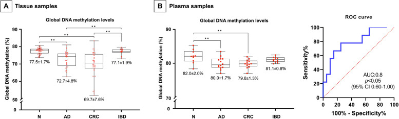
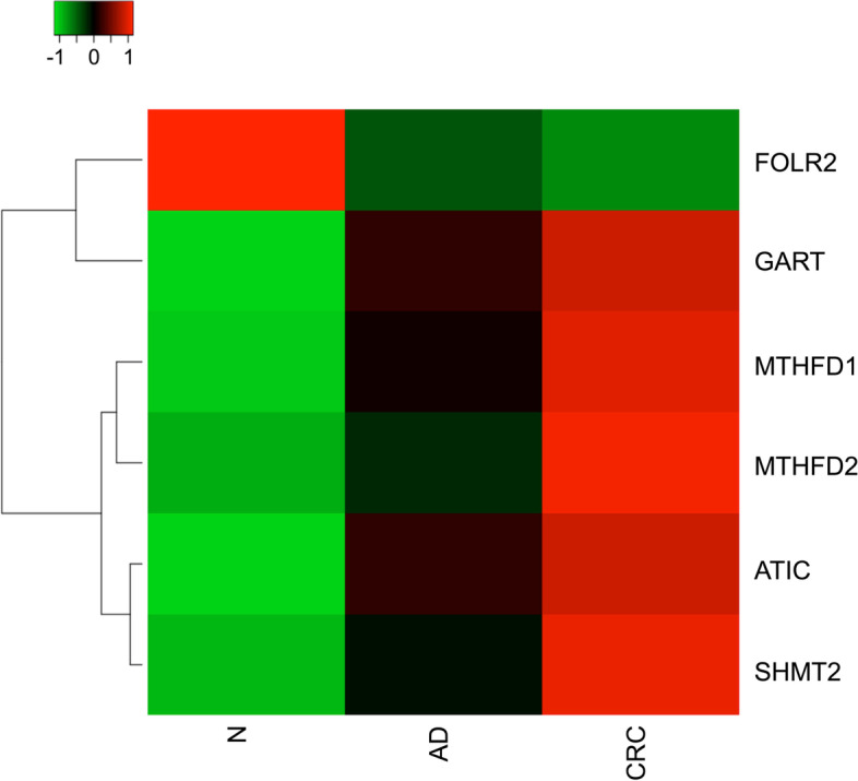
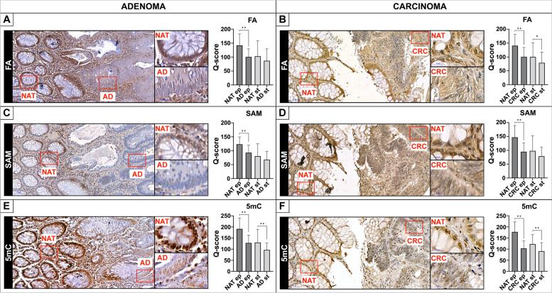
Results: Significantly decreased LINE-1 methylation level was observed in line with cancer progression both in tissue (adenoma: 72.7 ± 4.8%, and CRC: 69.7 ± 7.6% vs. normal: 77.5 ± 1.7%, p ≤ 0.01) and liquid biopsies (adenoma: 80.0 ± 1.7%, and CRC: 79.8 ± 1.3% vs. normal: 82.0 ± 2.0%, p ≤ 0.01). However, no significant changes were recognized in inflammatory bowel disease cases. According to in silico analysis of microarray data, altered mRNA levels of several DNA methylation-related enzymes were detected in tumours vs. healthy biopsies, namely one-carbon metabolism-related genes-which met our analysing criteria-showed upregulation, while FOLR2 was downregulated. Using immunohistochemistry, DNMTs, and FOLR2 expression were confirmed. Moreover, significantly diminished folic acid and S-adenosylmethionine levels were observed in parallel with decreasing 5-methylcytosine staining in tumours compared to normal adjacent to tumour tissues (p ≤ 0.05).

Conclusion: Our results suggest that LINE-1 hypomethylation may have a distinguishing value in precancerous stages compared to healthy samples in liquid biopsies. Furthermore, the reduction of global DNA methylation level could be linked to reduced methyl-donor availability with the contribution of decreased FOLR2 expression.

Keywords: Colorectal cancer; DNA methylation; Epigenetics; Folic acid; Liquid biopsy; S-adenosylmethionine.


Conflict of interest statement

The authors declare that they have no competing interest.

Figures

Fig. 1
Fig. 1
LINE-1 methylation of tissue, LINE-1 methylation, and receiver operating characteristic analysis of plasma specimens. A Significantly decreased LINE-1 methylation was observed in tumours compared to healthy and IBD tissue biopsies (**p ≤ 0.01). There were no significant methylation level alterations in IBD tissue samples in comparison with N biopsies. B Significant decrease of LINE-1 methylation was noticed in AD and CRC vs. N blood samples (**p ≤ 0.01). IBD specimens did not show significantly altered methylation level compared to healthy liquid biopsies (left). AD samples were separated from healthy controls with 66.7% sensitivity and 90.0% specificity (threshold 80.0%) (right). N: healthy, AD: colorectal adenoma, CRC: colorectal carcinoma, IBD: inflammatory bowel disease, ROC: receiver operating characteristic nalysis
Fig. 2
Fig. 2
Significant mRNA expression alterations of folate receptors and genes involved in one-carbon cycle. On the heatmap, distinct colours are coupled to different expression intensity values: green - low, black - intermediate, red - high intensity. Each row represents different genes, and each column indicates the investigated sample types. Only candidates owning significant expression changes (p ≤ 0.05) and fold change value greater or equal than |1.4| in both AD and CRC tissue samples compared to N were presented on the heatmap. One-carbon metabolism-related enzymes showed significantly elevated mRNA expression in AD and CRC samples compared to N. FOLR2 had a significantly lower transcript level in tumorous samples vs. healthy controls (p ≤ 0.05). N: healthy, AD: colorectal adenoma, CRC: colorectal carcinoma
Fig. 3
Fig. 3
Immunohistochemistry staining of methyl-donor molecules and 5mC in AD and CRC tissue samples. Significantly lower staining intensity of FA (A, B), SAM (C, D), and 5mC (E, F) was observed in epithelial cells of both AD and CRC regions compared to NAT areas (**p ≤ 0.01). Slightly reducing trend was noticed in stromal cells except for 5mC labelling in AD (E), and CRC (F) (**p ≤ 0.01), along with FA staining in CRC specimens (B) (*p ≤ 0.05) also possessing significantly lower immunostaining. Q-score method was applied to evaluate the staining intensity of immunohistochemistry. Scale bars on the left: 50 µm, on the right: 20 µm. FA: folic acid, SAM: S-adenosylmethionine, 5mC: 5-methylcytosine, NAT: normal adjacent to tumour tissue, AD: colorectal adenoma, CRC: colorectal carcinoma, ep: epithelial cells, st: stromal cell, Q-score: Quick-score method

Similar articles

Cited by

References

    1. Sung H, Ferlay J, Siegel RL, Laversanne M, Soerjomataram I, Jemal A, et al. Global Cancer Statistics 2020: GLOBOCAN Estimates of Incidence and Mortality Worldwide for 36 Cancers in 185 Countries. CA Cancer J Clin. 2021;71(3):209–49. doi: 10.3322/caac.21660. - DOI - PubMed
    1. Fearon ER, Vogelstein B. A genetic model for colorectal tumorigenesis. Cell. 1990;61(5):759–767. doi: 10.1016/0092-8674(90)90186-I. - DOI - PubMed
    1. Greenstein AJ, Sachar DB, Smith H, Janowitz HD, Aufses AH. Patterns of neoplasia in Crohn’s disease and ulcerative colitis. Cancer. 1980;46(2):403–407. doi: 10.1002/1097-0142(19800715)46:2<403::AID-CNCR2820460232>3.0.CO;2-6. - DOI - PubMed
    1. Gillen CD, Walmsley RS, Prior P, Andrews HA, Allan RN. Ulcerative colitis and Crohn’s disease: a comparison of the colorectal cancer risk in extensive colitis. Gut. 1994;35(11):1590. doi: 10.1136/gut.35.11.1590. - DOI - PMC - PubMed
    1. Ng SC, Shi HY, Hamidi N, Underwood FE, Tang W, Benchimol EI, et al. Worldwide incidence and prevalence of inflammatory bowel disease in the 21st century: a systematic review of population-based studies. Lancet. 2018;390(10114):2769–2778. doi: 10.1016/S0140-6736(17)32448-0. - DOI - PubMed