. 2022 Jun 2;11(1):34.
doi: 10.1186/s40164-022-00286-x.

IL-39 promotes chronic graft-versus-host disease by increasing T and B Cell pathogenicity

Affiliations

IL-39 promotes chronic graft-versus-host disease by increasing T and B Cell pathogenicity

Kangkang Lv et al. Exp Hematol Oncol. .

Abstract

Background: Chronic graft-versus-host disease (cGVHD) remains a major complication during the late phase of allogeneic hematopoietic stem cell transplantation (allo-HSCT). IL-39, a newly described pro-inflammatory cytokine belonging to the IL-12 family, plays a role in lupus development. Recently, IL-39 has been identified as a pathogenic factor in acute GVHD (aGVHD). However, the role of IL-39 in the pathogenesis of cGVHD remains unclear.

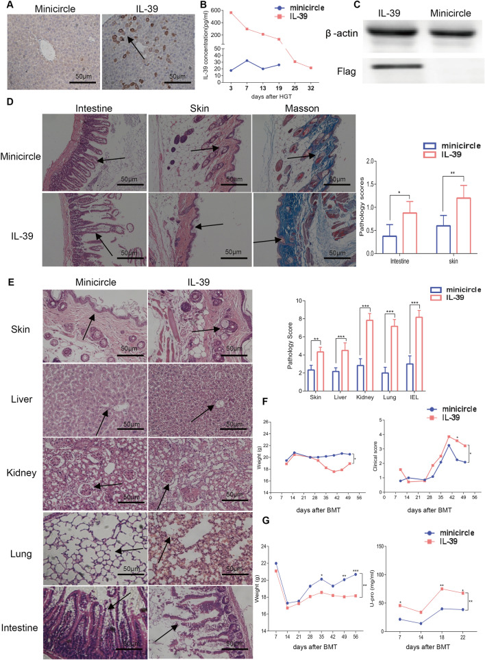
Methods: We constructed a recombinant IL-39 plasmid and established scleroderma and lupus-like cGVHD models. Quantitative PCR and enzyme-linked immunosorbent assay (ELISA) were used to detect IL-39 expression in mice and patients post transplantation, respectively. Hydrodynamic gene transfer (HGT) was performed to achieve IL-39 overexpression in vivo. Multiparameter flow cytometry, western blotting, and assays in vitro were performed to investigate the effect of IL-39 on cGVHD.

Results: The relative expression of IL-23p19 and EBi3 was significantly increased in the intestine of cGVHD mice on day 40 post allo-HSCT, and IL-39 levels were significantly elevated in the serum of patients following allo-HSCT. Overexpression of IL-39 significantly aggravated the severity of cGVHD. Increased IL-39 levels promoted T-cell activation and germinal center responses, and may exacerbate thymic damage. Consistently, blocking IL-39 markedly ameliorated immune dysregulation in the cGVHD mice. Furthermore, we found that IL-39 was produced by B cells, CD11b+ cells, and CD8+T cells after activation. Stimulation of IL-39 led to upregulation of the IL-39 receptor on CD4+T cells and further caused activation of the STAT1/STAT3 pathway, through which IL-39 may exert its pro-inflammatory effects.

Conclusion: Our study reveals a critical role for IL-39 in cGVHD pathogenesis and indicates that IL-39 may serve as a potential therapeutic target for cGVHD prevention.

Keywords: Allogeneic hematopoietic stem cell transplantation; Chronic graft-versus-host disease; IL-12; IL-39.


Conflict of interest statement

We declare that no competing interests exists.

Figures

Fig. 1
Fig. 1
IL-39 aggravates the development of murine cGVHD. BALB/c mice were hydrodynamically injected with minicircles or IL-39 plasmids (n = 6 per group). Three days after HGT, irradiated BALB/c recipients were infused with 1 × 107 bone marrow cells and 1 × 106 splenocytes from C57BL/6 mice, to establish a scleroderma-like cGVHD model. IL-39 expression in the liver 7 days after HGT treatment was detected by immunohistochemistry (A) and western blotting (C). IL-39 levels in the serum were detected weekly using enzyme-linked immunosorbent assay (B). Representative histopathological images and pathology scores of the skin, intestine, and Masson’s trichrome staining are shown (D). Body weights and GVHD scores in scleroderma-like mice are shown (F). Three days after HGT, irradiated recipients were infused with 5 × 106 bone marrow cells and 4 × 107 CD25 splenocytes from DBA/2 mice to establish a lupus-like cGVHD mouse model. Representative histopathological images and pathology scores of the skin, liver, intestine, kidney, and lung are shown (E). Body weights and urine protein levels in lupus-like mice are shown (G). The data are representative of at least three independent experiments. Values are presented as mean ± SD. *P < 0.05; **P < 0.01; ***P < 0.001
Fig. 2
Fig. 2
IL-39 promotes pro-inflammatory responses of T and B cells in the scleroderma-like cGVHD mice model. Irradiated BALB/c recipients were infused with 1 × 107 bone marrow cells and 1 × 106 splenocytes from the C57BL/6 mice. Splenocytes (n = 6 in each group) were collected and stained for FACS analysis eight weeks after transplantation. The percentages and numbers of CD4+, CD69+CD4+T, CD8+, and CD69+CD8+T cells in H2Kb+ cells from the spleens of recipients are shown (A). Lymphocytes were isolated from the spleens of recipients and treated with PMA, brefeldin A, and ionomycin for 4-6 h. The percentage and number of TNF-α-(B) and IL-4-(C) positive T cells in the spleens of recipients are shown. The percentage and number of Treg (CD4+CD25+Foxp3+) cells in the spleens of recipients are shown (D). The percentage and number of CD4+CD8+T cells and the CD4+/CD4+CD8+ and CD8+/CD4+CD8+ ratios in the thymus of recipients are shown (E). The percentage and number of Tfh (CD4+CXCR5+PD-1+) cells in the spleens of recipients are shown (F). The percentage and number of GC B (B220+GL-7+Fas+) cells in the spleens of recipients are shown (G). The data are representative of at least three independent experiments. Values are presented as mean ± SD. *P < 0.05; **P < 0.01
Fig. 3
Fig. 3
IL-39 promotes the pro-inflammatory responses of T and B cells in the lupus-like cGVHD mice model. Irradiated BALB/c recipients were infused with 5 × 106 bone marrow cells and 4 × 107 CD25 splenocytes from DBA/2 mice. Splenocytes (n = 6 per group) were collected and stained for FACS analysis 8 weeks post transplantation. The percentage and number of CD4+CD8+T cells and the CD4+/CD4+CD8+ and CD8+/CD4+CD8+ ratios in the thymuses of recipient mice are shown (A). The percentage and number of Tfh (CD4+CXCR5+PD-1+) cells in the spleens of recipients are shown (B). The percentage and number of GC B (B220+GL-7+Fas+) cells in the spleens of recipients are shown (C). The data are representative of at least three independent experiments. Values are presented as mean ± SD. *P < 0.05; **P < 0.01
Fig. 4
Fig. 4
Anti-IL-39 antibody ameliorates immune dysregulation in scleroderma-like cGVHD. Irradiated BALB/c recipients were infused with 1 × 107 bone marrow cells and 1 × 106 splenocytes from the C57BL/6 mice. Fourteen days after transplantation, each mouse in the antibody group received 100 μl (100 μg) of IL-39 antibody, while each mouse in the control group received 100 μl (100 μg) of isotype control antibody via intraperitoneal injection twice a week for 6 weeks. Splenocytes (n = 4 in each group) were collected and stained for FACS analysis eight weeks after transplantation. The percentages and numbers of CD4+, CD69+CD4+T, CD8+, and CD69+CD8+T cells in H2Kb+ cells from the spleens of recipients are shown (A). Lymphocytes were isolated from the spleen and treated with PMA, brefeldin A, and ionomycin for 4-6 h. The percentage and number of TNF-α-(B) and IL-4-(C) positive T cells in the spleens of recipients are shown. The percentage and number of Treg (CD4+CD25+Foxp3+) cells in the spleens of recipients are shown (D). The percentage and number of CD4+CD8+T cells and the CD4+/CD4+CD8+ and CD8+/CD4+CD8+ ratios in the thymuses of recipients are shown (E). The percentage and number of Tfh (CD4+CXCR5+PD-1+) cells in the spleens of recipients are shown (F). The percentage and number of GC B (B220+GL-7+Fas+) cells in the spleens of recipients are shown (G). The data are representative of at least three independent experiments. Values are presented as mean ± SD. *P < 0.05; **P < 0.01
Fig. 5
Fig. 5
Serum IL-39 levels were significantly elevated in patients with cGVHD. CXCL13 and IL-39 levels in serum were detected in non-GVHD, mild, and moderate/severe cGVHD patients (A). The prognostic value of CXCL13 and IL-39 in no-cGVHD and mild and moderate/severe cGVHD patients was evaluated using ROC curves (B). CD4+T cells, CD8+T cells, B cells, and CD11b+ cells were sorted from the splenocytes of C57BL/6 mice or PBMCs from healthy donors using a Cell Isolation Kit according to the manufacturer’s protocol. For CD4+ and CD8+T cell activation, plates were coated with 2 mg/ml anti-CD3 and 0.4 mg/ml anti-CD28 Abs overnight. Next, 2 × 105 T cells were cultured with recombinant mouse or human IL-39 proteins (20 ng/ml) for 72 h. For B cell and CD11b+ cell activation, 2 × 105 B cells and CD11b+ cells were cultured with LPS (5 ng/ml) and recombinant mouse or human IL-39 proteins (20 ng/ml) for 72 h. The relative expression of IL-39 and IL-39R genes was determined by real-time PCR in CD4+T cells, CD8+T cells, B cells, and CD11b+ cells from the splenocytes of C57BL/6 mice and PBMCs from healthy donors (C). In the following experiments, plates were coated with 2 mg/ml anti-CD3 and 0.4 mg/ml anti-CD28 Abs overnight. T cells (2 × 105 T cells were cultured with various concentrations of recombinant mouse or human IL-39 proteins for 72 h. Phosphorylation of STAT1/STAT3 detected by western blotting in sorted primary T cells from mice or healthy donors is shown (D, E). The data are representative of at least three independent experiments. Values are presented as mean ± SEM. *P < 0.05; ***P < 0.001

References

    1. Zeiser R, Blazar BR. Pathophysiology of chronic graft-versus-host disease and therapeutic targets. N Engl J Med. 2017;377(26):2565–2579. doi: 10.1056/NEJMra1703472. - DOI - PubMed
    1. Pidala J, Kurland B, Chai X, Majhail N, Weisdorf DJ, Pavletic S, et al. Patient-reported quality of life is associated with severity of chronic graft-versus-host disease as measured by NIH criteria: report on baseline data from the Chronic GVHD Consortium. Blood. 2011;117(17):4651–4657. doi: 10.1182/blood-2010-11-319509. - DOI - PMC - PubMed
    1. MacDonald KP, Hill GR, Blazar BR. Chronic graft-versus-host disease: biological insights from preclinical and clinical studies. Blood. 2017;129(1):13–21. doi: 10.1182/blood-2016-06-686618. - DOI - PMC - PubMed
    1. Socié G, Ritz J. Current issues in chronic graft-versus-host disease. Blood. 2014;124(3):374–384. doi: 10.1182/blood-2014-01-514752. - DOI - PMC - PubMed
    1. Forcade E, Kim HT, Cutler C, Wang K, Alho AC, Nikiforow S, et al. Circulating T follicular helper cells with increased function during chronic graft-versus-host disease. Blood. 2016;127(20):2489–2497. doi: 10.1182/blood-2015-12-688895. - DOI - PMC - PubMed