Case Reports
. 2022 May 24:2022:8474741.
doi: 10.1155/2022/8474741. eCollection 2022.

The Youngest Case of Metachronous Bilateral Acinic Cell Carcinoma of the Parotid Gland: A Case Report and Literature Review

Affiliations
Case Reports

The Youngest Case of Metachronous Bilateral Acinic Cell Carcinoma of the Parotid Gland: A Case Report and Literature Review

Raid Alhayaza et al. Case Rep Otolaryngol. .

Abstract

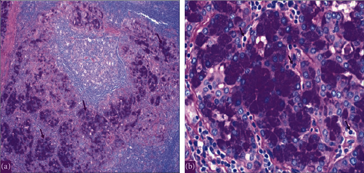
Introduction: Acinic cell carcinoma (ACC) is a low-grade malignant salivary neoplasm that represents 17% of all salivary gland malignancies. It has a tendency to affect young individuals, especially females. ACC mainly originates in the parotid gland and has a potential for recurrence and metastases. Rarely, ACC can affect both parotid glands in a single individual. A bilateral ACC of the parotid gland could either present as a synchronous or a metachronous tumor. Case Report. Our patient is a 19-year-old female known case of ACC of the right parotid gland. The tumor was resected in December 2017. After 3 years, she presented with a left parotid pain and swelling, which raised the suspicion of a contralateral metachronous tumor of the left parotid gland. In September 30, 2020 we proceeded with ultrasound-guided fine needle aspiration of the left intraparotid lesion, and the results turned out to be consistent with ACC. Here, we report a case of a 19-year-old female presenting with metachronous bilateral ACC of the parotid gland with an interval of 3 years, which is the 6th of its kind in the literature and the youngest amongst them.

Conclusion: Despite the rareness of metachronous occurrence of bilateral ACC of the parotid gland, it is still encountered in the medical practice. Here, we are highlighting the importance of follow-up with a periodic clinical and radiological examinations, bearing in mind the contralateral nonaffected parotid gland.


Conflict of interest statement

The authors declare that they have no conflicts of interest.

Figures

Figure 1
Figure 1
The CT scan showing a right parotid gland lesion in 2017.
Figure 2
Figure 2
The histopathology with hematoxylin and eosin (H&E) stain showing a multifocal acinic cell carcinoma of the right parotid gland in 2017. (a) Low power showing lymphoid-rich stroma with lymphoid follicles and prominent germinal centers within tumor. (b) Medium power showing tumor with acini and granular cytoplasm. (c) High power x60 showing the basophilic granular cytoplasm (zymogen granules of neoplastic serous acini).
Figure 3
Figure 3
The PET scan in November 2017. (a)–(c) There is mild background level FDG avidity (SUVmax 2.2) in the region of the right parotid. This uptake is most likely postsurgical. Also, there is mild FDG avidity (SUVmax 1.7) within a few prominent (up to 11 mm short axis) right upper cervical (levels II-III) lymph nodes. The exact etiology remains indeterminate (i.e., whether could be malignant or reactive due to her recent surgery). Also, small volume lymph nodes (measuring up to 8 mm short axis) are present in the left upper cervical/level II region, with minimal FDG uptake, as this is a common site for reactive lymphadenopathy. On the other hand, there are additional small volume bilateral submental and submandibular lymph nodes (up to 6 mm short axis) with no definite FDG uptake. Uptake of the tracer elsewhere is within physiological limits.
Figure 4
Figure 4
The PET scan in November 2018. The study was compared with the previous scan of November 20, 2017. Overall, the study ((a)–(c)) is stable without any evidence of local recurrence or distant metastasis.
Figure 5
Figure 5
The CT scan showing a left deep parotid gland lesion in 2020.
Figure 6
Figure 6
The PET scan in August 2020. The study showed an interval anatomic increase in size and metabolic progression in the known suspicious left deep parotid lesion ((a)–(c)). Measure approximately 2.0 × 1.9 × 2.2 cm in anteroposterior, transverse, and craniocaudal dimensions, respectively, with standard uptake value max (SUVmax) of 3.2. The SUVmax was previously 2.2. On the other hand, there is a more prominent left infraparotid subcentimeter node showing mild fluorodeoxyglucose (FDG) uptake. SUVmax 1.2 is indeterminate. Moreover, an anatomically stable mild FDG-avid left jugulodigastric subcentimeter node with SUVmax up to 1.4 and right supraclavicular node (SUVmax 1.0) are nonspecific, likely reactive. No other size significant FDG-avid cervical lymphadenopathy.
Figure 7
Figure 7
The histopathology with hematoxylin and eosin (H&E) stain showing acinic cell carcinoma with lymphoid background of the left parotid gland in 2020. (a) Low power showing lymphoid-rich stroma. (b) Medium power showing tumor with acini and granular cytoplasm. (c) High power x60 showing the basophilic granular cytoplasm (zymogen granules of neoplastic serous acini). The same tumor occurred metachronously in the left parotid gland.
Figure 8
Figure 8
The histopathology with periodic acid–Schiff plus diastase (PASD) stain showing acinic cell carcinoma of the left parotid gland in 2020. (a) Periodic acid–Schiff plus diastase (PASD) stain low power showing acinic cell carcinoma cells with PASD-positive granular cytoplasm. (b) Periodic acid–Schiff plus diastase (PASD) stain high power showing PAS + diastase resistant granules in Acinic cells.
Figure 9
Figure 9
Symptoms, investigations, radiotherapy course, and surgeries.
Figure 10
Figure 10
The PET scan in January 2022. The study ((a)–(c)) was compared with previous studies. The upper and lower cervical lymph nodes are showing mild FDG avidity with SUVmax of 1.8. However, there is no change morphologically, and it is most likely reactive. On the other hand, physiological radiotracer distribution is seen in the brain and there are no new suspicious FDG-avid lesions seen otherwise.
Figure 11
Figure 11
Representation of the bilateral metachronous occurrence of the parotid gland ACC in our case.

References

    1. Al-Zaher N., Obeid A., Al-Salam S., Al-Kayyali B. S. Acinic cell carcinoma of the salivary glands: a literature review. Hematology/Oncology and Stem Cell Therapy . 2009;2 - PubMed
    1. Bartos V. Metachronous bilateral acinic cell carcinoma of the parotid gland in a young woman. Sisli Etfal Hastanesi Tip Bulteni . 2019;53:306–309. doi: 10.14744/SEMB.2019.62134. - DOI - PMC - PubMed
    1. Eneroth C. M., Hamberger C. A., Jakobsson P. R. å. LXIII Malignancy of acinic cell carcinoma. The Annals of Otology, Rhinology and Laryngology . 1966;75 - PubMed
    1. Godwin J. T., Foote F. W., Frazell E. L. Acinic cell adenocarcinoma of the parotid gland; report of twenty-seven cases. The American Journal of Pathology . 1954;30(3):465–477. - PMC - PubMed
    1. Clarke J. S., Hentz E. C., Mahoney W. D. Bilateral acinic cell carcinoma of the parotid gland. Annals of Surgery . 1969;170 - PMC - PubMed

Publication types

LinkOut - more resources