

Quantitative assessment of left ventricular systolic function in patients with systemic lupus erythematosus: a non-invasive pressure-strain loop technique
- PMID: 35655829
- PMCID: PMC9131320
- DOI: 10.21037/qims-21-951
Quantitative assessment of left ventricular systolic function in patients with systemic lupus erythematosus: a non-invasive pressure-strain loop technique
Abstract
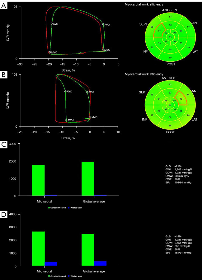
Background: Systemic lupus erythematosus (SLE) is associated with a variety of cardiovascular diseases, even in the early stage of disease development. The purpose of this study was to quantitatively evaluate left ventricular (LV) systolic function in patients with SLE using a novel non-invasive pressure-strain loop (PSL) technique.
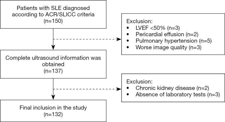
Methods: This prospective case-control study included 132 patients with SLE and 99 normal controls, all of whom underwent traditional transthoracic echocardiography. The LV myocardial work was evaluated with the PSL technique based on speckle tracking and brachial artery blood pressure. The differences among groups were compared, and the correlations between myocardial work, laboratory data, and disease activity were analyzed in the SLE group.
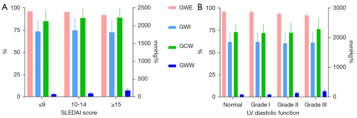
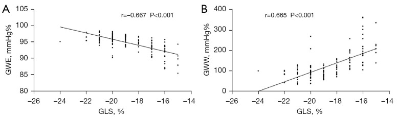
Results: Compared with the normal group, SLE patients had significantly higher global wasted work {GWW; SLE: 109 [82-150] mmHg%; controls: 66 [45-109] mmHg%; P<0.001} and impaired global work efficiency [GWE; SLE: 95% (94-97%); controls: 97% (96-98%); P<0.001]. Global work index (GWI) and global constructive work (GCW) did not show significant differences (P>0.05). Further subdivision analysis found that the increase of GWW and the damage of GWE were more obvious in SLE patients with high disease activity or severe diastolic dysfunction. Multivariate analysis revealed that increased erythrocyte sedimentation rate (ESR), C-reactive protein (CRP), anti-phospholipid antibodies, peak strain dispersion, and SLE Disease Activity Index (SLEDAI) were independently associated with increased GWW (β=0.189, 0.230, 0.444, 0.111, and 0.180, respectively; all P<0.05) and damaged GWE (β=-0.184, -0.130, -0.468, -0.149, and -0.191, respectively; all P<0.05).
Conclusions: The non-invasive PSL can quantitatively evaluate the LV systolic function in SLE patients. This technique may provide a new method for monitoring cardiac function in chronic diseases.
Keywords: Systemic lupus erythematosus (SLE); myocardial work; pressure-strain loop (PSL).
2022 Quantitative Imaging in Medicine and Surgery. All rights reserved.
Conflict of interest statement
Conflicts of Interest: All authors have completed the ICMJE uniform disclosure form (available at https://qims.amegroups.com/article/view/10.21037/qims-21-951/coif). The authors have no conflicts of interest to declare.
Figures
References
-
- Winau L, Hinojar Baydes R, Braner A, Drott U, Burkhardt H, Sangle S, D'Cruz DP, Carr-White G, Marber M, Schnoes K, Arendt C, Klingel K, Vogl TJ, Zeiher AM, Nagel E, Puntmann VO. High-sensitive troponin is associated with subclinical imaging biosignature of inflammatory cardiovascular involvement in systemic lupus erythematosus. Ann Rheum Dis 2018;77:1590-8. 10.1136/annrheumdis-2018-213661 - DOI - PubMed
-
- Unger ED, Dubin RF, Deo R, Daruwalla V, Friedman JL, Medina C, Beussink L, Freed BH, Shah SJ. Association of chronic kidney disease with abnormal cardiac mechanics and adverse outcomes in patients with heart failure and preserved ejection fraction. Eur J Heart Fail 2016;18:103-12. 10.1002/ejhf.445 - DOI - PMC - PubMed
LinkOut - more resources
Full Text Sources
Research Materials
Miscellaneous