Review
. 2022 Jun 1;13(3):773-786.
doi: 10.14336/AD.2021.1116. eCollection 2022 Jun.

Histone Deacetylase 3: A Potential Therapeutic Target for Atherosclerosis

Affiliations
Review

Histone Deacetylase 3: A Potential Therapeutic Target for Atherosclerosis

Li-Ping Jiang et al. Aging Dis. .

Abstract

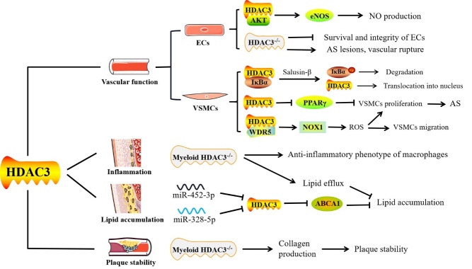
Atherosclerosis, the pathological basis of most cardiovascular disease, is characterized by plaque formation in the intima. Secondary lesions include intraplaque hemorrhage, plaque rupture, and local thrombosis. Vascular endothelial function impairment and smooth muscle cell migration lead to vascular dysfunction, which is conducive to the formation of macrophage-derived foam cells and aggravates inflammatory response and lipid accumulation that cause atherosclerosis. Histone deacetylase (HDAC) is an epigenetic modifying enzyme closely related to chromatin structure and gene transcriptional regulation. Emerging studies have demonstrated that the Class I member HDAC3 of the HDAC super family has cell-specific functions in atherosclerosis, including 1) maintenance of endothelial integrity and functions, 2) regulation of vascular smooth muscle cell proliferation and migration, 3) modulation of macrophage phenotype, and 4) influence on foam cell formation. Although several studies have shown that HDAC3 may be a promising therapeutic target, only a few HDAC3-selective inhibitors have been thoroughly researched and reported. Here, we specifically summarize the impact of HDAC3 and its inhibitors on vascular function, inflammation, lipid accumulation, and plaque stability in the development of atherosclerosis with the hopes of opening up new opportunities for the treatment of cardiovascular diseases.

Keywords: HDAC3; HDAC3 inhibitors; acetylation; atherosclerosis; cardiovascular diseases.


Conflict of interest statement

Competing interests The authors declare that there are no disputes of interest.

Figures

Figure 1.
Figure 1.
The location of HDAC3 within the cell. HDAC3 may shuttle between the cytoplasm and the nucleus. HDAC3 is maintained in the cytoplasm in combination with inhibitor α of nuclear factor-κB (IκBα) and enters the nucleus when IκBα is degraded. In contrast, when newly synthesized IκBα is present in the nucleus, it binds to nuclear HDAC3 and transfers HDAC3 to the cytoplasm, leading to a redistribution of subcellular HDAC3. Moreover, amino acids 180 through 313 in the central part of HDAC3 act as the nuclear export signal, and amino acids 312 through 428 in the C-terminus act as the nuclear localization signal.
Figure 2.
Figure 2.
The potential role of HDAC3 in atherosclerosis (AS). In endothelial cells (ECs), the interaction between HDAC3 and AKT is beneficial to the AKT-eNOS signaling pathway and nitric oxide (NO) production. Deletion of HDAC3 in ECs damages cell integrity and survival rates. ApoE-/- mice lacking HDAC3 in the aortas showed severe atherosclerotic lesions and ruptured blood vessels. Furthermore, the IκBα-HDAC3 complex exists in the cytoplasm of vascular smooth muscle cells (VSMCs). Salusin-β treatment induces repression of PPARγ expression due to the nuclear translocation of HDAC3, which may be attributed to the phosphorylation and degradation of IκBα. Moreover, the combination of HDAC3 and WD-40 repeat-containing protein 5 (WDR5) forms a complexe, which positively modulates nicotinamide adenine dinucleotide phosphate oxidase 1 (NOX1), thereby increasing reactive oxygen species (ROS) levels and promoting the transformation of VSMCs into a phenotype with increased cell migration and proliferation. Macrophages in mice lacking myeloid HDAC3 are converted to an anti-inflammatory phenotype and present an enhanced lipid efflux capacity. MiR-452-3p or miR-328-5p inhibits HDAC3 expression by directly targeting 3'UTR but increases the acetylation and expression levels of the ABCA1 gene, thereby reducing lipid accumulation in THP-1 macrophage-derived foam cells. Collagen production by VSMCs is increased in myeloid HDAC3-deficient mice, thereby exhibiting a stable plaque phenotype in the atherosclerotic lesions.

Similar articles

Cited by

References

    1. Libby P (2021). Inflammation in Atherosclerosis-No Longer a Theory. Clin chem, 67(1): 131-142. - PubMed
    1. Hoeksema MA, Gijbels MJ, Van den Bossche J, van der Velden S, Sijm A, Neele AE, et al.. (2014). Targeting macrophage Histone deacetylase 3 stabilizes atherosclerotic lesions. EMBO Mol Med, 6(9): 1124-32. - PMC - PubMed
    1. Asare Y, Campbell-James TA, Bokov Y, Yu LL, Prestel M, El Bounkari O, et al.. (2020). Histone Deacetylase 9 Activates IKK to Regulate Atherosclerotic Plaque Vulnerability. Circ Res, 127(6): 811-823. - PubMed
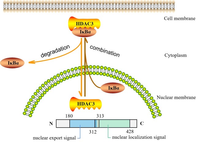
    1. Yang WM, Tsai SC, Wen YD, Fejer G, Seto E (2002). Functional domains of histone deacetylase-3. J Biol Chem, 277(11): 9447-54. - PubMed
    1. Ryu Y, Kee HJ, Sun S, Seok YM, Choi SY, Kim GR, et al.. (2019). Class I histone deacetylase inhibitor MS-275 attenuates vasoconstriction and inflammation in angiotensin II-induced hypertension. PloS one, 14(3): e0213186. - PMC - PubMed

LinkOut - more resources