Review
. 2022 Jun 1;13(3):712-731.
doi: 10.14336/AD.2021.1027. eCollection 2022 Jun.

Autophagy and Renal Fibrosis

Affiliations
Review

Autophagy and Renal Fibrosis

Shan Liang et al. Aging Dis. .

Abstract

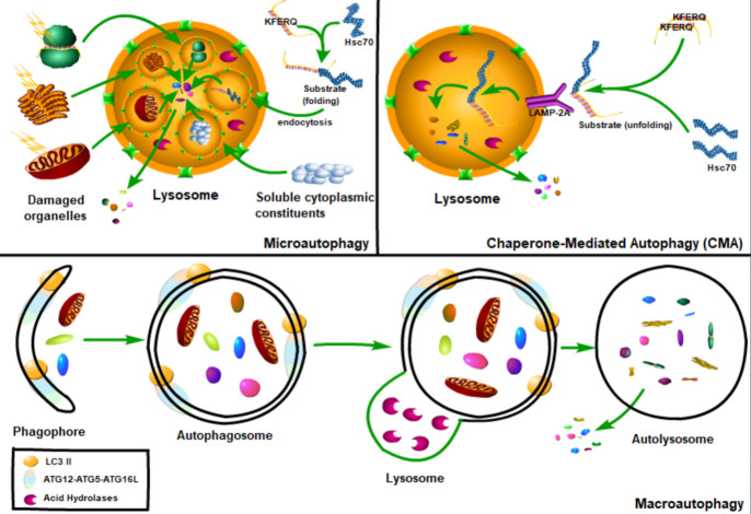
Renal fibrosis is a common process of almost all the chronic kidney diseases progressing to end-stage kidney disease. As a highly conserved lysosomal protein degradation pathway, autophagy is responsible for degrading protein aggregates, damaged organelles, or invading pathogens to maintain intracellular homeostasis. Growing evidence reveals that autophagy is involved in the progression of renal fibrosis, both in the tubulointerstitial compartment and in the glomeruli. Nevertheless, the specific role of autophagy in renal fibrosis has still not been fully understood. Therefore, in this review we will describe the characteristics of autophagy and summarize the recent advances in understanding the functions of autophagy in renal fibrosis. Moreover, the problem existing in this field and the possibility of autophagy as the potential therapeutic target for renal fibrosis have also been discussed.

Keywords: autophagy; cellular senescence; chronic kidney disease; renal fibrosis; senescence-associated secretory phenotype.


Conflict of interest statement

Competing interests All authors declare no conflict of interest.

Figures

Figure 1.
Figure 1.
The classification of autophagy. Autophagy can be divided into three types: macroautophagy, microautophagy and chaperone-mediated autophagy. Macroautophagy refers to the phagocytosis of large cytoplasmic materials by autophagosomes, and then fusion with lysosomes to degrade substrates. Microautophagy refers to the lysosome itself engulfing small components of the cytoplasm by invading the lysosome membrane. And the chaperone-mediated autophagy, the chaperone protein Hsc70 (heat shock cognate 70) and the accessory chaperone protein specifically recognize the cytoplasmic protein containing KFERQ-like pentapeptide, and then pass through the lysosomal-associated membrane glycoprotein 2A (LAMP2A) interaction, the unfolded protein is transported into the lysosome cavity through the multimeric translocation complex.
Figure 2.
Figure 2.
The formation of autophagy. Autophagy is a multi-step process involving initiation, nucleation, expansion, fusion and degradation. When starvation or treatment with rapamycin, ATG13 dephosphorylates and binds to ATG17 in a mTOR protein-dependent manner and activates ATG1 to induce autophagy. Then ATG1 and ATG13 interact with ATG17, ATG29, and ATG31 complexes to form a PAS scaffold complex, which is a prerequisite step for the assembly of ATG protein downstream of PAS. The ULK/ATG1 complex is recruited into the membrane structure independently of PI3P and its downstream ATG protein, and then it is stabilized in the membrane structure by PI3P. ATG9 vesicles, deriving from the Golgi apparatus, can provide lipids required for downstream protein assembly of PAS, recruit ULK/ATG1 complexes, initiate autophagy and serve as a source of autophagosome membranes. Class III PI3K complex I (PI3KC3-C1) is necessary for the nucleation of autophagosomes and is composed of Vps34/VPS34, Vps15/p150, Vps30/BECN1 and Atg14/ATG14L. During autophagy induction, PI3KC3-C1, which produces PI3P on PAS, is recruited into PAS. PI3P transmits the received signal to the downstream ATG proteins through ATG18/WIPI protein. Two ubiquitin-like binding systems, the Atg8/LC3 system and the ATG12-ATG5-ATG16L system regulate the expansion and completion of autophagosomes. when starvation, mTOR protein is inactivated and starts LC3II transcription. LC3 is transformed into LC3I under the processing of ATG4, and then binds to PE under the catalysis of E1-like enzyme ATG7 and E2-like enzyme ATG3 (activated by ATG12-ATG5-ATG16L) and participates in the expansion and completion of autophagosome. After the phagocytic vesicles are expanded and expanded to form autophagosomes, only after the outer membrane of the autophagosomes fuse with the lysosome and complete the degradation of the contents by lysosomal hydrolases. The degraded substrate is eventually released into the cytoplasm for reuse. E1, ubiquitin-activating enzyme; E2, ubiquitin-conjugating enzyme; E3, ubiquitin ligase; ER, endoplasmic reticulum; PE, phosphatidylethanolamine.
Figure 3.
Figure 3.
Nutrient dependent autophagy regulation. Nutrition-dependent autophagy regulation is mainly related to mammalian rapamycin target complex 1 (mTORC1), adenosine monophosphate-activated protein kinase (AMPK), and oxidized nicotinamide adenine dinucleotide-dependent histone deacetylase (Sirtuin1). mTOR is the major negative regulator of autophagy. mTOR signaling pathway was activated in nutrient abundance, while AMPK signaling pathway and Sirtuin1 signaling pathway were activated in nutrient deficiency. AMPK is phosphorylated and activated by several upstream kinases, including LKB1, calcium/calmodulin-dependent protein kinase (CAMKK) and mitogen-activated protein kinase 7 (TAK1). Once activated, on the one hand, AMPK can directly phosphorylate serine/threonine protein kinase ULK1 to promote autophagy and/or directly phosphorylate raptor (a scaffold protein used to recruit mTOR substrates) to mediate the binding of 14-3-3 (a cytosolic anchor protein) to inactivate mTOR, thereby indirectly activate autophagy. On the other hand, AMPK can phosphorylate TSC2 in the TSC1/TSC2 complex, disrupt the interaction between TSC1 and TSC2, thereby inhibiting mTORC1 and indirectly activating autophagy. Rheb is located downstream of the TSC1/TSC2 complex and upstream of mTOR and acts as an exciter of mTOR and is inhibited by the TSC1/TSC2 complex. However, in the PI3K/Akt signaling pathway activated by growth factors or cytokines, Akt acts to activate mTOR by inducing the phosphorylation of PRAS40 and mediating its binding to 14-3-3. Moreover, Akt can also inactive TSC2 or inhibit FoxO3 (a transcription factor that can positively regulate autophagy) to suppress autophagy. When starvation, Sirtuin1 and Sirtuin2 are activated due to increased NAD+. Sirtuin 1 can deacetylate forkhead box protein O1 (FoxO1) to promote autophagic flux and/or directly deacetylate several essential autophagy proteins such as ATG5, ATG7 and microtubule-associated protein 1 light chain 3 (LC3) to induce autophagy. In addition, FoxO1 is acetylated after separation from Sirtuin2, and the acetylated FoxO1 promotes autophagy by enhancing its interaction with ATG7.
Figure 4.
Figure 4.
Stress-induced regulation of autophagy. Depicted are the connections between autophagy and ER stress, hypoxia, and the ROS levels.

References

    1. Bikbov B, Purcell CA, Levey AS, Smith M, Abdoli A, Abebe M, et al.. (2020). Global, regional, and national burden of chronic kidney disease, 1990-2017: a systematic analysis for the Global Burden of Disease Study 2017. The Lancet, 395:709-733. - PMC - PubMed
    1. Keith DS, Nichols GA, Gullion CM, Brown JB, Smith DH (2004). Longitudinal follow-up and outcomes among a population with chronic kidney disease in a large managed care organization. Archives of internal medicine, 164:659-663. - PubMed
    1. Mizushima N, Levine B, Cuervo AM, Klionsky DJ (2008). Autophagy fights disease through cellular self-digestion. nature, 451:1069-1075. - PMC - PubMed
    1. Li W, He P, Huang Y, Li YF, Lu J, Li M, et al.. (2021). Selective autophagy of intracellular organelles: recent research advances. Theranostics, 11:222-256. - PMC - PubMed
    1. Mizushima N, Levine B (2020). Autophagy in Human Diseases. N Engl J Med, 383:1564-1576. - PubMed

LinkOut - more resources