

Stem Cell-Laden Hydrogel-Based 3D Bioprinting for Bone and Cartilage Tissue Engineering
- PMID: 35656197
- PMCID: PMC9152119
- DOI: 10.3389/fbioe.2022.865770
Stem Cell-Laden Hydrogel-Based 3D Bioprinting for Bone and Cartilage Tissue Engineering
Abstract
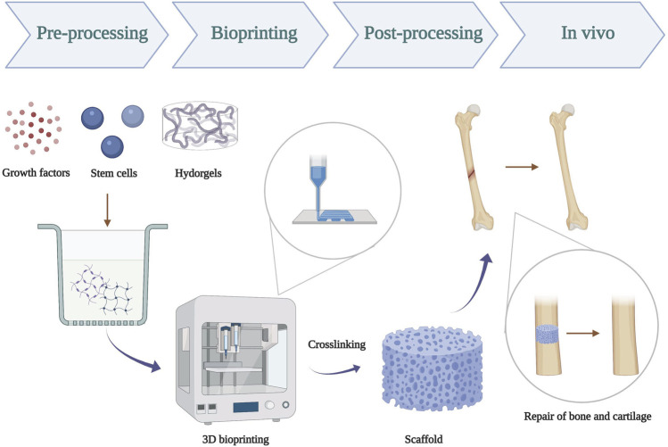
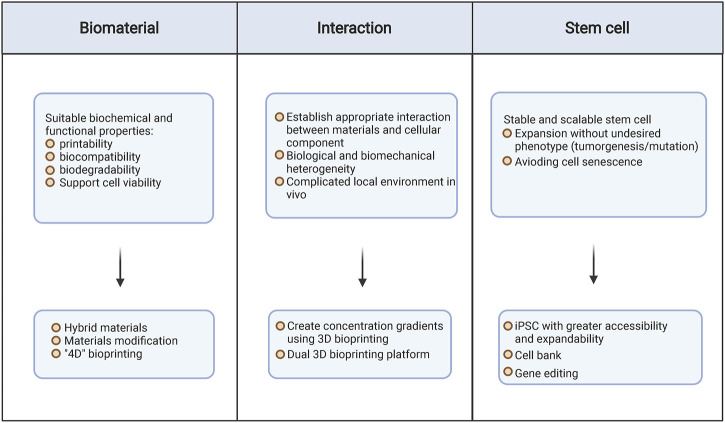
Tremendous advances in tissue engineering and regenerative medicine have revealed the potential of fabricating biomaterials to solve the dilemma of bone and articular defects by promoting osteochondral and cartilage regeneration. Three-dimensional (3D) bioprinting is an innovative fabrication technology to precisely distribute the cell-laden bioink for the construction of artificial tissues, demonstrating great prospect in bone and joint construction areas. With well controllable printability, biocompatibility, biodegradability, and mechanical properties, hydrogels have been emerging as an attractive 3D bioprinting material, which provides a favorable biomimetic microenvironment for cell adhesion, orientation, migration, proliferation, and differentiation. Stem cell-based therapy has been known as a promising approach in regenerative medicine; however, limitations arise from the uncontrollable proliferation, migration, and differentiation of the stem cells and fortunately could be improved after stem cells were encapsulated in the hydrogel. In this review, our focus was centered on the characterization and application of stem cell-laden hydrogel-based 3D bioprinting for bone and cartilage tissue engineering. We not only highlighted the effect of various kinds of hydrogels, stem cells, inorganic particles, and growth factors on chondrogenesis and osteogenesis but also outlined the relationship between biophysical properties like biocompatibility, biodegradability, osteoinductivity, and the regeneration of bone and cartilage. This study was invented to discuss the challenge we have been encountering, the recent progress we have achieved, and the future perspective we have proposed for in this field.
Keywords: 3D bioprinting; bone; cartilage; hydrogel; stem cell.
Copyright © 2022 Yang, Yi, Liu, Zhang, Mei, Feng, Tu and Li.
Conflict of interest statement
The authors declare that the research was conducted in the absence of any commercial or financial relationships that could be construed as a potential conflict of interest.
Figures
Similar articles
-
Cell-laden hydrogels for osteochondral and cartilage tissue engineering.Acta Biomater. 2017 Jul 15;57:1-25. doi: 10.1016/j.actbio.2017.01.036. Epub 2017 Jan 11. Acta Biomater. 2017. PMID: 28088667 Free PMC article. Review.
-
Bio-inspired hydrogel composed of hyaluronic acid and alginate as a potential bioink for 3D bioprinting of articular cartilage engineering constructs.Acta Biomater. 2020 Apr 1;106:114-123. doi: 10.1016/j.actbio.2020.01.046. Epub 2020 Feb 3. Acta Biomater. 2020. PMID: 32027992
-
Nanocomposite bioinks for 3D bioprinting.Acta Biomater. 2022 Oct 1;151:45-69. doi: 10.1016/j.actbio.2022.08.014. Epub 2022 Aug 13. Acta Biomater. 2022. PMID: 35970479 Review.
-
Generating adipose stem cell-laden hyaluronic acid-based scaffolds using 3D bioprinting via the double crosslinked strategy for chondrogenesis.Mater Sci Eng C Mater Biol Appl. 2021 May;124:112072. doi: 10.1016/j.msec.2021.112072. Epub 2021 Mar 26. Mater Sci Eng C Mater Biol Appl. 2021. PMID: 33947564
-
Extracellular Matrix/Amorphous Magnesium Phosphate Bioink for 3D Bioprinting of Craniomaxillofacial Bone Tissue.ACS Appl Mater Interfaces. 2020 May 27;12(21):23752-23763. doi: 10.1021/acsami.0c05311. Epub 2020 May 12. ACS Appl Mater Interfaces. 2020. PMID: 32352748 Free PMC article.
Cited by
-
Advances in the Treatment of Partial-Thickness Cartilage Defect.Int J Nanomedicine. 2022 Dec 13;17:6275-6287. doi: 10.2147/IJN.S382737. eCollection 2022. Int J Nanomedicine. 2022. PMID: 36536940 Free PMC article. Review.
-
Visualized trends and bibliometric analysis in ankle cartilage repair from 2004 to 2024.Front Med (Lausanne). 2024 Nov 20;11:1503707. doi: 10.3389/fmed.2024.1503707. eCollection 2024. Front Med (Lausanne). 2024. PMID: 39635584 Free PMC article.
-
Exploring the Potential of Artificial Intelligence for Hydrogel Development-A Short Review.Gels. 2023 Oct 25;9(11):845. doi: 10.3390/gels9110845. Gels. 2023. PMID: 37998936 Free PMC article. Review.
-
Emerging 3D bioprinting applications in plastic surgery.Biomater Res. 2023 Jan 3;27(1):1. doi: 10.1186/s40824-022-00338-7. Biomater Res. 2023. PMID: 36597149 Free PMC article. Review.
-
Enhancing 3D Printing of Gelatin/Siloxane-Based Cellular Scaffolds Using a Computational Model.Polymers (Basel). 2025 Jun 30;17(13):1838. doi: 10.3390/polym17131838. Polymers (Basel). 2025. PMID: 40647847 Free PMC article.
References
-
- Akiyama H., Chaboissier M.-C., Martin J. F., Schedl A., de Crombrugghe B. (2002). The Transcription Factor Sox9 Has Essential Roles in Successive Steps of the Chondrocyte Differentiation Pathway and Is Required for Expression of Sox5 and Sox6. Genes Dev. 16 (21), 2813–2828. 10.1101/gad.1017802 - DOI - PMC - PubMed
Publication types
LinkOut - more resources
Full Text Sources