Review
. 2022 Apr 7;3(5):519-538.
doi: 10.1039/d2cb00019a. eCollection 2022 May 11.

The chemistry and biology of natural ribomimetics and related compounds

Affiliations
Review

The chemistry and biology of natural ribomimetics and related compounds

Takeshi Tsunoda et al. RSC Chem Biol. .

Abstract

Natural ribomimetics represent an important group of specialized metabolites with significant biological activities. Many of the activities, e.g., inhibition of seryl-tRNA synthetases, glycosidases, or ribosomes, are manifestations of their structural resemblance to ribose or related sugars, which play roles in the structural, physiological, and/or reproductive functions of living organisms. Recent studies on the biosynthesis and biological activities of some natural ribomimetics have expanded our understanding on how they are made in nature and why they have great potential as pharmaceutically relevant products. This review article highlights the discovery, biological activities, biosynthesis, and development of this intriguing class of natural products.


Conflict of interest statement

There are no conflicts to declare.

Figures

Fig. 1
Fig. 1. Chemical structures of six-membered ring pseudosugars.
Fig. 2
Fig. 2. Chemical structures of clinically used ribomimetics.
Fig. 3
Fig. 3. Chemical structures of natural ribomimetics.
Fig. 4
Fig. 4. Chemical structures of natural S-ribomimetics.
Fig. 5
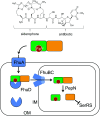
Fig. 5. Albomycin enters a bacterial cell using the bacterial iron uptake system. The siderophore unit is important for the compound to get into bacterial cells by utilizing the bacterial iron uptake system. First, the ferric siderophore passes the outer membrane through the ferrichrome outer membrane transporter FhuA. In the periplasm, the ferrichrome binds FhuD, which is then transported into the cytoplasm through the membrane channel FhuBC. In the cytoplasm, the amide bond is hydrolysed by the endogenous peptidase PepN releasing the active thioheptose nucleoside unit, which inhibits seryl-tRNA synthetase (SerRS). OM, outer membrane; IM, inner membrane.
Fig. 6
Fig. 6. Biosynthetic gene clusters of the albomycins and a putative albomycin analogue.
Fig. 7
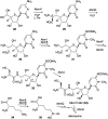
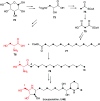
Fig. 7. Biosynthetic pathway to albomycins.
Fig. 8
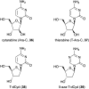
Fig. 8. Chemical structures of cytarabine and its thiofuranose analogues.
Fig. 9
Fig. 9. Chemical structures of plant-derived N-ribomimetics.
Fig. 10
Fig. 10. Chemical structures of DAB-1 analogues.
Fig. 11
Fig. 11. Crystal structures of glycosidases and DAB-1. (A) binding model of sucrose (Suc) with BmSUH (left; PDB code, 6LGF) and DAB-1 (DAB) with BmSUH (right; PDB code, 6LGD); (B) binding of arabinose (Ara) with α-l-arabinofuranosidase (left; PDB code, 6zpy) and DAB-1 with α-l-arabinofuranosidase (right; PDB code, 6zq1).
Fig. 12
Fig. 12. Chemical structures of DMDP and related compounds.
Fig. 13
Fig. 13. Chemical structures of fungal-derived N-ribomimetics.
Fig. 14
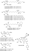
Fig. 14. Chemical structures of bacterial N-ribomimetics.
Fig. 15
Fig. 15. Biosynthetic origin of broussonetine J.
Fig. 16
Fig. 16. Biosynthetic origin of pramanicin.
Fig. 17
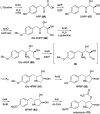
Fig. 17. Proposed biosynthetic pathway to DAB-1.
Fig. 18
Fig. 18. Biosynthesis of anisomycin.
Fig. 19
Fig. 19. Chemical structures of forodesine and galidesivir.
Fig. 20
Fig. 20. Chemical structures of C-ribomimetics.
Fig. 21
Fig. 21. Structures of the mammalian SAHH complexed with NAD+ and adenosine (Ado) (left; PDB code, 5AXA) and with NADH and 3KA (right; PDB code, 5AXC).
Fig. 22
Fig. 22. Structure of the T. thermophilus ribosome with bound pactamycin (PDB code, 1HNX). The red spheres show pactamycin.
Fig. 23
Fig. 23. Structures of allosamidin, trehazolin and their ribomimetic moieties.
Fig. 24
Fig. 24. Structures of caryose and calditol found in lipopolysaccharides or archaeal lipids.
Fig. 25
Fig. 25. Formation of the C-ribomimetics of epoxyqueuosine and queuosine in modified tRNA (oQ-tRNA and Q-tRNA, respectively).
Fig. 26
Fig. 26. Proposed calditol formation from a hexose via a radical mechanism.
Fig. 27
Fig. 27. Proposed formation of the C-ribomimetics of BHT cyclitol ether and pactamycin. ACP, acyl carrier protein.
Fig. 28
Fig. 28. Proposed tailoring steps in pactamycin biosynthesis. KS, ketosynthase; AT, acyltransferase; DH, dehydratase; KR, ketoreductase; ACP, acyl carrier protein.
Fig. 29
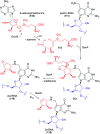
Fig. 29. Biosynthesis of neplanocin A and aristeromycin.
Fig. 30
Fig. 30. Catalytic mechanism of Ari2.
Fig. 31
Fig. 31. Structure–activity relationships of pactamycin.
None
Takeshi Tsunoda
None
Samuel Tanoeyadi
None
Philip J. Proteau
None
Taifo Mahmud

References

    1. Flatt P. M. Mahmud T. Nat. Prod. Rep. 2007;24:358–392. doi: 10.1039/B603816F. - DOI - PubMed
    1. Kudo F. Eguchi T. Chem. Rec. 2016;16:4–18. doi: 10.1002/tcr.201500210. - DOI - PubMed
    1. Scott L. J. Spencer C. M. Drugs. 2000;59:521–549. doi: 10.2165/00003495-200059030-00012. - DOI - PubMed
    1. Mahmud T. Nat. Prod. Rep. 2003;20:137–166. doi: 10.1039/B205561A. - DOI - PubMed
    1. McCormack P. L. Goa K. L. Drugs. 2003;63:2427–2434. doi: 10.2165/00003495-200363220-00006. - DOI - PubMed

LinkOut - more resources