Review
. 2022 May 17:10:901652.
doi: 10.3389/fcell.2022.901652. eCollection 2022.

Bioelectric Potential in Next-Generation Organoids: Electrical Stimulation to Enhance 3D Structures of the Central Nervous System

Affiliations
Review

Bioelectric Potential in Next-Generation Organoids: Electrical Stimulation to Enhance 3D Structures of the Central Nervous System

Michelle O'Hara-Wright et al. Front Cell Dev Biol. .

Abstract

Pluripotent stem cell-derived organoid models of the central nervous system represent one of the most exciting areas in in vitro tissue engineering. Classically, organoids of the brain, retina and spinal cord have been generated via recapitulation of in vivo developmental cues, including biochemical and biomechanical. However, a lesser studied cue, bioelectricity, has been shown to regulate central nervous system development and function. In particular, electrical stimulation of neural cells has generated some important phenotypes relating to development and differentiation. Emerging techniques in bioengineering and biomaterials utilise electrical stimulation using conductive polymers. However, state-of-the-art pluripotent stem cell technology has not yet merged with this exciting area of bioelectricity. Here, we discuss recent findings in the field of bioelectricity relating to the central nervous system, possible mechanisms, and how electrical stimulation may be utilised as a novel technique to engineer "next-generation" organoids.

Keywords: CNS; bioelectricity; brain; electrical stimulation; organoids model; pluripotenct stem cells; retina.


Conflict of interest statement

The authors declare that the research was conducted in the absence of any commercial or financial relationships that could be construed as a potential conflict of interest.

Figures

FIGURE 1
FIGURE 1
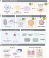
Generation of pluripotent stem cell-derived organoid models of the central nervous system (CNS). (A) Human pluripotent stem cells (hPSCs) can be isolated from the inner cell mass of an embryo- embryonic stem cells (ESCs). Alternatively, patient-dervied somatic cells can be reprogrammed to induced pluripotent stem cells (iPSCs). (B) Pluripotent stem cells can be differentiated into 3D aggregates which form embryoid bodies and neuroepithelial vesicles. Biomechanical and biochemical differentiation cues have been implemented to generate 3D organoid structures, but bioelectric cues of differentiation remain unexplored. (C) In unguided CNS organoid protocols, differentiation relies on the intrinsic self-organisation properties of PSCs, to generate cerebral brain organoids or retinal organoids. Meanwhile, guided protocols employ exogenous growth factors to generate brain-region specific organoids, spinal cord organoids, or retinal organoids.
FIGURE 2
FIGURE 2
Endogenous electrical fields and the transepithelial battery. (A) In vivo, all cells exhibit a resting membrane potential due to the segregation of ionic charges. Ion transport channels are asymmetrically distributed across apical and basal membranes. Ion flux occurs transcellularly and ions are selectively transported across the plasma membrane, whilst paracellular transport (between cells) must work against the resistance of tight junctions, adherens junctions and desmosomes. (B) The segregation of ionic charges creates a difference in membrane potential (Vmem) at each apical-basal membrane. All of the segregated chargers in epithelia amount to a transepithelial potential difference (VTEP). (C) When the epithelium is breached, the VTEP drops and ionic current flows out from the site of wound. Thus, the epithelial works as a battery to mediate wound repair.
FIGURE 3
FIGURE 3
Electrical stimulation of cells mediates cell-cycle related phenotypes. Electrical stimulation of CNS cells both in vivo and in vitro has been found to regulate the cell cycle at various phases. This may lead to an increase in cell survival, migration, proliferation, or alternatively exiting the cell cycle to increase cell differentiation and maturation.
FIGURE 4
FIGURE 4
Electrical stimulation of CNS cells: moving towards PSC-derived organoids. (A) Multipotent neural stem cells can be isolated from the primary brain and differentiated to neural progenitor cells and neurons. Alternatively, pluripotent stem cells can be differentiated into a neural lineage. Electrical stimulation studies have been undertaken on these two-dimensional systems. (B) Three-dimensional systems which have been employed to study electrical stimulation in vitro include primary brain slices (an ex vivo model of the CNS), and neurosphere aggregates derived from neural progenitor cells. However, the study of electrical stimulation on pluripotent stem cell-derived brain organoids remains unexplored. (C) In vivo, electrical stimulation of the brain is performed on patients either using invasive methods, whereby electrodes directly contact brain regions, or non-invasively. To mimic in vivo brain stimulation in in vitro models of the brain, conductive polymers within 3D hydrogels have been utilised. (D) However, to best apply electrical stimulation to PSC-derived CNS organoids, suitable delivery platforms must be established. A single well system allows controlled delivery of electrical stimulation, whilst a multi-well plate system is higher-throughput to allow in situ electrical stimulation of cells. Electrodes can be configured in arrays, or pairs, generating electrical fields between each cathode (+) and anode (−). (✔), electrical stimulation experimentation performed; (?), not yet explored.
FIGURE 5
FIGURE 5
Electrical stimulation to improve PSC-derived CNS organoids. (A) In current standard culture conditions, PSC-derived organoid systems, for example neural organoids, have several problems or shortcomings. Organoids may be heterogeneous and exhibit inter-differentiation or inter-cell line variability. Neural stem cells may have a low differentiation efficiency, and neural progenitor cells may have a variable growth rate. With increased time in culture, neural networks begin to form but neurite outgrowth and branching may be minimal, limiting functionality. As organoids continue to grow, oxygen transport to the centre of the organoid is limited, leading to the formation of a necrotic core. (B) Electrical stimulation of neural organoids may enhance phenotypes relating to the current problems in organoid cultures. Functioning as an additional physiological cue, electrical field modulation may reduce heterogeneity in differentiation and enhance neurogenesis. Neural stem cells and progenitor cells may have an increased capacity for differentiation and proliferation. Electrical fields may also promote neurite outgrowth and branching, creating more mature neural networks with more potential for functionality. Electrical fields may also prevent apoptosis and enhance cell survival, decreasing the formation of necrotic core in older organoids. (C) Many potential molecular mechanisms are implicated to respond to electrical field modulation, including transient oxidative or ionic signals, and canonical signaling networks capable of regulating a wide-range of cell cycle-related and metabolic processes. Electrical stimulation-induced signals may therefore act throughout organoid development to induce phenotypes that could improve organoid development, differentiation and maturation. BDNF, Brain derived neurotrophic factor; GDNF, glial cell line–derived neurotrophic factor; PI3K, phosphoinositide 3-kinase; ROS, reactive oxygen species.

Similar articles

Cited by

References

    1. Abasi S., Aggas J. R., Venkatesh N., Vallavanatt I. G., Guiseppi-Elie A. (2020). Design, Fabrication and Testing of an Electrical Cell Stimulation and Recording Apparatus (ECSARA) for Cells in Electroculture. Biosens. Bioelectron. 147, 111793. 10.1016/j.bios.2019.111793 - DOI - PubMed
    1. Achberger K., Probst C., Haderspeck J., Bolz S., Rogal J., Chuchuy J., et al. (2019). Merging Organoid and Organ-On-A-Chip Technology to Generate Complex Multi-Layer Tissue Models in a Human Retina-On-A-Chip Platform. eLife 8. 10.7554/eLife.46188 - DOI - PMC - PubMed
    1. Al-Sakere B., André F., Bernat C., Connault E., Opolon P., Davalos R. V., et al. (2007). Tumor Ablation with Irreversible Electroporation. PLOS ONE 2, e1135. 10.1371/journal.pone.0001135 - DOI - PMC - PubMed
    1. Allen S. J., Watson J. J., Shoemark D. K., Barua N. U., Patel N. K. (2013). GDNF, NGF and BDNF as Therapeutic Options for Neurodegeneration. Pharmacol. Ther. 138, 155–175. 10.1016/j.pharmthera.2013.01.004 - DOI - PubMed
    1. Ao Z., Cai H., Wu Z., Krzesniak J., Tian C., Lai Y. Y., et al. (2022). Human Spinal Organoid-On-A-Chip to Model Nociceptive Circuitry for Pain Therapeutics Discovery. Anal. Chem. 94, 1365–1372. 10.1021/acs.analchem.1c04641 - DOI - PMC - PubMed

LinkOut - more resources