. 2022 Jun 24;50(11):6398-6413.
doi: 10.1093/nar/gkac461.

Noncanonical prokaryotic X family DNA polymerases lack polymerase activity and act as exonucleases

Affiliations

Noncanonical prokaryotic X family DNA polymerases lack polymerase activity and act as exonucleases

Maria Prostova et al. Nucleic Acids Res. .

Abstract

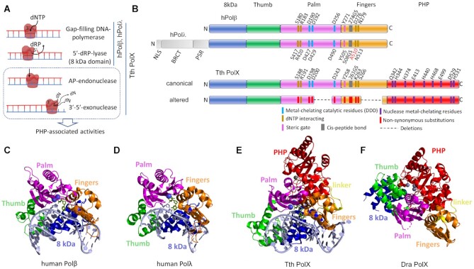
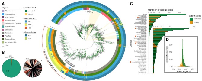
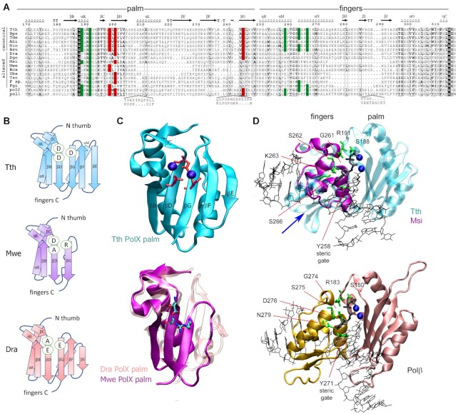
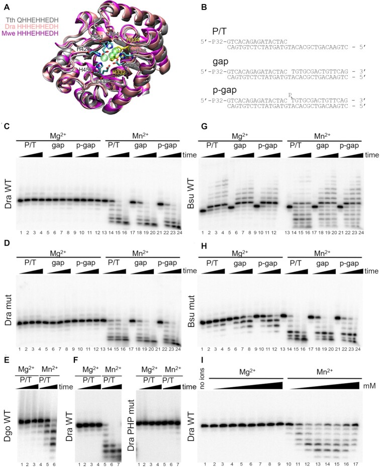
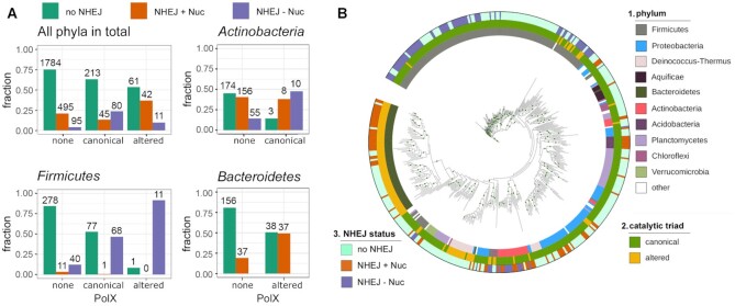
The X family polymerases (PolXs) are specialized DNA polymerases that are found in all domains of life. While the main representatives of eukaryotic PolXs, which have dedicated functions in DNA repair, were studied in much detail, the functions and diversity of prokaryotic PolXs have remained largely unexplored. Here, by combining a comprehensive bioinformatic analysis of prokaryotic PolXs and biochemical experiments involving selected recombinant enzymes, we reveal a previously unrecognized group of PolXs that seem to be lacking DNA polymerase activity. The noncanonical PolXs contain substitutions of the key catalytic residues and deletions in their polymerase and dNTP binding sites in the palm and fingers domains, but contain functional nuclease domains, similar to canonical PolXs. We demonstrate that representative noncanonical PolXs from the Deinococcus genus are indeed inactive as DNA polymerases but are highly efficient as 3'-5' exonucleases. We show that both canonical and noncanonical PolXs are often encoded together with the components of the non-homologous end joining pathway and may therefore participate in double-strand break repair, suggesting an evolutionary conservation of this PolX function. This is a remarkable example of polymerases that have lost their main polymerase activity, but retain accessory functions in DNA processing and repair.


Figures

Figure 1.
Figure 1.
The structure and functions of prokaryotic and human PolXs. (A) Activities of human Polβ, Polλ and T. thermophilus (Tth) PolX. (B) The overall domain structure of human Polβ, Polλ, canonical prokaryotic PolXs (illustrated for Tth PolX) and altered prokaryotic PolXs. The N-terminal lyase domain is shown in blue, the thumb domain is green, the palm domain is pink, the fingers domain is orange, the PHP domain is red. The N-terminal part of Polλ, including the nuclear localization signal (NLS), the BRCA1 C-terminal (BRCT) and proline-serine rich (PSR) domains, is shown in gray. The active site residues of the palm and PHP domains are marked in blue and violet in canonical PolXs (the numbering is for T. thermophilus PolX) and in red in altered PolXs. (C) Structure of human Polβ in complex with gapped DNA (PDB: 1BPX). (D) Structure of the catalytic core of human Polλ in complex with gapped DNA (PDB: 1XSN). (E) Structure ofT. thermophilus PolX in complex with gapped DNA (PDB: 3AU0). (F) Structure of D. radiodurans (Dra) PolX (PDB: 2W9M). PolXs are roughly aligned by their palm domains. The active site magnesium in the palm domain is shown with blue spheres; metal ions bound in the exonuclease PHP domain are shown in gray.
Figure 2.
Figure 2.
The diversity of prokaryotic PolXs. (A) The phylogenetic tree of PolX proteins based on the multiple alignment 1433 non-redundant PolX sequences. Positions of archaeal PolXs on the tree are marked by light ochre sectors. The features of PolX proteins are annotated as follows (from the inner to the outer rings): 1, phylum; 2, the status of the catalytic triad in the palm domain (canonical, green; altered, ochre); 3, the size of the palm domain (blue, ≥80 residues; orange, 65–80 residues; red, <65 residues); 4, the size of the fingers domain (dark blue, ≥35 residues; green, <35 residues). The main cluster of noncanonical PolXs is indicated. The green dots on the nodes correspond to bootstrap values of 98-100. (B) Distribution of catalytic triad residues in canonical PolXs (left) and altered PolXs (right, three most abundant variants are indicated; see Supplementary Table S1 for altered triad frequencies). (C) Distribution of canonical and altered PolX sequences among prokaryotic classes (archaeal classes are indicated with red circles). (D) Distribution of the lengths of canonical and altered PolXs.
Figure 3.
Figure 3.
The catalytic site of canonical and altered PolXs. (A) Alignment of the sequences of the palm and fingers domains in PolXs. The catalytic triad residues (aspartate or glutamate) are shown in red, the conserved dNTP binding residues are green; similar residues (similarity score >0.7 in the non-redundant collection of PolXs) are shown in bold, absolutely conserved residues are shown in black. The abbreviations of the species names are as follows: Tth, Thermus thermophilus; Bps, Burkholderia pseudomallei; Tde, Thiohalomonas denitrificans; Ahi, Actinomadura hibisca; Nin, Nonomuraea indica; Bsu, Bacillus subtilis; Dra – Deinococcus radiodurans; Dgo – Deinococcus gobiensis; Msi – Meiothermus silvanus; Mwe - Mesorhizobium wenxiniae, Bca - Bradyrhizobium canariense, Sba - Sphingobacteriaceae bacterium, Tso - Taibaiella soli, F. sp. - Flavisolibacter sp. X7X, polβ and polλ - human Polβ and Polλ. The protein secondary structure is shown above the alignment for T. thermophilus PolX. Amino acid numbering for T. thermophilus PolX is shown above and below the alignment. Sequence alignments of Dra and Dgo PolXs were manually curated according to the Dra PolX structure. (B) Schematic representation of the palm domain topology for T. thermophilus (top), M. wenxiniae (middle) and D. radiodurans (bottom) PolXs. The catalytic triad residues are indicated in ovals. (С) (Top) The structure of the palm domain of T. thermophilus PolX (PDB: 3AUO). The catalytic triad residues are shown in red as stick models, Mg2+ cations are shown as blue spheres. (Bottom) Superimposition of the structure of the palm domain of D. radiodurans PolX (pink, PDB: 2W9M) and a modeled structure of the palm domain of M. wenxiniae PolX (magenta). (D) Structures of the fingers domain in prokaryotic PolXs and human Polβ. (Top) Superimposition of the structures of T. thermophilus PolX (fingers and palm domains, turquoise) in complex with gapped DNA (PDB: 3AU0) and of the fingers domain of M. silvanus PolX (violet), based on structural modeling; the position of the deleted region is indicated with a blue arrow. (Bottom) Structure of human Polβ with gapped DNA (PDB: 1BPY). Mg2+ ions bound in the active site of the palm domain are shown as blue spheres, the incoming dNTP is black. Positions of functionally important residues (green) in the fingers domain are indicated.
Figure 4.
Figure 4.
Analysis of the catalytic activities of noncanonical PolXs. (A) Structure of the PHP domain of bacterial PolXs. Superposition of the structures of PHP domains of T. thermophilus PolX (gray, PDB: 3AUO), D. radiodurans PolX (pink, PDB: 2W9M) and a modeled structure of M. wenxiniae PolX (magenta) is shown. Zn2+ ions from the crystal structure of D. radiodurans PolX are shown as semitransparent green spheres. The active site residues are shown as stick models, the residues mutated in this study in D. radiodurans PolX are shown in yellow. The C-terminal α-helix is not shown for clarity. (B) Schemes of the DNA substrates: primer-template (P/T) and gapped substrates with 5′-OH or 5′-phosphorylated downstream oligonucleotide (gap and p-gap, respectively) used for analysis of PolX activities. (C, D) Analysis of the activities of D. radiodurans PolX in the presence of 200 μM dNTP substrates and 11 mM Mg2+ or 2 mM Mn2+ cations. The reactions were performed with 20 nM of wild-type PolX from D. radiodurans (C) or its palm domain mutant with substitutions in the polymerase active site (D) at 30°C for 0, 10, 30, 90 min. (E) Analysis of the activity of wild-type PolX from D. gobiensis on the P/T substrate in the same reaction conditions. (F) Comparison of the activities of wild-type D. radiodurans PolX and its PHP mutant with substitutions in the exonuclease site in the same reaction conditions. (G,H) Analysis of the activities of wild-type PolX from B. subtilis (G) and its mutant with a single substitution in the catalytic triad (H) in the same reaction conditions. (I) Determination of optimal divalent metal ion concentration for wild-type PolX from D. radiodurans. The concentrations of Mg2+ and Mn2+ were varied between 0.25 and 50 mM in the presence of 200 μM dNTPs. For all experiments, representative gels from two-three independent replicates are shown.
Figure 5.
Figure 5.
Co-occurrence of PolXs with components of the NHEJ pathway in bacterial genomes. (A) Distribution of the NHEJ genes in the non-redundant sample of 2826 complete bacterial genomes either lacking PolX, or containing canonical or altered PolXs, shown for all bacterial phyla or individually for Firmicutes, Actinobacteria and Bacteroidetes. For Actinobacteria, only canonical PolXs are shown due to the very small number of genomes with altered PolX in this phylum (3 genomes in the non-redundant sample used for analysis). For Bacteroidetes, only altered PolXs are shown due to the absence of genomes with canonical PolX in the non-redundant sample. The proportion of genome variants with each gene combination is shown on the ordinate axis. The numbers of genomes in each group are indicated. (B) Co-occurrence of PolXs with NHEJ genes in the non-redundant set of bacterial genomes, shown on a phylogenetic tree generated for the 452 PolX sequences found in these genomes (note that the tree topology is different from Figure 2A due to the much smaller number of PolX sequences used for analysis; this tree is used to illustrate solely the diversity of combinations of PolX and NHEJ in different phyla, and not the evolution of PolXs). The rings are annotated as follows: 1, phylum (the color code corresponds to Figure 2A); 2, catalytic triad status (green, canonical; ochre, altered); 3, NHEJ status (ligh green, no NHEJ; orange, NHEJ+Nuc; violet, NHEJ-Nuc). The green dots on the nodes correspond to bootstrap values of 98–100.

Similar articles

Cited by

References

    1. Raia P., Delarue M., Sauguet L.. An updated structural classification of replicative DNA polymerases. Biochem. Soc. Trans. 2019; 47:239–249. - PubMed
    1. Yang W., Gao Y.. Translesion and repair DNA polymerases: diverse structure and mechanism. Annu. Rev. Biochem. 2018; 87:239–261. - PMC - PubMed
    1. Hoitsma N.M., Whitaker A.M., Schaich M.A., Smith M.R., Fairlamb M.S., Freudenthal B.D.. Structure and function relationships in mammalian DNA polymerases. Cell. Mol. Life Sci. 2020; 77:35–59. - PMC - PubMed
    1. Jain R., Aggarwal A.K., Rechkoblit O.. Eukaryotic DNA polymerases. Curr. Opin. Struct. Biol. 2018; 53:77–87. - PubMed
    1. Baril E.F., Brown O.E., Jenkins M.D., Laszlo J.. Deoxyribonucleic acid polymerase with rat liver ribosomes and smooth membranes. Purification and properties of the enzymes. Biochemistry. 1971; 10:1981–1992. - PubMed

Publication types