Review
. 2022 Jun 3;79(6):337.
doi: 10.1007/s00018-022-04363-6.

Transcription factor networks in trophoblast development

Affiliations
Review

Transcription factor networks in trophoblast development

Henrieta Papuchova et al. Cell Mol Life Sci. .

Abstract

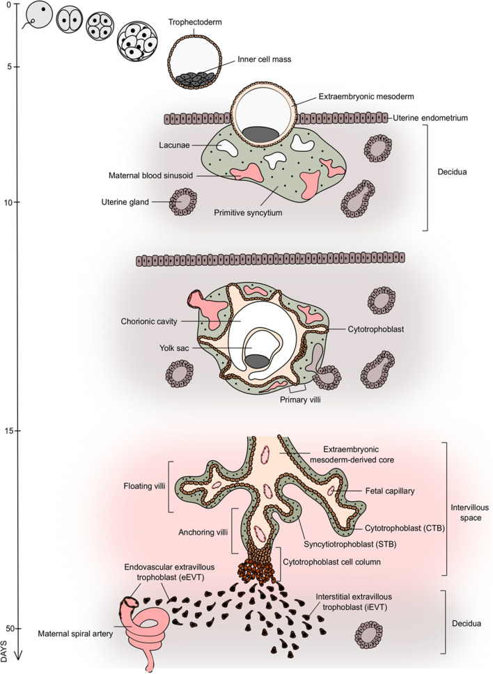
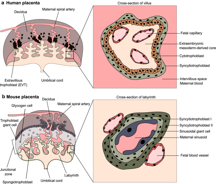
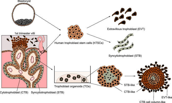
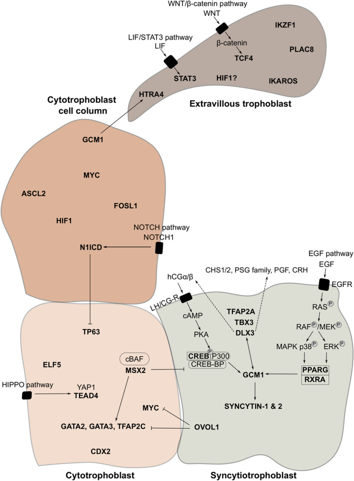
The placenta sustains embryonic development and is critical for a successful pregnancy outcome. It provides the site of exchange between the mother and the embryo, has immunological functions and is a vital endocrine organ. To perform these diverse roles, the placenta comprises highly specialized trophoblast cell types, including syncytiotrophoblast and extravillous trophoblast. The coordinated actions of transcription factors (TFs) regulate their emergence during development, subsequent specialization, and identity. These TFs integrate diverse signaling cues, form TF networks, associate with chromatin remodeling and modifying factors, and collectively determine the cell type-specific characteristics. Here, we summarize the general properties of TFs, provide an overview of TFs involved in the development and function of the human trophoblast, and address similarities and differences to their murine orthologs. In addition, we discuss how the recent establishment of human in vitro models combined with -omics approaches propel our knowledge and transform the human trophoblast field.

Keywords: Human placenta; Human trophoblast stem cells; Transcription factors; Trophoblast.


Conflict of interest statement

The authors declare no conflict of interest.

Figures

Fig. 1
Fig. 1
The development of the human placenta. Initiated by fertilization, the zygote undergoes multiple divisions and gives rise to a blastocyst 4–5 days post-fertilization (dpf). The blastocyst consists of the inner cell mass, which gives rise to the embryo proper and the trophectoderm, which gives rise to the trophoblast of the placenta. Upon implantation of the blastocyst into the uterine endometrium, the establishment of the multi-nucleated primitive syncytium (PS) and the cytotrophoblast (CTB) monolayer begins. Around day 9 dpf, lacunae form within the PS, fuse subsequently with uterine capillaries, and establish maternal sinusoids filled with maternal blood by day 13 dpf. Simultaneously, the CTB expands through the PS, extending towards the maternal decidua and forming primary villi. By the end of the third trimester, the main placental structure, the villous tree, is fully established. The villous tree consists of the extraembryonic mesoderm-derived core, fetal capillaries, the CTB monolayer and the multinucleated syncytiotrophoblast (STB) layer, arising from CTB fusion. The floating villi of the villous tree are located in the intervillous space filled with maternal blood, while the anchoring villi extend towards the decidua. At the tip of the anchoring villi, CTB forms a cytotrophoblast cell column, comprising the proliferative progenitor population of invasive extravillous trophoblast (EVT). The EVT can be divided into two subtypes; the endovascular EVT (eEVT), which remodels the maternal spiral arteries and the interstitial EVT (iEVT), which invades the decidua
Fig. 2
Fig. 2
Comparison of the human and murine placenta. a The main unit of the human placenta is a villous tree, comprising the extraembryonic mesoderm-derived core (ExM-dC), containing fetal capillaries. The ExM-dC is covered by the cytotrophoblast (CTB) monolayer and the multinucleated syncytiotrophoblast (STB) layer that is in contact with maternal blood. The inset shows a cross-section through the villi. At the tip of the villi, the cytotrophoblast cell column forms. It gives rise to the interstitial extravillous trophoblast (iEVT), invading the decidua and the endovascular extravillous trophoblast (eEVT), invading the maternal spiral arteries. b The murine placenta can be divided into the junctional zone and the labyrinth zone. The junctional zone consists of the spongiotrophoblasts and the glycogen cells. The trophoblast giant cells invade the maternal decidua and remodel maternal arteries and thus are considered to correspond to human EVT. In the labyrinth zone, the fetal blood is separated from the maternal blood sinusoids by fetal endothelial cells, two layers of STB (STB-I and STB-II) and sinusoidal giant cells. The labyrinth functionally relates to human villi
Fig. 3
Fig. 3
Human trophoblast stem cells and trophoblast organoids. The human trophoblast stem cells (hTSC) can be derived from both the blastocyst and the cytotrophoblast (CTB) of the first-trimester placenta. The hTSCs represent the proliferative CTB population and can be differentiated into extravillous trophoblast (EVT) and syncytiotrophoblast (STB) upon defined culture conditions. Moreover, trophoblast organoids (TOs) can be established from the first-trimester CTB. TOs have an STB-like core and CTB-like shell and thus provide an inside-out three-dimensional model. TOs can be further differentiated to form the cytotrophoblast cell column (CCC)-like and EVT-like populations
Fig. 4
Fig. 4
Overview of TFs operating in different types of human trophoblast. TFs are shown in bold. Arrows indicate interactions between TFs, signaling ligands and receptors. Dashed arrows indicate secreted components. The bar line indicates an inhibitory relationship between TFs

References

    1. Boyd JD, Hamilton WJ. The human placenta. Cambridge (England) Heffer. London: Palgrave Macmillan; 1970.
    1. Hertig AT, Rock J, Adams EC. A description of 34 human ova within the first 17 days of development. Am J Anat. 1956;98:435–493. doi: 10.1002/aja.1000980306. - DOI - PubMed
    1. Pijnenborg R, Dixon G, Robertson WB, Brosens I. Trophoblastic invasion of human decidua from 8 to 18 weeks of pregnancy. Placenta. 1980;1:3–19. doi: 10.1016/s0143-4004(80)80012-9. - DOI - PubMed
    1. Simmons DG, Fortier AL, Cross JC. Diverse subtypes and developmental origins of trophoblast giant cells in the mouse placenta. Dev Biol. 2007;304:567–578. doi: 10.1016/j.ydbio.2007.01.009. - DOI - PubMed
    1. Hemberger M, Hanna CW, Dean W. Mechanisms of early placental development in mouse and humans. Nat Rev Genet. 2020;21:27–43. doi: 10.1038/s41576-019-0169-4. - DOI - PubMed

Substances

LinkOut - more resources