Review
. 2022 Aug 1;323(2):H257-H275.
doi: 10.1152/ajpheart.00059.2022. Epub 2022 Jun 3.

Myocardial mesostructure and mesofunction

Affiliations
Review

Myocardial mesostructure and mesofunction

Alexander J Wilson et al. Am J Physiol Heart Circ Physiol. .

Abstract

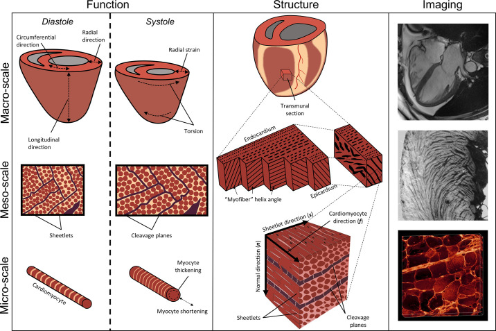
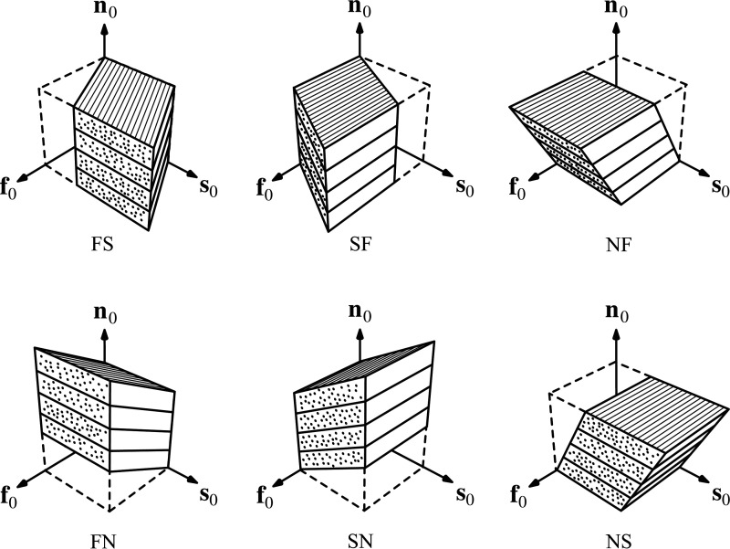
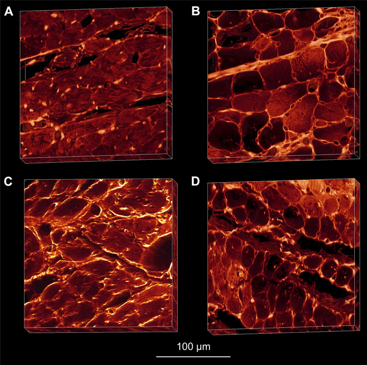
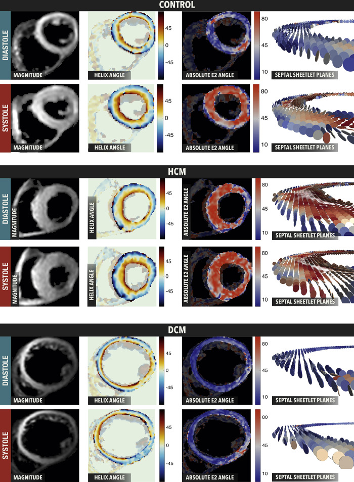
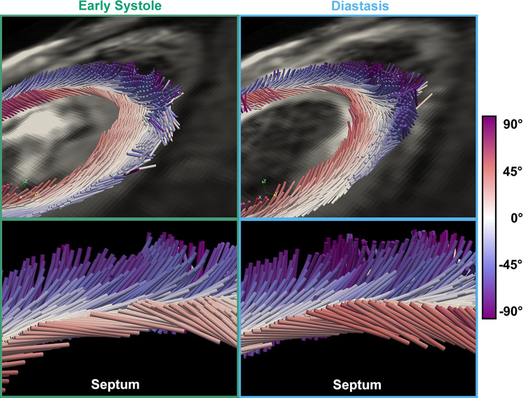
The complex and highly organized structural arrangement of some five billion cardiomyocytes directs the coordinated electrical activity and mechanical contraction of the human heart. The characteristic transmural change in cardiomyocyte orientation underlies base-to-apex shortening, circumferential shortening, and left ventricular torsion during contraction. Individual cardiomyocytes shorten ∼15% and increase in diameter ∼8%. Remarkably, however, the left ventricular wall thickens by up to 30-40%. To accommodate this, the myocardium must undergo significant structural rearrangement during contraction. At the mesoscale, collections of cardiomyocytes are organized into sheetlets, and sheetlet shear is the fundamental mechanism of rearrangement that produces wall thickening. Herein, we review the histological and physiological studies of myocardial mesostructure that have established the sheetlet shear model of wall thickening. Recent developments in tissue clearing techniques allow for imaging of whole hearts at the cellular scale, whereas magnetic resonance imaging (MRI) and computed tomography (CT) can image the myocardium at the mesoscale (100 µm to 1 mm) to resolve cardiomyocyte orientation and organization. Through histology, cardiac diffusion tensor imaging (DTI), and other modalities, mesostructural sheetlets have been confirmed in both animal and human hearts. Recent in vivo cardiac DTI methods have measured reorientation of sheetlets during the cardiac cycle. We also examine the role of pathological cardiac remodeling on sheetlet organization and reorientation, and the impact this has on ventricular function and dysfunction. We also review the unresolved mesostructural questions and challenges that may direct future work in the field.

Keywords: cardiac anatomy; diffusion tensor imaging; mechanics; mesostructured; sheetlets.


Conflict of interest statement

No conflicts of interest, financial or otherwise, are declared by the authors.

Figures

Figure 1.
Figure 1.
The multiscale structure and function of the cardiac ventricles. Left column: mechanical function of the whole heart (top) includes ejection fraction, torsion, radial, circumferential, and longitudinal strains. At the mesoscale (middle), mesofunction (middle left) includes transmural shear because of helix angle slope, helix angle reorientation during contraction, and sliding of sheetlets producing wall thickening. At the cellular level (bottom), cardiomyocyte function (bottom left) includes both cellular shortening and transverse thickening. Middle column: structure of the whole heart (top middle) with a transmural tissue block. Myocardial mesostructure (middle) shows the transmural block of myocardium with changing helix angle through the wall, as well as the right block showing cleavage planes giving rise to sheetlets. Microscale structure (bottom middle) shows cardiomyocytes with a cardiomyocyte longitudinal direction (f), which are bundled into sheetlets forming a plane along the both sheetlet (s) and f directions. Orthogonal to the sheetlet plane is the sheetlet-normal direction (n). Right column: real imaging data including a four-chamber cardiac magnetic resonance imaging (MRI, top right), a short-axis macrograph showing sheetlet structures across the left ventricular wall (middle right), and cardiomyocyte cross-sections and their organization into sheetlets (bottom right) using extended volume confocal microscopy.
Figure 2.
Figure 2.
Mesostructural coordinates and angles. From a reference tissue block (A), the myocardium can be defined by three orthogonal directions (B), the aggregate cardiomyocyte (f), sheetlet (s), and sheetlet-normal (n) directions. Helix angle (C) is the projection of the f onto the longitudinal-circumferential plane. Secondary eigenvector angle (E2A; D) is the projection of the sheetlet direction s onto the cross-myocyte plane. The transverse angle (E) is the projection of f onto the short axis plane (radial-circumferential plane). Sheetlet elevation and sheetlet azimuth (F) are projections of the n onto the longitudinal-radial plane and the short-axis plane, respectively.
Figure 3.
Figure 3.
Sheetlet structures imaged in cardiac long-axis (left) and short-axis (right) views. The macrographs (top) show light myocardium with dark cleavage planes, and schematic representations are also presented (bottom). Sheetlet elevation (middle left) is shown as the angle between the radial direction (R) and the projection of the sheetlet-normal direction (nproj) on the long-axis plane. Sheetlet azimuth (middle right) is shown as the angle between R and nproj on the short-axis plane. The long-axis view shows cleavage planes extending in a radial pattern toward the epicardium, with local connections as opposed to a full transmural span. In the short-axis view, cleavage planes have a herringbone or V-shaped structure, with structural discontinuities located approximately in the midwall. Intersecting populations of sheetlets are most visible in the subendocardial surface of the short-axis macrograph (top right).
Figure 4.
Figure 4.
Orthogonal sheetlet populations. Imaging of the myocardium using extended volume confocal microscopy allows for a virtual cut that shows the cross-myocyte plane at all locations. In the midwall, two populations of sheetlets are revealed, one with a positive sheetlet angle (secondary eigenvector angle, +E2A), and one with a negative sheetlet angle (–E2A).
Figure 5.
Figure 5.
Six modes of mesoscale shear strain. From an initial cube state, shear deformation in the form of myocyte-sheet (FS), sheet-myocyte (SF), normal-myocyte (NF), myocyte-sheet (FN), sheet-normal (SN), and normal-sheet (NS) modes are applied. Because of the laminar mesostructure of the myocardium, the different shear modes result in different shear stiffness measurements. In particular, the NF and NS shear modes have reduced shear stiffness compared with the FS, FN, SF, and SN modes. This provides evidence that sliding of sheetlets facilitates myocardial deformation. Reproduced with permission from Sommer et al. (85).
Figure 6.
Figure 6.
Extended volume confocal microscopy of laminar mesostructure in healthy (A), diseased (B) and treated (C and D) hearts. Sheetlets of the Wistar Kyoto (A) and treated spontaneously hypertensive rats (SHR, C and D) showed no collagen deposition between sheetlets, maintaining normal sheetlet organization. However SHR (B) myocardium showed marked deposition of collagen between sheetlets, and the loss of structural separation. Reproduced with permission from Wilson et al. (77).
Figure 7.
Figure 7.
In vivo sheetlet orientation in control, hypertrophic cardiomyopathy (HCM), and dilated cardiomyopathy (DCM) hearts. In control hearts, secondary eigenvector angle (E2A) is low (blue) in diastole and high (red) in systole. HCM hearts show high (red) E2A in both systole and diastole, whereas DCM hearts show low (blue) E2A in both systole and diastole. Reproduced under CC BY-NC-ND 4.0 from Nielles-vallespin et al. (62).
Figure 8.
Figure 8.
The reorientation of aggregate cardiomyocytes through the cardiac cycle. Cylinders represent the aggregate cardiomyocyte direction, with color indicating the helix angle. During early systole (left) to diastasis (right) subendocardial helix angle increases. Reproduced under CC BY 4.0 from Moulin et al. (15).

Similar articles

Cited by

References

    1. Spotnitz HM, Spotnitz WD, Cottrell TS, Spiro D, Sonnenblick EH. Cellular basis for volume related wall thickness changes in the rat left ventricle. J Mol Cell Cardiol 6: 317–331, 1974. doi:10.1016/0022-2828(74)90074-1. - DOI - PubMed
    1. Grant RP. Notes on the muscular architecture of the left ventricle. Circulation 32: 301–308, 1965. doi:10.1161/01.CIR.32.2.301. - DOI - PubMed
    1. Auchampach J, Han L, Huang GN, Kühn B, Lough JW, O'Meara CC, Payumo AY, Rosenthal NA, Sucov HM, Yutzey KE, Patterson M. Measuring cardiomyocyte cell-cycle activity and proliferation in the age of heart regeneration. Am J Physiol Heart Circ Physiol 322: H579–H596, 2022. doi:10.1152/ajpheart.00666.2021. - DOI - PMC - PubMed
    1. Rienks M, Papageorgiou AP, Frangogiannis NG, Heymans S. Myocardial extracellular matrix: an ever-changing and diverse entity. Circ Res 114: 872–888, 2014. doi:10.1161/CIRCRESAHA.114.302533. - DOI - PubMed
    1. Pope AJ, Sands GB, Smaill BH, LeGrice IJ. Three-dimensional transmural organization of perimysial collagen in the heart. Am J Physiol Heart Circ Physiol 295: 1243–1252, 2008. doi:10.1152/ajpheart.00484.2008. - DOI - PMC - PubMed

Publication types

LinkOut - more resources